Articles related to anatomy include:
The parietal bones are two bones in the skull which, when joined at a fibrous joint known as a cranial suture, form the sides and roof of the neurocranium. In humans, each bone is roughly quadrilateral in form, and has two surfaces, four borders, and four angles. It is named from the Latin paries (-ietis), wall.
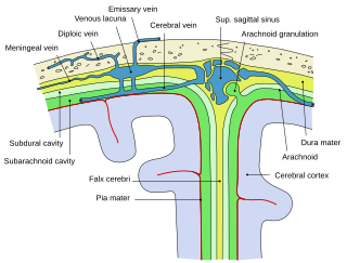
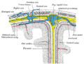
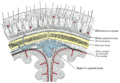
Arachnoid granulations are small outpouchings of the arachnoid mater and subarachnoid space into the dural venous sinuses of the brain. The granulations are thought to mediate the draining of cerebrospinal fluid (CSF) from the subarachnoid space into the venous system.

The dura mater, is the outermost of the three meningeal membranes. The dura mater has two layers, an outer periosteal layer closely adhered to the neurocranium, and an inner meningeal layer known as the dural border cell layer. The two dural layers are for the most part fused together forming a thick fibrous tissue membrane that covers the brain and the vertebrae of the spinal column. But the layers are separated at the dural venous sinuses to allow blood to drain from the brain. The dura covers the arachnoid mater and the pia mater the other two meninges in protecting the central nervous system.

The great cerebral vein is one of the large blood vessels in the skull draining the cerebrum of the brain. It is also known as the vein of Galen, named for its discoverer, the Greek physician Galen.

Cerebral circulation is the movement of blood through a network of cerebral arteries and veins supplying the brain. The rate of cerebral blood flow in an adult human is typically 750 milliliters per minute, or about 15% of cardiac output. Arteries deliver oxygenated blood, glucose and other nutrients to the brain. Veins carry "used or spent" blood back to the heart, to remove carbon dioxide, lactic acid, and other metabolic products. The neurovascular unit regulates cerebral blood flow so that activated neurons can be supplied with energy in the right amount and at the right time. Because the brain would quickly suffer damage from any stoppage in blood supply, the cerebral circulatory system has safeguards including autoregulation of the blood vessels. The failure of these safeguards may result in a stroke. The volume of blood in circulation is called the cerebral blood flow. Sudden intense accelerations change the gravitational forces perceived by bodies and can severely impair cerebral circulation and normal functions to the point of becoming serious life-threatening conditions.

In anatomy, the epidural space is the potential space between the dura mater and vertebrae (spine).
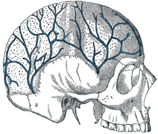
The diploic veins are large, thin-walled valveless veins that channel in the diploë between the inner and outer layers of the cortical bone in the skull, first identified in dogs by the anatomist Guillaume Dupuytren. A single layer of endothelium lines these veins supported by elastic tissue. They develop fully by the age of two years. The diploic veins drain this area into the dural venous sinuses. The four major trunks of the diploic veins found on each side of the head are frontal, anterior temporal, posterior temporal, and occipital diploic veins. They tend to be symmetrical, with the same pattern of large veins on each side of the skull. It has been suggested that the venous patterns they form resemble fingerprints in their individuality.

The falx cerebri is a large, crescent-shaped fold of dura mater that descends vertically into the longitudinal fissure to separate the cerebral hemispheres. It supports the dural sinuses that provide venous and CSF drainage from the brain. It is attached to the crista galli anteriorly, and blends with the tentorium cerebelli posteriorly.

The arachnoid mater is one of the three meninges, the protective membranes that cover the brain and spinal cord. It is so named because of its resemblance to a spider web. The arachnoid mater is a derivative of the neural crest mesoectoderm in the embryo.

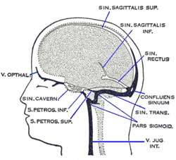
The dural venous sinuses are venous sinuses (channels) found between the periosteal and meningeal layers of dura mater in the brain. They receive blood from the cerebral veins, and cerebrospinal fluid (CSF) from the subarachnoid space via arachnoid granulations. They mainly empty into the internal jugular vein. Cranial venous sinuses communicate with veins outside the skull through emissary veins. These communications help to keep the pressure of blood in the sinuses constant.

The straight sinus, also known as tentorial sinus or the sinus rectus, is an area within the skull beneath the brain. It receives blood from the inferior sagittal sinus and the great cerebral vein, and drains into the confluence of sinuses.
The inferior sagittal sinus, within the human head, is an area beneath the brain which allows blood to drain outwards posteriorly from the center of the head. It drains to the straight sinus, which connects to the transverse sinuses. See diagram : labeled in the brain as "SIN. SAGITTALIS INF.".
The transverse sinuses, within the human head, are two areas beneath the brain which allow blood to drain from the back of the head. They run laterally in a groove along the interior surface of the occipital bone. They drain from the confluence of sinuses to the sigmoid sinuses, which ultimately connect to the internal jugular vein. See diagram : labeled under the brain as "SIN. TRANS.".

The occipital vein is a vein of the scalp. It originates from a plexus around the external occipital protuberance and superior nuchal line to the back part of the vertex of the skull. It usually drains into the internal jugular vein, but may also drain into the posterior auricular vein. It drains part of the scalp.

The squamous part of the frontal bone is the superior portion when viewed in standard anatomical orientation. There are two surfaces of the squamous part of the frontal bone: the external surface, and the internal surface.

The superior cerebral veins are several cerebral veins that drain the superolateral and superomedial surfaces of the cerebral hemispheres into the superior sagittal sinus. There are 8-12 cerebral veins. They are predominantly found in the sulci between the gyri, but can also be found running across the gyri.

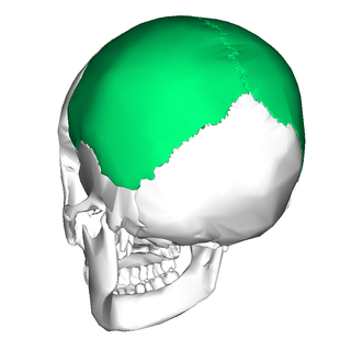
The calvaria is the top part of the skull. It is the superior part of the neurocranium and covers the cranial cavity containing the brain. It forms the main component of the skull roof.

The following outline is provided as an overview of and topical guide to human anatomy:

The middle cerebral veins - the superficial middle cerebral vein and the deep middle cerebral vein - are two veins running along the lateral sulcus. The superficial middle cerebral vein is also known as the superficial Sylvian vein, and the deep middle cerebral vein is also known as the deep Sylvian vein. The lateral sulcus is also known as the Sylvian fissure.