Falx cerebri

Last updated
Falx cerebri
Falxcerebri.jpg
Dura mater and its processes exposed by removing part of the right half of the skull and the brain.
Gray769.png
Diagrammatic representation of a section across the top of the skull, showing the membranes of the brain, etc. (Falx cerebri is yellow line running down center.)
Details
Part of Meninges
Identifiers
Latin falx cerebri
NeuroNames 1237
TA98 A14.1.01.103
TA2 5374
FMA 83967
Anatomical terms of neuroanatomy

The falx cerebri (also known as the cerebral falx) is a large, crescent-shaped fold of dura mater that descends vertically into the longitudinal fissure between the cerebral hemispheres of the human brain, [1] separating the two hemispheres and supporting dural sinuses that provide venous and CSF drainage to the brain. [2] It is attached to the crista galli anteriorly, and blends with the tentorium cerebelli posteriorly. [3]

Contents

The falx cerebri is often subject to age-related calcification, and a site of falcine meningiomas. [2]

The falx cerebri is named for its sickle-like shape.[ citation needed ]

Anatomy

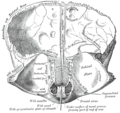
The falx cerebri is a strong, crescent-shaped sheet lying in the sagittal plane between the two hemispheres. [3] It is a dural formation (one of four dural partitions of the brain along with the falx cerebelli, tentorium cerebelli, and diaphragma sellae); it is formed through invagination of the dura mater into the longitudinal fissure between the cerebral hemispheres. [2]

Anteriorly, the falx cerebri is narrower, thinner, and may have a number of perforations. It is broader posteriorly. [3]

Attachments

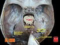
The falx cerebri attaches anteriorly at the crista galli (proximally to the cribriform plate and to the frontal and ethmoid sinuses). [1]

Posteriorly, it blends into the upper surface of the cerebellar tentorium. [3]

Its convex superior margin is attached to the internal surface of the skull on either side of the midline. This attachment runs as far back as the internal occipital protuberance (the latter representing its posterior-most point of attachment [2] ); the superior sagittal sinus runs in the cranial groove between the falx cerebri's two attachments. [3]

The (concave) inferior margin of the falx cerebri is free. [3]

Vascular supply

The falx cerebri receives its blood supply primarily from two vessels; the anterior portion receives blood supply from the anterior meningeal artery (a.k.a. anterior falx artery, or anterior falcine artery) (a branch of the anterior ethmoidal artery), and the posterior portion from the posterior meningeal artery (a branch of the ascending pharyngeal artery). [2]

Lymphatic drainage of the falx cerebri occurs mostly via meningeal lymphatic vessels that run parallel to the dural sinuses and that eventually exit the cranial vault through the jugular foramen to empty into deep cervical lymph nodes. A minority of lymph from the falx cerebri is drained anteriorly through the cribiform plate into the lymphatics of the nasal mucosa. [2]

Innervation

The falx cerebri receives innervaton from all three branches of the trigeminal nerve. It receives symphatetic innervation predominantly from the superior cervical ganglia. It may receive additional innervation from dorsal rami of CN 1 and CN 2, the hypoglossal nerve, and recurrent branches of the vagus nerve. [2]

Anatomical relations

The falx cerebri is situated in the longitudinal fissure, in between the cerebral hemispheres. [3] The corpus callosum lies immediately inferior to the lower (free) margin of falx cerebri. [2]

Dural venous sinuses

The superior sagittal sinus is contained in the superior margin of the falx cerebri and overlies the longitudinal fissure of the brain. [1]

The inferior sagittal sinus is contained in the inferior [1] free [3] margin of the falx cerebri and arches over the corpus callosum, deep within the longitudinal fissure. [1]

The straight sinus courses along the juncture of the falx cerebri and cerebellar tentorium. [3]

Anatomical variation

Total or partial agenesis of the falx cerebri may occur, and may result in adherence of the cerebral hemispheres across the midline. Agenesis is usually associated with other developmental complications; falx cerebri agenesis in absence of other neural symptoms is exceedingly rare. [2]

Microanatomy

The falx cerebri contains blood vessels, and nerves. [2]

Clinical significance

Calcification

Calcification of the falx cerebri is more prevalent in older patients, often without a determinable cause, and without pathogenic symptoms. [4]

Meningioma

Falcine meningioma is a meningioma arising from the falx cerebri and completely concealed by the overlying cortex. Falcine meningioma tends to grow predominantly into one cerebral hemisphere but is often bilateral, and in some patients the tumor grows into the inferior edge of the sagittal sinus. However, although much information is available regarding meningiomas, little is known about falcine meningiomas. [5]

Surgical landmark

The falx cerebri is a significant surgical landmark for access of the lateral ventricles via the interhemispheric transcallosal approach; agenesis (complete or partial) of the falx cerebri results in the adherence of the cerebral hemispheres, blocking midline transcallosal surgical access to the ventricles. [2]

Subfalcine brain herniation

Subfalcine herniation of the cingulate gyrus may occur following traumatic brain injury. [2]

Additional images

See also

Related Research Articles

Articles related to anatomy include:

<span class="mw-page-title-main">Meninges</span> Three membranes that envelop the brain and spinal cord

In anatomy, the meninges are the three membranes that envelop the brain and spinal cord. In mammals, the meninges are the dura mater, the arachnoid mater, and the pia mater. Cerebrospinal fluid is located in the subarachnoid space between the arachnoid mater and the pia mater. The primary function of the meninges is to protect the central nervous system.

<span class="mw-page-title-main">Internal carotid artery</span> Artery of the human brain

The internal carotid artery is an artery in the neck which supplies the anterior circulation of the brain.

<span class="mw-page-title-main">Dura mater</span> Outermost layer of the protective tissues around the central nervous system (meninges)

In neuroanatomy, dura mater is a thick membrane made of dense irregular connective tissue that surrounds the brain and spinal cord. It is the outermost of the three layers of membrane called the meninges that protect the central nervous system. The other two meningeal layers are the arachnoid mater and the pia mater. It envelops the arachnoid mater, which is responsible for keeping in the cerebrospinal fluid. It is derived primarily from the neural crest cell population, with postnatal contributions of the paraxial mesoderm.

<span class="mw-page-title-main">Great cerebral vein</span>

The great cerebral vein is one of the large blood vessels in the skull draining the cerebrum of the brain. It is also known as the vein of Galen, named for its discoverer, the Greek physician Galen.

<span class="mw-page-title-main">Cerebral circulation</span> Brain blood supply

Cerebral circulation is the movement of blood through a network of cerebral arteries and veins supplying the brain. The rate of cerebral blood flow in an adult human is typically 750 milliliters per minute, or about 15% of cardiac output. Arteries deliver oxygenated blood, glucose and other nutrients to the brain. Veins carry "used or spent" blood back to the heart, to remove carbon dioxide, lactic acid, and other metabolic products. The neurovascular unit regulates cerebral blood flow so that activated neurons can be supplied with energy in the right amount and at the right time. Because the brain would quickly suffer damage from any stoppage in blood supply, the cerebral circulatory system has safeguards including autoregulation of the blood vessels. The failure of these safeguards may result in a stroke. The volume of blood in circulation is called the cerebral blood flow. Sudden intense accelerations change the gravitational forces perceived by bodies and can severely impair cerebral circulation and normal functions to the point of becoming serious life-threatening conditions.

<span class="mw-page-title-main">Intracranial hemorrhage</span> Hemorrhage, or bleeding, within the skull

Intracranial hemorrhage (ICH), also known as intracranial bleed, is bleeding within the skull. Subtypes are intracerebral bleeds, subarachnoid bleeds, epidural bleeds, and subdural bleeds.

<span class="mw-page-title-main">Epidural space</span> Space between the dura mater and vertebrae

In anatomy, the epidural space is the potential space between the dura mater and vertebrae (spine).

<span class="mw-page-title-main">Cerebellar tentorium</span> Vertebrate brain structure separating the cerebellum from the occipital lobes

The cerebellar tentorium or tentorium cerebelli is an extension of the dura mater between the inferior aspect of the occipital lobes and the superior aspect of the cerebellum. The free border of the tentorium gives passage to the midbrain.

<span class="mw-page-title-main">Ophthalmic nerve</span> Sensory nerve of the face

The ophthalmic nerve (CN V1) is a sensory nerve of the head. It is one of three divisions of the trigeminal nerve (CN V), a cranial nerve. It has three major branches which provide sensory innervation to the eye, and the skin of the upper face and anterior scalp, as well as other structures of the head.

<span class="mw-page-title-main">Straight sinus</span> Venous sinus draining the brain

The straight sinus, also known as tentorial sinus or the sinus rectus, is an area within the skull beneath the brain. It receives blood from the inferior sagittal sinus and the great cerebral vein, and drains into the confluence of sinuses.

<span class="mw-page-title-main">Superior sagittal sinus</span> Anatomical structure of the brain

The superior sagittal sinus, within the human head, is an unpaired area along the attached margin of the falx cerebri. It allows blood to drain from the lateral aspects of anterior cerebral hemispheres to the confluence of sinuses. Cerebrospinal fluid drains through arachnoid granulations into the superior sagittal sinus and is returned to venous circulation.

<span class="mw-page-title-main">Inferior sagittal sinus</span> Anatomical structure of the brain

The inferior sagittal sinus, within the human head, is an area beneath the brain which allows blood to drain outwards posteriorly from the center of the head. It drains to the straight sinus, which connects to the transverse sinuses. See diagram : labeled in the brain as "SIN. SAGITTALIS INF.".

<span class="mw-page-title-main">Falx cerebelli</span> Skull anatomy

The falx cerebelli is a small sickle-shaped fold of dura mater projecting forwards into the posterior cerebellar notch as well as projecting into the vallecula of the cerebellum between the two cerebellar hemispheres.

<span class="mw-page-title-main">Lesser wing of sphenoid bone</span>

The lesser wings of the sphenoid or orbito-sphenoids are two thin triangular plates, which arise from the upper and anterior parts of the body, and, projecting lateralward, end in sharp points [Fig. 1].

<span class="mw-page-title-main">Transverse sinuses</span>

The transverse sinuses, within the human head, are two areas beneath the brain which allow blood to drain from the back of the head. They run laterally in a groove along the interior surface of the occipital bone. They drain from the confluence of sinuses to the sigmoid sinuses, which ultimately connect to the internal jugular vein. See diagram : labeled under the brain as "SIN. TRANS.".

<span class="mw-page-title-main">Squamous part of occipital bone</span>

The squamous part of occipital bone is situated above and behind the foramen magnum, and is curved from above downward and from side to side.

<span class="mw-page-title-main">Middle cranial fossa</span>

The middle cranial fossa is formed by the sphenoid bones, and the temporal bones. It lodges the temporal lobes, and the pituitary gland. It is deeper than the anterior cranial fossa, is narrow medially and widens laterally to the sides of the skull. It is separated from the posterior cranial fossa by the clivus and the petrous crest.

<span class="mw-page-title-main">Anterior cranial fossa</span>

The anterior cranial fossa is a depression in the floor of the cranial base which houses the projecting frontal lobes of the brain. It is formed by the orbital plates of the frontal, the cribriform plate of the ethmoid, and the small wings and front part of the body of the sphenoid; it is limited behind by the posterior borders of the small wings of the sphenoid and by the anterior margin of the chiasmatic groove. The lesser wings of the sphenoid separate the anterior and middle fossae.

<span class="mw-page-title-main">Outline of human anatomy</span> Overview of and topical guide to human anatomy

The following outline is provided as an overview of and topical guide to human anatomy:

References

PD-icon.svgThis article incorporates text in the public domain from page 873 of the 20th edition of Gray's Anatomy (1918)

  1. 1 2 3 4 5 Saladin K. "Anatomy & Physiology: The Unity of Form and Function. New York: McGraw Hill, 2014. Print. pp 512, 770-773
  2. 1 2 3 4 5 6 7 8 9 10 11 12 Bair, Michael M.; Munakomi, Sunil (2022), "Neuroanatomy, Falx Cerebri", StatPearls, Treasure Island (FL): StatPearls Publishing, PMID   31424888 , retrieved 2022-04-26
  3. 1 2 3 4 5 6 7 8 9 Standring, Susan (2020). Gray's Anatomy: The Anatomical Basis of Clinical Practice (42th ed.). New York. p. 398. ISBN   978-0-7020-7707-4. OCLC   1201341621.{{cite book}}: CS1 maint: location missing publisher (link)
  4. Daghighi MH, Rezaei V, Zarrintan S, Pourfathi H (2007). "Intracranial physiological calcifications in adults on computed tomography in Tabriz, Iran." Folia Morphol (Warsz).66 (2):115-9. PMID   17594669
  5. Chung SB, Kim CY, Park CK, Kim DG, Jung HW (2007). "Falx Meningiomas: Surgical Results and Lessons Learned from 68 Cases." J Korean Neurosurg Soc. 42 (4): 276-280. PMC   2588203