| Arachnoid granulation | |
|---|---|
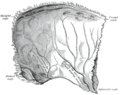
 Diagrammatic representation of a section across the top of the skull, showing the membranes of the brain, etc. ("Arachnoid granulation" label is at top right.) | |
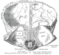
 Arachnoid granulations seen on autopsy, where the dura mater has been removed but the arachnoid mater is left in place. | |
| Details | |
| Identifiers | |
| Latin | granulationes arachnoideae |
| TA98 | A14.1.01.205 |
| TA2 | 5389 |
| FMA | 77760 |
| Anatomical terminology | |
Arachnoid granulations (also arachnoid villi, and Pacchionian granulations or bodies) are small outpouchings of the arachnoid mater and subarachnoid space into the dural venous sinuses of the brain. The granulations are thought to mediate the draining of cerebrospinal fluid (CSF) from the subarachnoid space into the venous system. [1]
Contents
- Anatomy
- Structure
- Development
- Function
- Cerebrospinal fluid resorption
- Subarachnoid systolic overpressure dampening
- Clinical significance
- Eponym
- References
- Additional images
The largest and most numerous granulations lie along the superior sagittal sinus; they are however present along other dural sinuses as well.