Subarachnoid cisterns

Last updated
Subarachnoid cisterns
Gray768.png
Diagram showing the positions of the three principal subarachnoid cisterns, cisterna magna is shown as cisterna cerebellomedullaris
Details
Identifiers
Latin cisterna subarachnoideum
Anatomical terminology

The subarachnoid cisterns are spaces formed by openings in the subarachnoid space, an anatomic space in the meninges of the brain. [1] The space is situated between the two meninges, the arachnoid mater and the pia mater. These cisterns are filled with cerebrospinal fluid (CSF). [1]

Contents

Structure

Although the pia mater adheres to the surface of the brain, closely following the contours of its gyri and sulci, the arachnoid mater only covers its superficial surface, bridging across the gyri. This leaves wider spaces between the pia and arachnoid and the cavities are known as the subarachnoid cisterns.

Although they are often described as distinct compartments, the subarachnoid cisterns are not truly anatomically distinct. Rather, these subarachnoid cisterns are separated from each other by a trabeculated porous wall with various-sized openings.

Cisterns

There are many cisterns in the brain with several large ones noted with their own name. At the base of the spinal cord is another subarachnoid cistern: the lumbar cistern which is the site for a lumbar puncture. Some major subarachnoid cisterns:

  1. The vertebral artery and the origin of the posterior inferior cerebellar artery (PICA)
  2. The ninth (IX), tenth (X), eleventh (XI) and twelfth (XII) cranial nerves
  1. The basilar artery and the origin of the anterior inferior cerebellar artery (AICA)
  2. The origin of the superior cerebellar arteries
  3. The sixth (VI) cranial nerve
  1. The optic chiasm
  2. The bifurcation of the basilar artery
  3. Peduncular segments of the posterior cerebral arteries (PCA)
  4. Peduncular segments of the superior cerebellar arteries
  5. Perforating branches of the PCA
  6. The posterior communicating arteries (PCoA)
  7. The basal vein
  8. The third (III) cranial nerve, which passes between the posterior cerebral and superior cerebellar arteries
  1. The seventh (VII) and eighth (VIII) cranial nerves
  2. The anteroinferior cerebellar artery (AICA)
  3. The fifth (V) cranial nerve and the petrosal vein
  1. The great cerebral vein
  2. The posterior pericallosal arteries
  3. The third portion of the superior cerebellar arteries
  4. Perforating branches of the posterior cerebral and superior cerebellar arteries
  5. The third portion of the posterior cerebral arteries
Its supratentorial portion contains:
  1. The basal vein
  2. The posterior cerebral artery
Its infratentorial portion contains:
  1. The superior cerebellar artery
  2. The fourth (IV) nerve
  1. The anterior choroidal artery
  2. The medial posterior choroidal artery
  3. The basal vein
  1. The internal carotid artery
  2. The origin of the anterior choroidal artery
  3. The origin of the posterior communicating artery
  1. The middle cerebral artery
  2. The middle cerebral veins
  3. The fronto-orbital veins
  4. Collaterals to the basal vein
  1. The anterior cerebral arteries (A1 and proximal A2)
  2. The anterior communicating artery
  3. Heubner's artery
  4. The hypothalamic arteries
  5. The origin of the fronto-orbital arteries

Clinical significance

It is clinically significant that cerebral arteries, veins, and cranial nerves traverse through the subarachnoid space, maintaining their meningeal covering until they exit the skull.

See also

Related Research Articles

<span class="mw-page-title-main">Cerebrospinal fluid</span> Clear, colorless bodily fluid found in the brain and spinal cord

Cerebrospinal fluid (CSF) is a clear, colorless body fluid found within the tissue that surrounds the brain and spinal cord of all vertebrates.

Articles related to anatomy include:

<span class="mw-page-title-main">Brainstem</span> Posterior part of the brain, adjoining and structurally continuous

The brainstem is the stalk-like part of the brain that interconnects the cerebrum and diencephalon with the spinal cord. In the human brain, the brainstem is composed of the midbrain, the pons, and the medulla oblongata. The midbrain is continuous with the thalamus of the diencephalon through the tentorial notch.

<span class="mw-page-title-main">Oculomotor nerve</span> Cranial nerve III, for eye movements

The oculomotor nerve, also known as the third cranial nerve, cranial nerve III, or simply CN III, is a cranial nerve that enters the orbit through the superior orbital fissure and innervates extraocular muscles that enable most movements of the eye and that raise the eyelid. The nerve also contains fibers that innervate the intrinsic eye muscles that enable pupillary constriction and accommodation. The oculomotor nerve is derived from the basal plate of the embryonic midbrain. Cranial nerves IV and VI also participate in control of eye movement.
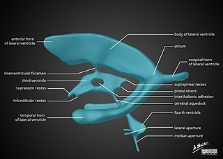
<span class="mw-page-title-main">Internal carotid artery</span> Artery supplying the brain

The internal carotid artery is an artery in the neck which supplies the anterior and middle cerebral circulation.

<span class="mw-page-title-main">Internal capsule</span> White matter structure situated in the inferomedial part of each cerebral hemisphere of the brain

The internal capsule is a white matter structure situated in the inferomedial part of each cerebral hemisphere of the brain. It carries information past the basal ganglia, separating the caudate nucleus and the thalamus from the putamen and the globus pallidus. The internal capsule contains both ascending and descending axons, going to and coming from the cerebral cortex. It also separates the caudate nucleus and the putamen in the dorsal striatum, a brain region involved in motor and reward pathways.

<span class="mw-page-title-main">Dura mater</span> Outermost layer of the protective tissues around the central nervous system (meninges)

In neuroanatomy, dura mater is a thick membrane made of dense irregular connective tissue that surrounds the brain and spinal cord. It is the outermost of the three layers of membrane called the meninges that protect the central nervous system. The other two meningeal layers are the arachnoid mater and the pia mater. It envelops the arachnoid mater, which is responsible for keeping in the cerebrospinal fluid. It is derived primarily from the neural crest cell population, with postnatal contributions of the paraxial mesoderm.

<span class="mw-page-title-main">Fourth ventricle</span> One of four central brain cavities filled with cerebrospinal fluid

The fourth ventricle is one of the four connected fluid-filled cavities within the human brain. These cavities, known collectively as the ventricular system, consist of the left and right lateral ventricles, the third ventricle, and the fourth ventricle. The fourth ventricle extends from the cerebral aqueduct to the obex, and is filled with cerebrospinal fluid (CSF).

<span class="mw-page-title-main">Cisterna magna</span> Space in the brain

The cisterna magna is the largest of the subarachnoid cisterns. It occupies the space created by the angle between the caudal/inferior surface of the cerebellum, and the dorsal/posterior surface of the medulla oblongata. The fourth ventricle communicates with the cistern via the unpaired midline median aperture. It is continuous inferiorly with the subarachnoid space of the spinal canal.

<span class="mw-page-title-main">Posterior cerebral artery</span> Artery which supplies blood to the occipital lobe of the brain

The posterior cerebral artery (PCA) is one of a pair of cerebral arteries that supply oxygenated blood to the occipital lobe, part of the back of the human brain. The two arteries originate from the distal end of the basilar artery, where it bifurcates into the left and right posterior cerebral arteries. These anastomose with the middle cerebral arteries and internal carotid arteries via the posterior communicating arteries.

<span class="mw-page-title-main">Cerebellopontine angle</span> Structure between the cerebellum and pons

The cerebellopontine angle (CPA) is located between the cerebellum and the pons. The cerebellopontine angle is the site of the cerebellopontine angle cistern.

<span class="mw-page-title-main">Middle cranial fossa</span>

The middle cranial fossa is formed by the sphenoid bones, and the temporal bones. It lodges the temporal lobes, and the pituitary gland. It is deeper than the anterior cranial fossa, is narrow medially and widens laterally to the sides of the skull. It is separated from the posterior cranial fossa by the clivus and the petrous crest.

<span class="mw-page-title-main">Tela choroidea</span>

The tela choroidea is a region of meningeal pia mater that adheres to the underlying ependyma, and gives rise to the choroid plexus in each of the brain’s four ventricles. Tela is Latin for woven and is used to describe a web-like membrane or layer. The tela choroidea is a very thin part of the loose connective tissue of pia mater overlying and closely adhering to the ependyma. It has a rich blood supply. The ependyma and vascular pia mater – the tela choroidea, form regions of minute projections known as a choroid plexus that projects into each ventricle. The choroid plexus produces most of the cerebrospinal fluid of the central nervous system that circulates through the ventricles of the brain, the central canal of the spinal cord, and the subarachnoid space. The tela choroidea in the ventricles forms from different parts of the roof plate in the development of the embryo.

<span class="mw-page-title-main">Pontine cistern</span> Subarachnoid cistern in front of the pons

The pontine cistern is a subarachnoid cistern situated ventrally/anteriorly to the pons. It contains the basilar artery. Each lateral aperture opens into the pontine cistern just posterior to the cranial nerve VIII.

<span class="mw-page-title-main">Interpeduncular cistern</span> Subarachnoid cistern above and in front of the pons

The interpeduncular cistern is the subarachnoid cistern situated between the dorsum sellae (anteriorly) and the two cerebral peduncles of the mesencephalon (midbrain). Its roof is represented by the floor of the third ventricle. Its floor is formed by the arachnoid membrane extending between the temporal lobes of either side. Anteriorly, it extends to the optic chiasm.

The quadrigeminal cistern is a subarachnoid cistern situated between splenium of corpus callosum, and the superior surface of the cerebellum. It contains a part of the great cerebral vein, the posterior cerebral artery, quadrigeminal artery, glossopharyngeal nerve, and the pineal gland.

The ambient cistern is a bilaterally paired subarachnoid cistern situated at either lateral aspect of the mesencephalon (midbrain). Each ambient cistern has a supratentorial compartment and an infratentorial compartment. Each is continuous anteriorly with the interpeduncular cistern, and posteriorly with the quadrigeminal cistern.

<span class="mw-page-title-main">Outline of human anatomy</span> Overview of and topical guide to human anatomy

The following outline is provided as an overview of and topical guide to human anatomy:

<span class="mw-page-title-main">Tentorial notch</span> Brain structure in humans and some mammals

The tentorial notch refers to the anterior opening between the free edge of the cerebellar tentorium and the clivus for the passage of the brainstem.

References

  1. 1 2 Purves, Dale (2011). Neuroscience (5th ed.). Sunderland, Mass.: Sinauer. p. 742. ISBN   978-087893-695-3.