
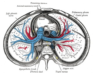
The thoracic cavity is the chamber of the body of vertebrates that is protected by the thoracic wall. The central compartment of the thoracic cavity is the mediastinum. There are two openings of the thoracic cavity, a superior thoracic aperture known as the thoracic inlet and a lower inferior thoracic aperture known as the thoracic outlet.

A body cavity is any space or compartment, or potential space in the animal body. Cavities accommodate organs and other structures; cavities as potential spaces contain fluid.

The pleural cavity also known as the pleural space, is the thin fluid-filled space between the two pulmonary pleurae of each lung. A pleura is a serous membrane which folds back onto itself to form a two-layered membranous pleural sac. The outer pleura is attached to the chest wall, but is separated from it by the endothoracic fascia. The inner pleura covers the lungs and adjoining structures, including blood vessels, bronchi and nerves. The pleural cavity can be viewed as a potential space because the two pleurae adhere to each other under all normal conditions.

The pericardium is a double-walled sac containing the heart and the roots of the great vessels. The pericardial sac has two layers, a serous layer and a fibrous layer. It encloses the pericardial cavity which contains pericardial fluid.

The retroperitoneal space (retroperitoneum) is the anatomical space in the abdominal cavity behind (retro) the peritoneum. It has no specific delineating anatomical structures. Organs are retroperitoneal if they have peritoneum on their anterior side only. Structures that are not suspended by mesentery in the abdominal cavity and that lie between the parietal peritoneum and abdominal wall are classified as retroperitoneal.
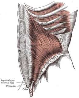
A fascia is a band or sheet of connective tissue, primarily collagen, beneath the skin that attaches, stabilizes, encloses, and separates muscles and other internal organs. Fascia is classified by layer, as superficial fascia, deep fascia, and visceral or parietal fascia, or by its function and anatomical location.

Dura mater is a thick membrane made of dense irregular connective tissue that surrounds the brain and spinal cord. It is the outermost of the three layers of membrane called the meninges that protect the central nervous system. The other two meningeal layers are the arachnoid mater and the pia mater. The dura surrounds the brain and the spinal cord and is responsible for keeping in the cerebrospinal fluid. It is derived primarily from the neural crest cell population, with postnatal contributions of the paraxial mesoderm.
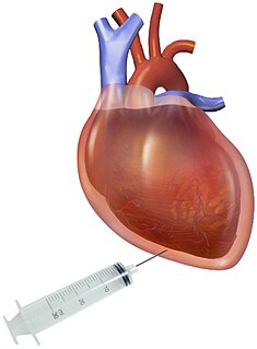
In medicine, pericardiocentesis (PCC) is a procedure where fluid is aspirated from the pericardium.
The peritoneal cavity is a true space between the parietal peritoneum and visceral peritoneum. Both the parietal and visceral peritonea are not different but the same peritoneum given two names depending on their function/location. It is one of the spaces derived from the coelomic cavity of the embryo, the others being the pleural cavities around the lungs and the pericardial cavity around the heart.

In anatomy, serous membrane is a smooth tissue membrane consisting of two layers of mesothelium, which secrete serous fluid. The inner layer that covers organs (viscera) in body cavities is called the visceral membrane. A second layer of epithelial cells of the serous membrane, called the parietal layer, lines the body wall. Between the two layers is a potential space, mostly empty except for a few milliliters of lubricating serous fluid that is secreted by the two serous membranes.

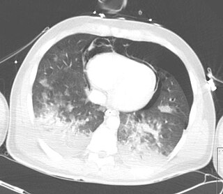
A hemothorax is an accumulation of blood within the pleural cavity. The symptoms of a hemothorax include chest pain and difficulty breathing, while the clinical signs include reduced breath sounds on the affected side and a rapid heart rate. Hemothoraces are usually caused by an injury but may occur spontaneously: due to cancer invading the pleural cavity, as a result of a blood clotting disorder, as an unusual manifestation of endometriosis, in response to a collapsed lung, or rarely in association with other conditions.

Pericardial effusion is an abnormal accumulation of fluid in the pericardial cavity. Because of the limited amount of space in the pericardial cavity, fluid accumulation leads to an increased intrapericardial pressure which can negatively affect heart function. A pericardial effusion with enough pressure to adversely affect heart function is called cardiac tamponade. Pericardial effusion usually results from a disturbed equilibrium between the production and re-absorption of pericardial fluid, or from a structural abnormality that allows fluid to enter the pericardial cavity.
Fremitus is a vibration transmitted through the body. In common medical usage, it usually refers to assessment of the lungs by either the vibration intensity felt on the chest wall and/or heard by a stethoscope on the chest wall with certain spoken words, although there are several other types.
Pericardial fluid is the serous fluid secreted by the serous layer of the pericardium into the pericardial cavity. The pericardium consists of two layers, an outer fibrous layer and the inner serous layer. This serous layer has two membranes which enclose the pericardial cavity into which is secreted the pericardial fluid. The fluid is similar to the cerebrospinal fluid of the brain which also serves to cushion and allow some movement of the LUND/ LUND # * {{}}''''

Lateral plate mesoderm is a type of mesoderm that is found at the periphery of the embryo.
In anatomy, a potential space is a space between two adjacent structures that are normally pressed together. The pleural space, between the visceral and parietal pleura of the lung, is a potential space. Though it only contains a small amount of fluid normally, it can sometimes accumulate fluid or air that widens the space. The pericardial space is another potential space that may fill with fluid (effusion) in certain disease states (e.g. pericarditis; a large pericardial effusion may result in cardiac tamponade.
Pneumopericardium is a medical condition where air enters the pericardial cavity. This condition has been recognized in preterm neonates, in which it is associated with severe lung pathology, after vigorous resuscitation, or in the presence of assisted ventilation. This is a serious complication, which if untreated may lead to cardiac tamponade and death. Pneumomediastinum, which is the presence of air in the mediastinum, may mimic and also coexist with pneumopericardium.

The excretory system of gastropods removes nitrogenous waste and maintains the internal water balance of these creatures, commonly referred to as snails and slugs. The primary organ of excretion is a nephridium.
A pericardial window is a cardiac surgical procedure to create a fistula – or "window" – from the pericardial space to the pleural cavity. The purpose of the window is to allow a pericardial effusion to drain from the space surrounding the heart into the chest cavity – where the fluid is not as dangerous; an untreated pericardial effusion can lead to cardiac tamponade and death.

Anatomical terminology is a form of scientific terminology used by anatomists, zoologists, and health professionals such as doctors.