The danger triangle of the face consists of the area from the corners of the mouth to the bridge of the nose, including the nose and maxilla. Due to the special nature of the blood supply to the human nose and surrounding area, it is possible for retrograde infection from the nasal area to spread to the brain, causing cavernous sinus thrombosis, meningitis, or brain abscess.
Articles related to anatomy include:

The levator labii superioris is a muscle of the human body used in facial expression. It is a broad sheet, the origin of which extends from the side of the nose to the zygomatic bone.

The facial artery, formerly called the external maxillary artery, is a branch of the external carotid artery that supplies blood to superficial structures of the medial regions of the face.

The cavernous sinus within the human head is one of the dural venous sinuses creating a cavity called the lateral sellar compartment bordered by the temporal bone of the skull and the sphenoid bone, lateral to the sella turcica.
In facial anatomy, the modiolus is a dense, compact, mobile, fibromuscular tissue mass of facial muscles formed by the interlacing of a number of muscles just lateral to the angle of the mouth opposite the second upper premolar tooth.

The superior ophthalmic vein is a vein of the orbit that drains venous blood from structures of the upper orbit. It is formed by the union of the angular vein, and supraorbital vein. It passes backwards within the orbit alongside the ophthalmic artery, then exits the orbit through the superior orbital fissure to drain into the cavernous sinus.

The facial vein is a relatively large vein in the human face. It commences at the side of the root of the nose and is a direct continuation of the angular vein where it also receives a small nasal branch.

The infraorbital artery is a small artery in the head that arises from the maxillary artery and passes through the inferior orbital fissure to enter the orbit, then passes forward along the floor of the orbit, finally exiting the orbit through the infraorbital foramen to reach the face.

The buccal branches of the facial nerve, are of larger size than the rest of the branches, pass horizontally forward to be distributed below the orbit and around the mouth.

The buccal space is a fascial space of the head and neck. It is a potential space in the cheek, and is paired on each side. The buccal space is superficial to the buccinator muscle and deep to the platysma muscle and the skin. The buccal space is part of the subcutaneous space, which is continuous from head to toe.

The following outline is provided as an overview of and topical guide to human anatomy:
Mouth infections, also known as oral infections, are a group of infections that occur around the oral cavity. They include dental infection, dental abscess, and Ludwig's angina. Mouth infections typically originate from dental caries at the root of molars and premolars that spread to adjacent structures. In otherwise healthy patients, removing the offending tooth to allow drainage will usually resolve the infection. In cases that spread to adjacent structures or in immunocompromised patients, surgical drainage and systemic antibiotics may be required in addition to tooth extraction. Since bacteria that normally reside in the oral cavity cause mouth infections, proper dental hygiene can prevent most cases of infection. As such, mouth infections are more common in populations with poor access to dental care or populations with health-related behaviors that damage one's teeth and oral mucosa. This is a common problem, representing nearly 36% of all encounters within the emergency department related to dental conditions.

The sublingual space is a fascial space of the head and neck. It is a potential space located below the mouth and above the mylohyoid muscle, and is part of the suprahyoid group of fascial spaces.

The submental space is a fascial space of the head and neck. It is a potential space located between the mylohyoid muscle superiorly, the platysma muscle inferiorly, under the chin in the midline. The space coincides with the anatomic region termed the submental triangle, part of the anterior triangle of the neck.

The mental space is a fascial space of the head and neck. It is a potential space, bilaterally located in the chin, between the mentalis muscle superiorly and the platysma muscle inferiorly. These spaces may be created by pathology, e.g., the spread of odontogenic infection. Commonly the origin of the infection is an anterior mandibular tooth with associated periapical abscess which erodes through the buccal cortical plate of the mandibular at a level below the attachment of the mentalis muscle. The mental space also has the mental foramen located laterally on both the right and left sides. This is important as many mandibular nerves pass through these foremens.
Fascial spaces are potential spaces that exist between the fasciae and underlying organs and other tissues. In health, these spaces do not exist; they are only created by pathology, e.g. the spread of pus or cellulitis in an infection. The fascial spaces can also be opened during the dissection of a cadaver. The fascial spaces are different from the fasciae themselves, which are bands of connective tissue that surround structures, e.g. muscles. The opening of fascial spaces may be facilitated by pathogenic bacterial release of enzymes which cause tissue lysis. The spaces filled with loose areolar connective tissue may also be termed clefts. Other contents such as salivary glands, blood vessels, nerves and lymph nodes are dependent upon the location of the space. Those containing neurovascular tissue may also be termed compartments.

The submasseterric space is a fascial space of the head and neck. It is a potential space in the face over the angle of the jaw, and is paired on each side. It is located between the lateral aspect of the mandible and the medial aspect of the masseter muscle and its investing fascia. The term is derived from sub- meaning "under" in Latin and masseteric which refers to the masseter muscle. The submasseteric space is one of the four compartments of the masticator space. Sometimes the submasseteric space is described as a series of spaces, created because the masseter muscle has multiple insertions that cover most of the lateral surface of the ramus of the mandible.
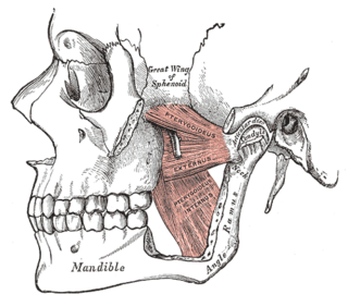
The pterygomandibular space is a fascial space of the head and neck. It is a potential space in the head and is paired on each side. It is located between the medial pterygoid muscle and the medial surface of the ramus of the mandible. The pterygomandibular space is one of the four compartments of the masticator space.

The infratemporal space is a fascial space of the head and neck. It is a potential space in the side of the head, and is paired on either side. It is located posterior to the maxilla, between the lateral pterygoid plate of the sphenoid bone medially and by the base of skull superiorly. The term is derived from infra- meaning below and temporal which refers to the temporalis muscle.