The parotid duct is formed when several interlobular ducts, the largest ducts inside the parotid gland, join. It emerges from the parotid gland. It runs forward along the lateral side of the masseter muscle for around 7 cm.[2] In this course, the duct is surrounded by the buccal fat pad.[2][3] It takes a steep turn at the border of the masseter muscle and passes through the buccinator muscle, opening into the vestibule of the mouth, the region of the mouth between the cheek and the gums, at the parotid papilla, which lies opposite to the second maxillary (upper) molar tooth. The parotid papillae can be palpated as small raised tissue area (papillae) on both sides of the mouth and protects the opening of the parotid duct. [4]
The buccinator acts as a valve that prevents air forcing into the duct, which would cause pneumoparotitis.[5]
Koplik's spots which are pathognomonic of measles are found near the opening of the parotid duct.
The parotid duct may be cannulated by inserting a tube through the internal orifice in the mouth.[2] Dye may be injected to allow for imaging of the parotid duct.[2]
History
Niels Stensen (also known as Nicolas Steno), a Danishanatomist (albeit best known as a geologist) is credited with the first detailed description of the duct in 1660,[6] hence the origin of its alternative name "Stensen duct".[2][6]
Additional images
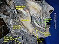
Outline of side of face, showing chief surface markings.
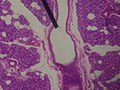
Microscopic slide of a human interlobular duct.
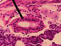
Microscopic slide of a human striated duct.
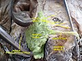
The left papilla (soft tissue protuberance at the exit) of the parotid duct is clearly visible on the cheek in the right of the photo.
Stensen N (1662). Observationes anatomicae, quibus varia oris, oculorum & narium vas describuntur novique salivae, lacrymarum & muci fontes deteguntur. Lugduni Batavorum: J. Chouët.
This page is based on this Wikipedia article Text is available under the CC BY-SA 4.0 license; additional terms may apply. Images, videos and audio are available under their respective licenses.