
The facial nerve, also known as the seventh cranial nerve, cranial nerve VII, or simply CN VII, is a cranial nerve that emerges from the pons of the brainstem, controls the muscles of facial expression, and functions in the conveyance of taste sensations from the anterior two-thirds of the tongue. The nerve typically travels from the pons through the facial canal in the temporal bone and exits the skull at the stylomastoid foramen. It arises from the brainstem from an area posterior to the cranial nerve VI and anterior to cranial nerve VIII.

The great auricular nerve is a cutaneous (sensory) nerve of the head. It originates from the second and third cervical (spinal) nerves (C2-C3) of the cervical plexus. It provides sensory innervation to the skin over the parotid gland and the mastoid process, parts of the outer ear, and to the parotid gland and its fascia.

In neuroanatomy, the mandibular nerve (V3) is the largest of the three divisions of the trigeminal nerve, the fifth cranial nerve (CN V). Unlike the other divisions of the trigeminal nerve (ophthalmic nerve, maxillary nerve) which contain only afferent fibers, the mandibular nerve contains both afferent and efferent fibers. These nerve fibers innervate structures of the lower jaw and face, such as the tongue, lower lip, and chin. The mandibular nerve also innervates the muscles of mastication.

The buccinator is a thin quadrilateral muscle occupying the interval between the maxilla and the mandible at the side of the face. It forms the anterior part of the cheek or the lateral wall of the oral cavity.

The inferior alveolar nerve (IAN) (also the inferior dental nerve) is a sensory branch of the mandibular nerve (CN V3) (which is itself the third branch of the trigeminal nerve (CN V)). The nerve provides sensory innervation to the lower/mandibular teeth and their corresponding gingiva as well as a small area of the face (via its mental nerve).

The auriculotemporal nerve is a sensory branch of the mandibular nerve (CN V3) that runs with the superficial temporal artery and vein, and provides sensory innervation to parts of the external ear, scalp, and temporomandibular joint. The nerve also conveys post-ganglionic parasympathetic fibres from the otic ganglion to the parotid gland.

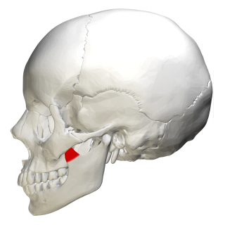
The depressor labii inferioris is a facial muscle. It helps to lower the bottom lip.
The lingual nerve carries sensory innervation from the anterior two-thirds of the tongue. It contains fibres from both the mandibular division of the trigeminal nerve (CN V3) and from the facial nerve (CN VII). The fibres from the trigeminal nerve are for touch, pain and temperature (general sensation), and the ones from the facial nerve are for taste (special sensation).
The mental nerve is a sensory nerve of the face. It is a branch of the posterior trunk of the inferior alveolar nerve, itself a branch of the mandibular nerve (CN V3), itself a branch of the trigeminal nerve (CN V). It provides sensation to the front of the chin and the lower lip, as well as the gums of the anterior mandibular (lower) teeth. It can be blocked with local anaesthesia for procedures on the chin, lower lip, and mucous membrane of the inner cheek. Problems with the nerve cause chin numbness.
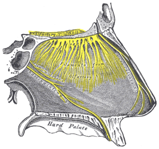
The nasopalatine nerve (also long sphenopalatine nerve) is a nerve of the head. It is a sensory branch of the maxillary nerve (CN V2) that passes through the pterygopalatine ganglion (without synapsing) and then through the sphenopalatine foramen to enter the nasal cavity, and finally out of the nasal cavity through the incisive canal and then the incisive fossa to enter the hard palate. It provides sensory innervation to the posteroinferior part of the nasal septum, and gingiva just posterior to the upper incisor teeth.

In human anatomy, the mandibular canal is a canal within the mandible that contains the inferior alveolar nerve, inferior alveolar artery, and inferior alveolar vein. It runs obliquely downward and forward in the ramus, and then horizontally forward in the body, where it is placed under the alveoli and communicates with them by small openings.
The mylohyoid nerve is a mixed nerve of the head. It is a branch of the inferior alveolar nerve. It provides motor innervation the mylohyoid muscle, and the anterior belly of the digastric muscle. It provides sensory innervation to part of the submental area, and sometimes also the mandibular (lower) molar teeth, requiring local anaesthesia for some oral procedures.

The infratemporal fossa is an irregularly shaped cavity that is a part of the skull. It is situated below and medial to the zygomatic arch. It is not fully enclosed by bone in all directions. It contains superficial muscles, including the lower part of the temporalis muscle, the lateral pterygoid muscle, and the medial pterygoid muscle. It also contains important blood vessels such as the middle meningeal artery, the pterygoid plexus, and the retromandibular vein, and nerves such as the mandibular nerve (CN V3) and its branches.

The buccal branches of the facial nerve, are of larger size than the rest of the branches, pass horizontally forward to be distributed below the orbit and around the mouth.

The buccal space is a fascial space of the head and neck. It is a potential space in the cheek, and is paired on each side. The buccal space is superficial to the buccinator muscle and deep to the platysma muscle and the skin. The buccal space is part of the subcutaneous space, which is continuous from head to toe.
In human anatomy, the mandible's coronoid process is a thin, triangular eminence, which is flattened from side to side and varies in shape and size. Its anterior border is convex and is continuous below with the anterior border of the ramus. Its posterior border is concave and forms the anterior boundary of the mandibular notch. The lateral surface is smooth, and affords insertion to the temporalis and masseter muscles. Its medial surface gives insertion to the temporalis, and presents a ridge which begins near the apex of the process and runs downward and forward to the inner side of the last molar tooth.
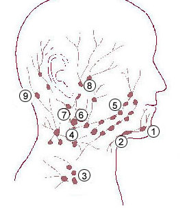
The submandibular lymph nodes, are some 3-6 lymph nodes situated at the inferior border of the ramus of mandible.
Inferior alveolar nerve block is a nerve block technique which induces anesthesia (numbness) in the areas of the mouth and face innervated by one of the inferior alveolar nerves which are paired on the left and right side. These areas are the skin and mucous membranes of the lower lip, the skin of the chin, the lower teeth and the labial gingiva of the anterior teeth, all unilaterally to the midline of the side on which the block is administered. However, depending on technique, the long buccal nerve may not be anesthetized by an IANB and therefore an area of buccal gingiva adjacent to the lower posterior teeth will retain normal sensation unless that nerve is anesthetized separately, via a (long) buccal nerve block. The inferior alveolar nerve is a branch of the mandibular nerve, the third division of the trigeminal nerve. This procedure attempts to anaesthetise the inferior alveolar nerve prior to it entering the mandibular foramen on the medial surface of the mandibular ramus.

In human anatomy, the mouth is the first portion of the alimentary canal that receives food and produces saliva. The oral mucosa is the mucous membrane epithelium lining the inside of the mouth.
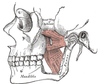
The pterygomandibular space is a fascial space of the head and neck. It is a potential space in the head and is paired on each side. It is located between the lateral pterygoid muscle and the medial surface of the ramus of the mandible. The pterygomandibular space is one of the four compartments of the masticator space.


