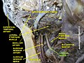
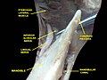
Lingual nerve

Last updated
Lingual nerve
Gray778.png
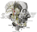
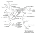
Distribution of the maxillary and mandibular nerves, and the submaxillary ganglion.
Gray1202.png
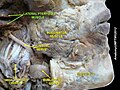
The mouth cavity. The apex of the tongue is turned upward, and on the right side a superficial dissection of its under surface has been made.
Details
From mandibular nerve
Innervates tongue
Identifiers
Latin nervus lingualis
MeSH D008036
TA98 A14.2.01.081
TA2 6267
FMA 53218
Anatomical terms of neuroanatomy

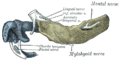
The lingual nerve carries sensory innervation from the anterior two-thirds of the tongue. It contains fibres from both the mandibular division of the trigeminal nerve (CN V3) and from the facial nerve (CN VII). The fibres from the trigeminal nerve are for touch, pain and temperature (general sensation), and the ones from the facial nerve are for taste (special sensation).

Contents

Structure

Origin

The lingual nerve arises from the posterior trunk of mandibular nerve (CN V3) [1] within the infratemporal fossa.[ citation needed ]

Course

The lingual nerve first courses deep to the lateral pterygoid muscle and superior to the tensor veli palatini muscle; while passing between these two muscles, it is joined by the chorda tympani, and often by a communicating branch from the inferior alveolar nerve. [1]

The nerve then comes to pass inferoanteriorly upon the medial pterygoid muscle towards the medial aspect of the ramus of mandible, eventually meeting the mandible at the junction of the ramus and body of mandible. Here, the lingual nerve is anterior and somewhat medial (deep) to the inferior alveolar nerve. [1]

It crosses obliquely to the side of the tongue beneath the constrictor pharyngis superior and styloglossus, and then between the hyoglossus and deep part of the submandibular gland; it finally runs from laterally to medially inferiorly crossing the duct of the submandibular gland, and along the tongue to its tip becoming the sublingual nerve, lying immediately beneath the mucous membrane.[ citation needed ]

The submandibular ganglion is suspended by two nerve filaments from the lingual nerve.[ citation needed ]

Distribution

General sensory

The lingual nerve supplies general somatic afferent (i.e. general sensory) innervation to the mucous membrane of the anterior two-thirds of the tongue (i.e. body of tongue) (whereas the posterior one-third (i.e. root of tongue) is innervated via the glossopharyngeal nerve (CN IX) [ citation needed ]), the floor of the oral cavity, and the mandibular/inferior lingual gingiva. [1]

Special sensory and parasymathetic autonomic

The lingual nerve also comes to convey fibres of the chorda tympani (which are derived from the facial nerve (CN VII)), which provide special sensation (taste) to the anterior two-thirds of the tongue as well as parasympathetic and sympathetic innervation.[ citation needed ]

Clinical significance

Lingual nerve injuries

The most common cause of lingual nerve injuries is third molar (wisdom tooth) surgery, less commonly the lingual nerve can be injured by local anaesthetic dental injections (particularly inferior dental block injections) and sublingual or submandibular surgery. [2]

Any injury to sensory nerves can result in pain, altered sensation and/or numbness, but usually a combination of all three symptoms arises. This can have a significant negative effect on the patient's quality of life affecting their daily function and psychological health. [3]

Patients should be routinely warned about lingual nerve injuries prior to wisdom tooth and floor of mouth surgery. The risk associated with wisdom tooth surgery is commonly accepted to be 2% temporary and 0.2% permanent. [4]

Warning patients of nerve injury prior to administration of deep dental injections has a risk of injury in approximately 1:14,000 with 25% of these remaining persistent.[ citation needed ] Preoperative warning about these injuries is routinely undertaken in the US and Germany. This reflects good practice recommended by the Royal College of Anaesthetists (prior warning of potential nerve injury in relation to spinal and epidural blocks 1 on 24–57,000 risk). [5] [ failed verification ]

Infiltration dentistry is a technique that may reduce the possibility of lingual nerve injuries by avoiding deep injections. [6]

Avoiding lingual access when undertaking wisdom tooth surgery will also avoid unnecessary lingual nerve injury [7] [8] [9]

See also

Additional images

Related Research Articles

<span class="mw-page-title-main">Facial nerve</span> Cranial nerve VII, for the face and tasting

The facial nerve, also known as the seventh cranial nerve, cranial nerve VII, or simply CN VII, is a cranial nerve that emerges from the pons of the brainstem, controls the muscles of facial expression, and functions in the conveyance of taste sensations from the anterior two-thirds of the tongue. The nerve typically travels from the pons through the facial canal in the temporal bone and exits the skull at the stylomastoid foramen. It arises from the brainstem from an area posterior to the cranial nerve VI and anterior to cranial nerve VIII.

<span class="mw-page-title-main">Mandibular nerve</span> Branch of the trigeminal nerve responsible for the lower face and jaw

In neuroanatomy, the mandibular nerve (V3) is the largest of the three divisions of the trigeminal nerve, the fifth cranial nerve (CN V). Unlike the other divisions of the trigeminal nerve (ophthalmic nerve, maxillary nerve) which contain only afferent fibers, the mandibular nerve contains both afferent and efferent fibers. These nerve fibers innervate structures of the lower jaw and face, such as the tongue, lower lip, and chin. The mandibular nerve also innervates the muscles of mastication.

<span class="mw-page-title-main">Submandibular gland</span> Human salivary gland

The paired submandibular glands are major salivary glands located beneath the floor of the mouth. In adult humans, they each weigh about 15 grams and contribute some 60–67% of unstimulated saliva secretion; on stimulation their contribution decreases in proportion as parotid gland secretion rises to 50%. The average length of the normal adult human submandibular salivary gland is approximately 27 mm, while the average width is approximately 14.3 mm.

<span class="mw-page-title-main">Digastric muscle</span> Small muscle located under the jaw in mammals

The digastric muscle is a bilaterally paired suprahyoid muscle located under the jaw. Its posterior belly is attached to the mastoid notch of temporal bone, and its anterior belly is attached to the digastric fossa of mandible; the two bellies are united by an intermediate tendon which is held in a loop that attaches to the hyoid bone. The anterior belly is innervated via the mandibular nerve, and the posterior belly is innervated via the facial nerve. It may act to depress the mandible or elevate the hyoid bone.

<span class="mw-page-title-main">Inferior alveolar nerve</span> Branch of the mandibular nerve

The inferior alveolar nerve (IAN) (also the inferior dental nerve) is a sensory branch of the mandibular nerve (CN V3) (which is itself the third branch of the trigeminal nerve (CN V)). The nerve provides sensory innervation to the lower/mandibular teeth and their corresponding gingiva as well as a small area of the face (via its mental nerve).

<span class="mw-page-title-main">Chorda tympani</span> Nerve carrying taste sensations

Chorda tympani is a branch of the facial nerve that carries gustatory (taste) sensory innervation from the front of the tongue and parasympathetic (secretomotor) innervation to the submandibular and sublingual salivary glands.

<span class="mw-page-title-main">Mylohyoid muscle</span> Paired muscle of the neck

The mylohyoid muscle or diaphragma oris is a paired muscle of the neck. It runs from the mandible to the hyoid bone, forming the floor of the oral cavity of the mouth. It is named after its two attachments near the molar teeth. It forms the floor of the submental triangle. It elevates the hyoid bone and the tongue, important during swallowing and speaking.

<span class="mw-page-title-main">Dental extraction</span> Operation to remove a tooth

A dental extraction is the removal of teeth from the dental alveolus (socket) in the alveolar bone. Extractions are performed for a wide variety of reasons, but most commonly to remove teeth which have become unrestorable through tooth decay, periodontal disease, or dental trauma, especially when they are associated with toothache. Sometimes impacted wisdom teeth cause recurrent infections of the gum (pericoronitis), and may be removed when other conservative treatments have failed. In orthodontics, if the teeth are crowded, healthy teeth may be extracted to create space so the rest of the teeth can be straightened.

<span class="mw-page-title-main">Buccal nerve</span> Nerve in the human face

The buccal nerve is a sensory nerve of the face arising from the mandibular nerve. It conveys sensory information from the skin of the cheek, and parts of the oral mucosa, periodontium, and gingiva.

<span class="mw-page-title-main">Inferior alveolar artery</span>

The inferior alveolar artery is an artery of the head. It is a branch of the maxillary artery. It descends through the infratemporal fossa as part of a neurovascular bundle with the inferior alveolar nerve and vein to the mandibular foramen where it enters and passes anteriorly inside the mandible, suplying the body of mandible and the dental pulp of the lower molar and premolar teeth. Its terminal incisor branch supplies the rest of the lower teeth. Its mental branch exits the mandibula anteriorly through the mental foramen to supply adjacent lip and skin.

<span class="mw-page-title-main">Mandibular canal</span>

In human anatomy, the mandibular canal is a canal within the mandible that contains the inferior alveolar nerve, inferior alveolar artery, and inferior alveolar vein. It runs obliquely downward and forward in the ramus, and then horizontally forward in the body, where it is placed under the alveoli and communicates with them by small openings.

<span class="mw-page-title-main">Mylohyoid nerve</span> Nerve of the head

The mylohyoid nerve is a mixed nerve of the head. It is a branch of the inferior alveolar nerve. It provides motor innervation the mylohyoid muscle, and the anterior belly of the digastric muscle. It provides sensory innervation to part of the submental area, and sometimes also the mandibular (lower) molar teeth, requiring local anaesthesia for some oral procedures.

<span class="mw-page-title-main">Infratemporal fossa</span> Cavity that is part of the skull

The infratemporal fossa is an irregularly shaped cavity that is a part of the skull. It is situated below and medial to the zygomatic arch. It is not fully enclosed by bone in all directions. It contains superficial muscles, including the lower part of the temporalis muscle, the lateral pterygoid muscle, and the medial pterygoid muscle. It also contains important blood vessels such as the middle meningeal artery, the pterygoid plexus, and the retromandibular vein, and nerves such as the mandibular nerve (CN V3) and its branches.

<span class="mw-page-title-main">Marginal mandibular branch of the facial nerve</span>

The marginal mandibular branch of the facial nerve arises from the facial nerve in the parotid gland at the parotid plexus. It passes anterior-ward deep to the platysma and depressor anguli oris muscles. It provides motor innervation to muscles of the lower lip and chin: the depressor labii inferioris muscle, depressor anguli oris muscle, and mentalis muscle. It communicates with the mental branch of the inferior alveolar nerve.

<span class="mw-page-title-main">Coronoid process of the mandible</span>

In human anatomy, the mandible's coronoid process is a thin, triangular eminence, which is flattened from side to side and varies in shape and size. Its anterior border is convex and is continuous below with the anterior border of the ramus. Its posterior border is concave and forms the anterior boundary of the mandibular notch. The lateral surface is smooth, and affords insertion to the temporalis and masseter muscles. Its medial surface gives insertion to the temporalis, and presents a ridge which begins near the apex of the process and runs downward and forward to the inner side of the last molar tooth.

Dental anesthesia is the application of anesthesia to dentistry. It includes local anesthetics, sedation, and general anesthesia.

Inferior alveolar nerve block is a nerve block technique which induces anesthesia (numbness) in the areas of the mouth and face innervated by one of the inferior alveolar nerves which are paired on the left and right side. These areas are the skin and mucous membranes of the lower lip, the skin of the chin, the lower teeth and the labial gingiva of the anterior teeth, all unilaterally to the midline of the side on which the block is administered. However, depending on technique, the long buccal nerve may not be anesthetized by an IANB and therefore an area of buccal gingiva adjacent to the lower posterior teeth will retain normal sensation unless that nerve is anesthetized separately, via a (long) buccal nerve block. The inferior alveolar nerve is a branch of the mandibular nerve, the third division of the trigeminal nerve. This procedure attempts to anaesthetise the inferior alveolar nerve prior to it entering the mandibular foramen on the medial surface of the mandibular ramus.

<span class="mw-page-title-main">Mandible</span> Lower jaw bone

In jawed vertebrates, the mandible, lower jaw, or jawbone is a bone that makes up the lower – and typically more mobile – component of the mouth.

<span class="mw-page-title-main">Human mouth</span> Part of human anatomy

In human anatomy, the mouth is the first portion of the alimentary canal that receives food and produces saliva. The oral mucosa is the mucous membrane epithelium lining the inside of the mouth.

<span class="mw-page-title-main">Pterygomandibular space</span>

The pterygomandibular space is a fascial space of the head and neck. It is a potential space in the head and is paired on each side. It is located between the lateral pterygoid muscle and the medial surface of the ramus of the mandible. The pterygomandibular space is one of the four compartments of the masticator space.

References

PD-icon.svgThis article incorporates text in the public domain from page 895 of the 20th edition of Gray's Anatomy (1918)

  1. 1 2 3 4 Mancall, Elliott L.; Brock, David G.; Gray, Henry, eds. (2011). Grays Clinical Neuroanatomy: The Anatomic Basis for Clinical Neuroscience. Philadelphia, PA: Elsevier Saunders. pp. 192–194. ISBN   978-1-4160-4705-6.
  2. Br Dent J. 2013 Oct;215(8):393-9. doi: 10.1038/sj.bdj.2013.993.
  3. "Archived copy" (PDF). www.quintpub.com. Archived from the original (PDF) on 15 February 2022. Retrieved 30 June 2022.{{cite web}}: CS1 maint: archived copy as title (link)
  4. sitecore\lewis.ashman@rcseng.ac.uk. "Recovering from Surgery — Royal College of Surgeons". Royal College of Surgeons. Retrieved 20 April 2018.
  5. "Risks associated with your anaesthetic, section 12: nerve damage associated with peripheral nerve block" (PDF). Archived from the original (PDF) on 2014-04-16. Retrieved 2014-04-15.
  6. J Am Dent Assoc. 2011 Sep;142 Suppl 3:19S-24S. The use of the mandibular infiltration anesthetic technique in adults. Meechan JG.
  7. J Oral Maxillofac Surg. 1995 Oct;53(10):1178-81. The relationship of the lingual nerve to the mandibular third molar region: an anatomic study. Pogrel MA1, Renaut A, Schmidt B, Ammar A.
  8. Br Dent J. 1996 Jun 22;180(12):456-61. Lingual nerve damage during lower third molar removal: a comparison of two surgical methods. Robinson PP1, Smith KG
  9. Eur J Dent Educ. 1999 May;3(2):52-5.The effect of surgical technique on lingual nerve damage during lower 3rd molar removal by dental students.Robinson PP, Loescher AR, Smith KG.