Related Research Articles

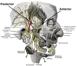
The facial nerve, also known as the seventh cranial nerve, cranial nerve VII, or simply CN VII, is a cranial nerve that emerges from the pons of the brainstem, controls the muscles of facial expression, and functions in the conveyance of taste sensations from the anterior two-thirds of the tongue. The nerve typically travels from the pons through the facial canal in the temporal bone and exits the skull at the stylomastoid foramen. It arises from the brainstem from an area posterior to the cranial nerve VI and anterior to cranial nerve VIII.

The great auricular nerve is a cutaneous (sensory) nerve of the head. It originates from the second and third cervical (spinal) nerves (C2-C3) of the cervical plexus. It provides sensory innervation to the skin over the parotid gland and the mastoid process, parts of the outer ear, and to the parotid gland and its fascia.

The digastric muscle is a bilaterally paired suprahyoid muscle located under the jaw. Its posterior belly is attached to the mastoid notch of temporal bone, and its anterior belly is attached to the digastric fossa of mandible; the two bellies are united by an intermediate tendon which is held in a loop that attaches to the hyoid bone. The anterior belly is innervated via the mandibular nerve, and the posterior belly is innervated via the facial nerve. It may act to depress the mandible or elevate the hyoid bone.
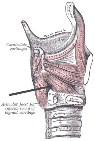
The lateral cricoarytenoid is an intrinsic muscle of the larynx. It attaches at the cricoid cartilage anteriorly, and at the arytenoid cartilage of the same side posteriorly. It is innervated by the recurrent laryngeal nerve. It acts to close the rima glottidis, thus closing the airway.

The corrugator supercilii muscle is a small, narrow, pyramidal muscle of the face. It arises from the medial end of the superciliary arch; it inserts into the deep surface of the skin of the eyebrow.

The stylohyoid muscle is one of the suprahyoid muscles. Its originates from the styloid process of the temporal bone; it inserts onto hyoid bone. It is innervated by a branch of the facial nerve. It acts draw the hyoid bone upwards and backwards.
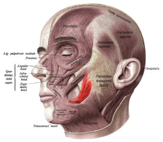
The risorius muscle is a highly variable muscle of facial expression. It has numerous and very variable origins, and inserts into the angle of the mouth. It receives motor innervation from branches of facial nerve. It may be absent or asymmetrical in some people. It pulls the angle of the mouth sidewise, such as during smiling.

The nasociliary nerve is a branch of the ophthalmic nerve (CN V1) (which is in turn a branch of the trigeminal nerve (CN V)). It is intermediate in size between the other two branches of the ophthalmic nerve, the frontal nerve and lacrimal nerve.

The posterior auricular artery is a small artery that arises from the external carotid artery. It ascends along the side of the head. It supplies several muscles of the neck and several structures of the head.
The deep temporal nerves are typically two nerves (one anterior and one posterior) which arise from the mandibular nerve (CN V3) and provide motor innervation to the temporalis muscle.

The infratrochlear nerve is a branch of the nasociliary nerve (itself a branch of the ophthalmic nerve (CN V1)) in the orbit. It exits the orbit inferior to the trochlea of superior oblique. It provides sensory innervation to structures of the orbit and skin of adjacent structures.

The zygomaticotemporal nerve (zygomaticotemporal branch, temporal branch) is a cutaneous (sensory) nerve of the head. It is a branch of the zygomatic nerve (itself a branch of the maxillary nerve (CN V2)). It arises in the orbit and exits the orbit through the zygomaticotemporal foramen in the zygomatic bone to enter the temporal fossa. It is distributed to the skin of the side of the forehead. It also contains a parasympathetic secretomotor component for the lacrimal gland which it confers to the lacrimal nerve (which then delivers it to the gland).
The zygomaticofacial nerve (or zygomaticofacial branch of zygomatic nerve or malar branch of zygomatic nerve) is a cutaneous (sensory) branch of the maxillary nerve (CN V2) that arises within the orbit. The zygomaticofacial nerve penetrates the inferolateral angle of the orbit, emerging into the face through the zygomaticofacial foramen, then penetrates the orbicularis oculi muscle to reach and innervate the skin of the prominence of the cheek.

The facial canal is a Z-shaped canal in the temporal bone of the skull. It extends between the internal acoustic meatus and stylomastoid foramen. It transmits the facial nerve.

The masseteric nerve is a nerve of the face. It is a branch of the mandibular nerve (CN V3). It passes through the mandibular notch to reach masseter muscle. It provides motor innervation the masseter muscle, and sensory innervation to the temporomandibular joint.

The infraorbital nerve is a branch of the maxillary nerve. It arises in the pterygopalatine fossa. It passes through the inferior orbital fissure to enter the orbit. It travels through the orbit, then enters and traverses the infraorbital canal, exiting the canal at the infraorbital foramen to reach the face. It provides sensory innervation to the skin and mucous membranes around the middle of the face.

The pyramidal eminence is a hollow conical projection upon the posterior wall of the tympanic cavity of the middle ear. The stapedius muscle arises in the hollow of the eminence and its tendon exits through its apex.

The digastric branch of facial nerve provides motor innervation to the posterior belly of the digastric muscle. It branches from the facial nerve near to the stylomastoid foramen as the CN VII exits the facial canal. It commonly arises in common with the stylohyoid branch of facial nerve.
The stylohyoid branch of facial nerve provides motor innervation to the stylohyoid muscle. It frequently arises from the facial nerve in common with the digastric branch of facial nerve.

The superior deep cervical lymph nodes are the deep cervical lymph nodes that are situated adjacent to the superior portion of the internal jugular vein. They drain either to the inferior deep cervical lymph nodes or into the jugular trunk.
References
- ↑ The Big Picture: Gross Anatomy, Medical Course & Step 1 Review. David A. Morton, K. Bo Foreman, Kurt H. Albertine (2nd ed.). New York. 2018. p. 222. ISBN 978-1-259-86264-9. OCLC 1044772257.
{{cite book}}: CS1 maint: location missing publisher (link) CS1 maint: others (link) - ↑ Standring, Susan (2020). Gray's Anatomy: The Anatomical Basis of Clinical Practice (42th ed.). New York. p. 749. ISBN 978-0-7020-7707-4. OCLC 1201341621.
{{cite book}}: CS1 maint: location missing publisher (link) - ↑ Gray, Henry (1918). Gray's Anatomy (20th ed.). p. 904.