| Chorda tympani | |
|---|---|
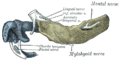
 The right tympanic membrane with the malleus and the chorda tympani, viewed from within the tympanic cavity (medial). | |
| Details | |
| From | Facial nerve |
| Innervates | Taste (anterior 2/3 of tongue) Sublingual gland |
| Identifiers | |
| Latin | nervus chorda tympani |
| MeSH | D002814 |
| TA98 | A14.2.01.084 A14.2.01.118 |
| TA2 | 6292 |
| FMA | 53228 |
| Anatomical terms of neuroanatomy | |
Chorda tympani is a branch of the facial nerve that carries gustatory (taste) sensory innervation from the front of the tongue and parasympathetic (secretomotor) innervation to the submandibular and sublingual salivary glands. [1]
Contents
- Structure
- Function
- Taste
- Chorda tympani transection
- Dysfunction
- Additional images
- References
- External links
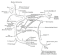
Chorda tympani has a complex course from the brainstem, through the temporal bone and middle ear, into the infratemporal fossa, and ending in the oral cavity. [2]


