
Tears are a clear liquid secreted by the lacrimal glands found in the eyes of all land mammals. Their functions include lubricating the eyes, removing irritants, and aiding the immune system. Tears also occur as a part of the body's natural pain response. Humans are the only mammals known to produce tears as part of an emotional response, such as out of joy or grief. Tears have symbolic significance among humans. Emotional secretion of tears may serve a biological function by excreting stress-inducing hormones built up through times of emotional distress. Tears are made up of water, electrolytes, proteins, lipids, and mucins that form layers on the surface of eyes. The different types of tears—basal, reflex, and emotional—vary significantly in composition..

The parasympathetic nervous system (PSNS) is one of the two divisions of the autonomic nervous system, the other being the sympathetic nervous system. The enteric nervous system (ENS) is now usually referred to as separate from the autonomic nervous system since it has its own independent reflex activity. The autonomic nervous system is responsible for regulating the body's unconscious actions. The parasympathetic system is responsible for stimulation of "rest-and-digest" or "feed and breed" activities that occur when the body is at rest, especially after eating, including sexual arousal, salivation, lacrimation (tears), urination, digestion and defecation. Its action is described as being complementary to that of the sympathetic nervous system, which is responsible for stimulating activities associated with the fight-or-flight response.

The facial nerve is the seventh cranial nerve, or simply CN VII. It emerges from the pons of the brainstem, controls the muscles of facial expression, and functions in the conveyance of taste sensations from the anterior two-thirds of the tongue. The nerves typically travels from the pons through the facial canal in the temporal bone and exits the skull at the stylomastoid foramen. It arises from the brainstem from an area posterior to the cranial nerve VI and anterior to cranial nerve VIII.
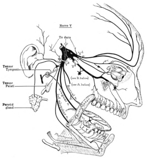
The trigeminal nerve (the fifth cranial nerve, or simply CN V) is a nerve responsible for sensation in the face and motor functions such as biting and chewing; it is the most complex of the cranial nerves. Its name ("trigeminal" = tri-, or three, and - geminus, or twin: thrice-twinned) derives from the fact that each of the two nerves (one on each side of the pons) has three major branches: the ophthalmic nerve (V1), the maxillary nerve (V2), and the mandibular nerve (V3). The ophthalmic and maxillary nerves are purely sensory, whereas the mandibular nerve supplies motor as well as sensory (or "cutaneous") functions. Adding to the complexity of this nerve is the fact that autonomic nerve fibers as well as special sensory fibers (taste) are contained within it.

The glossopharyngeal nerve, known as the ninth cranial nerve, is a mixed nerve that carries afferent sensory and efferent motor information. It exits the brainstem out from the sides of the upper medulla, just anterior to the vagus nerve. The motor division of the glossopharyngeal nerve is derived from the basal plate of the embryonic medulla oblongata, while the sensory division originates from the cranial neural crest.

The scalp is the anatomical area bordered by the human face at the front, and by the neck at the sides and back.

In anatomy, the orbit is the cavity or socket of the skull in which the eye and its appendages are situated. Anatomical term created by Gerard of Cremona. "Orbit" can refer to the bony socket, or it can also be used to imply the contents. In the adult human, the volume of the orbit is 30 millilitres, of which the eye occupies 6.5 ml. The orbital contents comprise the eye, the orbital and retrobulbar fascia, extraocular muscles, cranial nerves II, III, IV, V, and VI, blood vessels, fat, the lacrimal gland with its sac and nasolacrimal duct, the eyelids, medial and lateral palpebral ligaments, check ligaments, the suspensory ligament, septum, ciliary ganglion and short ciliary nerves.

In human physiology, the lacrimal glands are paired, almond-shaped exocrine glands, one for each eye, that secrete the aqueous layer of the tear film. They are situated in the upper lateral region of each orbit, in the lacrimal fossa of the orbit formed by the frontal bone. Inflammation of the lacrimal glands is called dacryoadenitis. The lacrimal gland produces tears which then flow into canals that connect to the lacrimal sac. From that sac, the tears drain through the lacrimal duct into the nose.

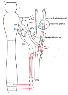
The otic ganglion is a small parasympathetic ganglion located immediately below the foramen ovale in the infratemporal fossa and on the medial surface of the mandibular nerve. It is functionally associated with the glossopharyngeal nerve and innervates the parotid gland for salivation.

The pterygopalatine ganglion is a parasympathetic ganglion found in the pterygopalatine fossa. It is largely innervated by the greater petrosal nerve ; and its axons project to the lacrimal glands and nasal mucosa. The flow of blood to the nasal mucosa, in particular the venous plexus of the conchae, is regulated by the pterygopalatine ganglion and heats or cools the air in the nose. It is one of four parasympathetic ganglia of the head and neck, the others being the submandibular ganglion, otic ganglion, and ciliary ganglion.
The auriculotemporal nerve is a branch of the mandibular nerve (V3) that runs with the superficial temporal artery and vein, and provides sensory innervation to various regions on the side of the head.
The greater petrosal nerve is a nerve in the skull that branches from the facial nerve; it forms part of a chain of nerves that innervate the lacrimal gland. The preganglionic parasympathetic axons of this nerve synapse in the pterygopalatine ganglion.

The ophthalmic nerve (V1) is one of three divisions of the trigeminal nerve (CN V). It has three branches that provide sensory innervation to the eye, skin of the upper face and anterior scalp.

The lacrimal nerve is the smallest branch of the ophthalmic nerve (V1), itself a branch of the trigeminal nerve (CN V). The other branches of the ophthalmic nerve are the frontal nerve and nasociliary nerve.
The zygomatic nerve is a branch of the maxillary nerve, itself a branch of the trigeminal nerve. It travels through the orbit and divides into branches that provide sensory innervation to skin over the zygomatic and temporal bones.

The submandibular ganglion is part of the human autonomic nervous system. It is one of four parasympathetic ganglia of the head and neck..
The lacrimal apparatus is the physiological system containing the orbital structures for tear production and drainage.
It consists of:

The nerve of the pterygoid canal is formed by the junction of the greater petrosal nerve and deep petrosal nerve, which passes from the foramen lacerum to the pterygopalatine fossa through the pterygoid canal.
The salivatory nuclei are the superior salivatory nucleus, and the inferior salivatory nucleus that innervate the salivary glands. They are located in the pontine tegmentum in the brainstem. They both are examples of cranial nerve nuclei.