| Trigeminal ganglion | |
|---|---|
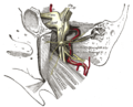
 Nerves of the orbit. Seen from above. (Trigeminal ganglion, labeled Semilunar Ganglion, visible near bottom.) | |
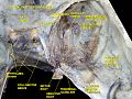
 Distribution of the maxillary and mandibular nerves, and the submaxillary ganglion. (Trigeminal ganglion, labeled Semilunar Ganglion, visible in upper left.) | |
| Details | |
| Identifiers | |
| Latin | ganglion trigeminale, ganglion semilunare (Gasseri) |
| MeSH | D012668 |
| TA98 | A14.2.01.014 |
| TA2 | 6194 |
| FMA | 52618 |
| Anatomical terms of neuroanatomy | |
The trigeminal ganglion (also known as: Gasserian ganglion, semilunar ganglion, or Gasser's ganglion) is the sensory ganglion of each trigeminal nerve (CN V). The trigeminal ganglion is located within the trigeminal cave (Meckel's cave), a cavity formed by dura mater.


