
The vagus nerve, also known as the tenth cranial nerve, cranial nerve X, or simply CN X, is a cranial nerve that carries sensory fibers that create a pathway that interfaces with the parasympathetic control of the heart, lungs, and digestive tract. It comprises two nerves—the left and right vagus nerves—but they are typically referred to collectively as a single subsystem.

The facial nerve, also known as the seventh cranial nerve, cranial nerve VII, or simply CN VII, is a cranial nerve that emerges from the pons of the brainstem, controls the muscles of facial expression, and functions in the conveyance of taste sensations from the anterior two-thirds of the tongue. The nerve typically travels from the pons through the facial canal in the temporal bone and exits the skull at the stylomastoid foramen. It arises from the brainstem from an area posterior to the cranial nerve VI and anterior to cranial nerve VIII.
Articles related to anatomy include:
The glossopharyngeal nerve, also known as the ninth cranial nerve, cranial nerve IX, or simply CN IX, is a cranial nerve that exits the brainstem from the sides of the upper medulla, just anterior to the vagus nerve. Being a mixed nerve (sensorimotor), it carries afferent sensory and efferent motor information. The motor division of the glossopharyngeal nerve is derived from the basal plate of the embryonic medulla oblongata, whereas the sensory division originates from the cranial neural crest.

In neuroanatomy, the mandibular nerve (V3) is the largest of the three divisions of the trigeminal nerve, the fifth cranial nerve (CN V). Unlike the other divisions of the trigeminal nerve (ophthalmic nerve, maxillary nerve) which contain only afferent fibers, the mandibular nerve contains both afferent and efferent fibers. These nerve fibers innervate structures of the lower jaw and face, such as the tongue, lower lip, and chin. The mandibular nerve also innervates the muscles of mastication.

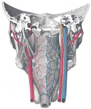
The internal carotid artery is an artery in the neck which supplies the anterior circulation of the brain.

In neuroanatomy, dura mater is a thick membrane made of dense irregular connective tissue that surrounds the brain and spinal cord. It is the outermost of the three layers of membrane called the meninges that protect the central nervous system. The other two meningeal layers are the arachnoid mater and the pia mater. It envelops the arachnoid mater, which is responsible for keeping in the cerebrospinal fluid. It is derived primarily from the neural crest cell population, with postnatal contributions of the paraxial mesoderm.

The otic ganglion is a small parasympathetic ganglion located immediately below the foramen ovale in the infratemporal fossa and on the medial surface of the mandibular nerve. It is functionally associated with the glossopharyngeal nerve and innervates the parotid gland for salivation.

The auriculotemporal nerve is a sensory branch of the mandibular nerve (CN V3) that runs with the superficial temporal artery and vein, and provides sensory innervation to parts of the external ear, scalp, and temporomandibular joint. The nerve also conveys post-ganglionic parasympathetic fibres from the otic ganglion to the parotid gland.

Chorda tympani is a branch of the facial nerve that carries gustatory (taste) sensory innervation from the front of the tongue and parasympathetic (secretomotor) innervation to the submandibular and sublingual salivary glands.
The auricular branch of the vagus nerve is often termed the Alderman's nerve or Arnold's nerve. The latter name is an eponym for Friedrich Arnold. The auricular branch of the vagus nerve supplies sensory innervation to the skin of the ear canal, tragus, and auricle.

The greater petrosal nerve is a nerve of the head mainly containing pre-ganglionic parasympathetic fibres which ultimately synapse in the pterygopalatine ganglion. It branches from the facial nerve and is derived from the parasympathetic part of the nervus intermedius component of CN VII, with its cell bodies located in the superior salivary nucleus. In the connective tissue substance of the foramen lacerum, the greater petrosal nerve unites with the (sympathetic) deep petrosal nerve to form the nerve of the pterygoid canal which proceeds to the pterygopalatine ganglion.

The geniculate ganglion is a collection of pseudounipolar sensory neurons of the facial nerve located in the facial canal of the head. It receives fibers from the facial nerve. It sends fibers that supply the lacrimal glands, submandibular glands, sublingual glands, tongue, palate, pharynx, external auditory meatus, stapedius muscle, posterior belly of the digastric muscle, stylohyoid muscle, and muscles of facial expression.

The petrous part of the temporal bone is pyramid-shaped and is wedged in at the base of the skull between the sphenoid and occipital bones. Directed medially, forward, and a little upward, it presents a base, an apex, three surfaces, and three angles, and houses in its interior, the components of the inner ear. The petrous portion is among the most basal elements of the skull and forms part of the endocranium. Petrous comes from the Latin word petrosus, meaning "stone-like, hard". It is one of the densest bones in the body. In other mammals, it is a separate bone, the petrosal bone.
The inferior ganglion of the glossopharyngeal nerve is a sensory ganglion. It is larger than and inferior to the superior ganglion of the glossopharyngeal nerve. It is located within the jugular foramen.
The superior ganglion of the glossopharyngeal nerve is a sensory ganglion of the peripheral nervous system. It is located within the jugular foramen where the glossopharyngeal nerve exits the skull. It is smaller than and superior to the inferior ganglion of the glossopharyngeal nerve.
The inferior ganglion of the vagus nerve is one of the two sensory ganglia of each vagus nerve. It contains neuron cell bodies of general visceral afferent fibers and special visceral afferent fibers. It is situated within the jugular fossa just below the skull. It is situated just below the superior ganglion of vagus nerve.

The dorsal nucleus of vagus nerve is a cranial nerve nucleus of the vagus nerve situated in the medulla oblongata of the brainstem ventral to the floor of the fourth ventricle. It contains nerve cell bodies of parasympathetic neurons of CN X that provide parasympathetic innervation to the gastrointestinal tract and lungs as well as other thoracic and abdominal organs. These functions include, among others, bronchoconstriction and gland secretion.
The pharyngeal plexus is a nerve plexus located upon the outer surface of the pharynx. It contains a motor component, a sensory component, and sympathetic component.
The meningeal branch of the vagus nerve is one of the first branches of the vagus nerve at the level of the superior ganglion. The neuron cell bodies reside within the superior ganglion and innervate the dura mater in the posterior cranial fossa of the base of the skull. The meningeal branch passes back into the skull through the jugular foramen.

