Eardrum

Last updated
Eardrum
Blausen 0328 EarAnatomy.png
View-normal-tympanic-membrane.png
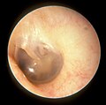
Right eardrum as seen through a speculum
Details
Identifiers
Latin membrana tympanica; myringa
MeSH D014432
TA98 A15.3.01.052
TA2 6870
FMA 9595
Anatomical terminology

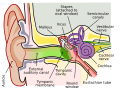
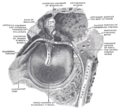
In the anatomy of humans and various other tetrapods, the eardrum, also called the tympanic membrane or myringa, is a thin, cone-shaped membrane that separates the external ear from the middle ear. Its function is to transmit sound from the air to the ossicles inside the middle ear, and thence to the oval window in the fluid-filled cochlea. The ear thereby converts and amplifies vibration in the air to vibration in cochlear fluid. [1] The malleus bone bridges the gap between the eardrum and the other ossicles. [2]

Contents

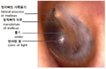
Rupture or perforation of the eardrum can lead to conductive hearing loss. Collapse or retraction of the eardrum can cause conductive hearing loss or cholesteatoma.

Structure

Orientation and relations

The tympanic membrane is oriented obliquely in the anteroposterior, mediolateral, and superoinferior planes. Consequently, its superoposterior end lies lateral to its anteroinferior end.[ citation needed ]

Anatomically, it relates superiorly to the middle cranial fossa, posteriorly to the ossicles and facial nerve, inferiorly to the parotid gland, and anteriorly to the temporomandibular joint.[ citation needed ]

Regions

The eardrum is divided into two general regions: the pars flaccida and the pars tensa. [3] The relatively fragile pars flaccida lies above the lateral process of the malleus between the Notch of Rivinus and the anterior and posterior malleal folds. Consisting of two layers and appearing slightly pinkish in hue, it is associated with[ vague ] Eustachian tube dysfunction and cholesteatomas. [4]

The larger pars tensa consists of three layers: skin, fibrous tissue, and mucosa. Its thick periphery forms a fibrocartilaginous ring called the annulus tympanicus or Gerlach's ligament. [5] while the central umbo tents inward at the level of the tip of malleus. The middle fibrous layer, containing radial, circular, and parabolic fibers, encloses the handle of malleus. Though comparatively robust, the pars tensa is the region more commonly associated with[ vague ] perforations. [6]

Umbo

The manubrium (Latin for "handle") of the malleus is firmly attached to the medial surface of the membrane as far as its center, drawing it toward the tympanic cavity. The lateral surface of the membrane is thus concave. The most depressed aspect of this concavity is termed the umbo (Latin for "shield boss"). [7]

Nerve supply

Sensation of the outer surface of the tympanic membrane is supplied mainly by the auriculotemporal nerve, a branch of the mandibular nerve (cranial nerve V3), with contributions from the auricular branch of the vagus nerve (cranial nerve X), the facial nerve (cranial nerve VII), and possibly the glossopharyngeal nerve (cranial nerve IX). The inner surface of the tympanic membrane is innervated by the glossopharyngeal nerve. [8]

Clinical significance

Examination

When the eardrum is illuminated during a medical examination, a cone of light radiates from the tip of the malleus to the periphery in the anteroinferior quadrant, this is what is known clinically as 5 o'clock.[ citation needed ]

Rupture

Unintentional perforation (rupture) has been described in blast injuries [9] and air travel, typically in patients experiencing upper respiratory congestion or general Eustachian tube dysfunction that prevents equalization of pressure in the middle ear. [10] It is also known to occur in swimming, diving (including scuba diving), [11] and martial arts. [12]

Patients with tympanic membrane rupture may experience bleeding, tinnitus, hearing loss, or disequilibrium (vertigo). However, they rarely require medical intervention, as between 80 and 95 percent of ruptures recover completely within two to four weeks. [13] [14] [15] The prognosis becomes more guarded as the force of injury increases. [15]

Surgical puncture for treatment of middle ear infections

In some cases, the pressure of fluid in an infected middle ear is great enough to cause the eardrum to rupture naturally. Usually, this consists of a small hole (perforation), from which fluid can drain out of the middle ear. If this does not occur naturally, a myringotomy (tympanotomy, tympanostomy) can be performed. A myringotomy is a surgical procedure in which a tiny incision is created in the eardrum to relieve pressure caused by excessive buildup of fluid, or to drain pus from the middle ear. The fluid or pus comes from a middle ear infection (otitis media), which is a common problem in children. A tympanostomy tube is inserted into the eardrum to keep the middle ear aerated for a prolonged time and to prevent reaccumulation of fluid. Without the insertion of a tube, the incision usually heals spontaneously in two to three weeks. Depending on the type, the tube is either naturally extruded in 6 to 12 months or removed during a minor procedure. [16]

Those requiring myringotomy usually have an obstructed or dysfunctional eustachian tube that is unable to perform drainage or ventilation in its usual fashion. Before the invention of antibiotics, myringotomy without tube placement was also used as a major treatment of severe acute otitis media. [16]

Society and culture

The Bajau people of the Pacific intentionally rupture their eardrums at an early age to facilitate diving and hunting at sea. Many older Bajau therefore have difficulties hearing. [17]

See also

Additional images

Related Research Articles

<span class="mw-page-title-main">Middle ear</span> Portion of the ear internal to the eardrum, and external to the oval window of the inner ear

The middle ear is the portion of the ear medial to the eardrum, and distal to the oval window of the cochlea.

<span class="mw-page-title-main">Cholesteatoma</span> Medical condition

Cholesteatoma is a destructive and expanding growth consisting of keratinizing squamous epithelium in the middle ear and/or mastoid process. Cholesteatomas are not cancerous as the name may suggest, but can cause significant problems because of their erosive and expansile properties. This can result in the destruction of the bones of the middle ear (ossicles), as well as growth through the base of the skull into the brain. They often become infected and can result in chronically draining ears. Treatment almost always consists of surgical removal.

The ossicles are three bones in either middle ear that are among the smallest bones in the human body. They serve to transmit sounds from the air to the fluid-filled labyrinth (cochlea). The absence of the auditory ossicles would constitute a moderate-to-severe hearing loss. The term "ossicle" literally means "tiny bone". Though the term may refer to any small bone throughout the body, it typically refers to the malleus, incus, and stapes of the middle ear.

<span class="mw-page-title-main">Otitis media</span> Inflammation of the middle ear

Otitis media is a group of inflammatory diseases of the middle ear. One of the two main types is acute otitis media (AOM), an infection of rapid onset that usually presents with ear pain. In young children this may result in pulling at the ear, increased crying, and poor sleep. Decreased eating and a fever may also be present. The other main type is otitis media with effusion (OME), typically not associated with symptoms, although occasionally a feeling of fullness is described; it is defined as the presence of non-infectious fluid in the middle ear which may persist for weeks or months often after an episode of acute otitis media. Chronic suppurative otitis media (CSOM) is middle ear inflammation that results in a perforated tympanic membrane with discharge from the ear for more than six weeks. It may be a complication of acute otitis media. Pain is rarely present. All three types of otitis media may be associated with hearing loss. If children with hearing loss due to OME do not learn sign language, it may affect their ability to learn.

<span class="mw-page-title-main">Eustachian tube</span> Tube connecting middle ear to throat

The Eustachian tube, also called the auditory tube or pharyngotympanic tube, is a tube that links the nasopharynx to the middle ear, of which it is also a part. In adult humans, the Eustachian tube is approximately 35 mm (1.4 in) long and 3 mm (0.12 in) in diameter. It is named after the sixteenth-century Italian anatomist Bartolomeo Eustachi.

<span class="mw-page-title-main">Ear</span> Organ of hearing and balance

An ear is the organ that enables hearing and body balance using the vestibular system. In mammals, the ear is usually described as having three parts: the outer ear, the middle ear and the inner ear. The outer ear consists of the pinna and the ear canal. Since the outer ear is the only visible portion of the ear in most animals, the word "ear" often refers to the external part alone. The middle ear includes the tympanic cavity and the three ossicles. The inner ear sits in the bony labyrinth, and contains structures which are key to several senses: the semicircular canals, which enable balance and eye tracking when moving; the utricle and saccule, which enable balance when stationary; and the cochlea, which enables hearing. The ear is a self cleaning organ through its relationship with earwax and the ear canals. The ears of vertebrates are placed somewhat symmetrically on either side of the head, an arrangement that aids sound localization.

<span class="mw-page-title-main">Conductive hearing loss</span> Medical condition

Conductive hearing loss (CHL) occurs when there is a problem transferring sound waves anywhere along the pathway through the outer ear, tympanic membrane (eardrum), or middle ear (ossicles). If a conductive hearing loss occurs in conjunction with a sensorineural hearing loss, it is referred to as a mixed hearing loss. Depending upon the severity and nature of the conductive loss, this type of hearing impairment can often be treated with surgical intervention or pharmaceuticals to partially or, in some cases, fully restore hearing acuity to within normal range. However, cases of permanent or chronic conductive hearing loss may require other treatment modalities such as hearing aid devices to improve detection of sound and speech perception.

A myringotomy is a surgical procedure in which an incision is created in the eardrum to relieve pressure caused by excessive buildup of fluid, or to drain pus from the middle ear. A tympanostomy tube may be inserted through the eardrum to keep the middle ear aerated for a prolonged time and to prevent reaccumulation of fluid. Without the insertion of a tube, the incision usually heals spontaneously within two to three weeks. Depending on the type, the tube is either naturally extruded in 6 to 12 months or removed during a minor procedure.

<span class="mw-page-title-main">Tympanoplasty</span> Surgical operation on the ear

Tympanoplasty is the surgical operation performed to reconstruct hearing mechanism of middle ear.

<span class="mw-page-title-main">Ear pain</span> Pain in the ear

Ear pain, also known as earache or otalgia, is pain in the ear. Primary ear pain is pain that originates from the ear. Secondary ear pain is a type of referred pain, meaning that the source of the pain differs from the location where the pain is felt.

<span class="mw-page-title-main">Tympanostomy tube</span> Medical device inserted into the eardrum

Tympanostomy tube, also known as a grommet,myringotomy tube, or pressure equalizing tube, is a small tube inserted into the eardrum via a surgical procedure called myringotomy to keep the middle ear aerated for a prolonged period of time, typically to prevent accumulation of fluid in the middle ear. The tube itself is made in a variety of designs, most often shaped like a grommet for short-term use, or with long flanges and sometimes resembling a T-shape for long-term use. Materials used to manufacture the tubes are often made from fluoroplastic or silicone, which have largely replaced the use of metal tubes made from stainless steel, titanium, or gold.

<span class="mw-page-title-main">Mastoiditis</span> Middle ear disease

Mastoiditis is the result of an infection that extends to the air cells of the skull behind the ear. Specifically, it is an inflammation of the mucosal lining of the mastoid antrum and mastoid air cell system inside the mastoid process. The mastoid process is the portion of the temporal bone of the skull that is behind the ear. The mastoid process contains open, air-containing spaces. Mastoiditis is usually caused by untreated acute otitis media and used to be a leading cause of child mortality. With the development of antibiotics, however, mastoiditis has become quite rare in developed countries where surgical treatment is now much less frequent and more conservative, unlike former times.

<span class="mw-page-title-main">Tympanic cavity</span> Small cavity surrounding the bones of the middle ear

The tympanic cavity is a small cavity surrounding the bones of the middle ear. Within it sit the ossicles, three small bones that transmit vibrations used in the detection of sound.

<span class="mw-page-title-main">Tympanometry</span> Acoustic evaluation of the condition of the middle ear

Tympanometry is an acoustic evaluation of the condition of the middle ear eardrum and the conduction bones by creating variations of air pressure in the ear canal.

<span class="mw-page-title-main">Perforated eardrum</span> Injury leading to a hole in the eardrum

A perforated eardrum is a prick in the eardrum. It can be caused by infection, trauma, overpressure, inappropriate ear clearing, and changes in middle ear pressure. An otoscope can be used to view the eardrum to diagnose a perforation. Perforations may heal naturally or require surgery.

Hearing loss with craniofacial syndromes is a common occurrence. Many of these multianomaly disorders involve structural malformations of the outer or middle ear, making a significant hearing loss highly likely.

Myringoplasty is the closure of the perforation of pars tensa of the tympanic membrane. When myringoplasty is combined with removal of scar tissue, it is called tympanoplasty. The operation is performed with the patient supine and face turned to one side. The graft material most commonly used for the surgery is temporalis fascia. The tragal cartilage and tragal perichondrium are also used as the graft by some surgeons.

<span class="mw-page-title-main">Tympanic membrane retraction</span> Medical condition

Tympanic membrane retraction describes a condition in which a part of the eardrum lies deeper within the ear than its normal position.

<i>Malleus</i> Bone of the middle ear

The malleus, or hammer, is a hammer-shaped small bone or ossicle of the middle ear. It connects with the incus, and is attached to the inner surface of the eardrum. The word is Latin for 'hammer' or 'mallet'. It transmits the sound vibrations from the eardrum to the incus (anvil).

Middle ear barotrauma (MEBT), also known to underwater divers as ear squeeze and reverse ear squeeze, is an injury caused by a difference in pressure between the external ear canal and the middle ear. It is common in underwater divers and usually occurs when the diver does not equalise sufficiently during descent or, less commonly, on ascent. Failure to equalise may be due to inexperience or eustachian tube dysfunction, which can have many possible causes. Unequalised ambient pressure increase during descent causes a pressure imbalance between the middle ear air space and the external auditory canal over the eardrum, referred to by divers as ear squeeze, causing inward stretching, serous effusion and haemorrhage, and eventual rupture. During ascent internal over-pressure is normally passively released through the eustachian tube, but if this does not happen the volume expansion of middle ear gas will cause outward bulging, stretching and eventual rupture of the eardrum known to divers as reverse ear squeeze. This damage causes local pain and hearing loss. Tympanic rupture during a dive can allow water into the middle ear, which can cause severe vertigo from caloric stimulation. This may cause nausea and vomiting underwater, which has a high risk of aspiration of vomit or water, with possibly fatal consequences.

References

PD-icon.svgThis article incorporates text in the public domain from page 1039 of the 20th edition of Gray's Anatomy (1918)

  1. Hilal, Fathi; Liaw, Jeffrey; Cousins, Joseph P.; Rivera, Arnaldo L.; Nada, Ayman (2023-04-01). "Autoincudotomy as an uncommon etiology of conductive hearing loss: Case report and review of literature". Radiology Case Reports. 18 (4): 1461–1465. doi:10.1016/j.radcr.2022.10.097. ISSN   1930-0433. PMC   9925837 . PMID   36798057.
  2. Purves, D; Augustine, G; Fitzpatrick, D; Hall, W; LaMantia, A; White, L; et al., eds. (2012). Neuroscience. Sunderland: Sinauer. ISBN   9780878936953.
  3. Gilberto, Nelson; Santos, Ricardo; Sousa, Pedro; O’Neill, Assunção; Escada, Pedro; Pais, Diogo (August 2019). "Pars tensa and tympanicomalleal joint: proposal for a new anatomic classification". European Archives of Oto-Rhino-Laryngology. 276 (8): 2141–2148. doi:10.1007/s00405-019-05434-4. PMID   31004197. S2CID   123959777.
  4. Jain, Shraddha (2019). "Role of Eustachian Dysfunction and Primary Sclerotic Mastoid Pneumatisation Pattern in Aetiology of Squamous Chronic Otitis Media: A Correlative Study". Indian Journal of Otolaryngology and Head and Neck Surgery. 71 (Suppl 2): 1190–1196. doi:10.1007/s12070-018-1259-x. PMC   6841851 . PMID   31750149.
  5. Mansour, Salah; Magnan, Jacques; Ahmad, Hassan Haidar; Nicolas, Karen; Louryan, Stéphane (2019). Comprehensive and Clinical Anatomy of the Middle Ear. Springer. ISBN   9783030153632.
  6. Marchioni D, Molteni G, Presutti L (February 2011). "Endoscopic Anatomy of the Middle Ear". Indian J Otolaryngol Head Neck Surg. 63 (2): 101–13. doi:10.1007/s12070-011-0159-0. PMC   3102170 . PMID   22468244.
  7. Gray's Anatomy (1918)
  8. Drake, Richard L., A. Wade Vogl, and Adam Mitchell. Gray's Anatomy For Students. 3rd ed. Philadelphia: Churchill Livingstone, 2015. Print. pg. 969
  9. Ritenour AE, Wickley A, Retinue JS, Kriete BR, Blackbourne LH, Holcomb JB, Wade CE (February 2008). "Tympanic membrane perforation and hearing loss from blast overpressure in Operation Enduring Freedom and Operation Iraqi Freedom wounded". J Trauma. 64 (2 Suppl): S174-8. doi:10.1097/ta.0b013e318160773e. PMID   18376162.
  10. Mirza S, Richardson H (May 2005). "Otic barotrauma from air travel". J Laryngol Otol. 119 (5): 366–70. doi:10.1258/0022215053945723. PMID   15949100. S2CID   45256115.
  11. Green SM; Rothrock SG; Green EA= (October 1993). "Tympanometric evaluation of middle ear barotrauma during recreational scuba diving". Int J Sports Med. 14 (7): 411–5. doi:10.1055/s-2007-1021201. PMID   8244609.
  12. Fields JD, McKeag DB, Turner JL (February 2008). "Traumatic tympanic membrane rupture in a mixed martial arts competition". Current Sports Med Rep. 7 (1): 10–11. doi: 10.1097/01.CSMR.0000308672.53182.3b . PMID   18296937. S2CID   205388185.
  13. Kristensen S (December 1992). "Spontaneous healing of traumatic tympanic membrane perforations in man: a century of experience". J Laryngol Otol. 106 (12): 1037–50. doi:10.1017/s0022215100121723. PMID   1487657. S2CID   21899785.
  14. Lindeman P, Edström S, Granström G, Jacobsson S, von Sydow C, Westin T, Aberg B (December 1987). "Acute traumatic tympanic membrane perforations. Cover or observe?". Arch Otolaryngol Head Neck Surg. 113 (12): 1285–7. doi:10.1001/archotol.1987.01860120031002. PMID   3675893.
  15. 1 2 Garth RJ (July 1995). "Blast injury of the ear: an overview and guide to management". Injury. 26 (6): 363–6. doi:10.1016/0020-1383(95)00042-8. PMID   7558254.
  16. 1 2 Smith N, Greinwald JR (2011). "To tube or not to tube: indications for myringotomy with tube placement". Current Opinion in Otolaryngology & Head and Neck Surgery. 19 (5): 363–366. doi:10.1097/MOO.0b013e3283499fa8. PMID   21804383. S2CID   3027628.
  17. Langenheim, Johnny (18 September 2010). "The last of the sea nomads". The Guardian . Retrieved 15 February 2016.