The inner ear is the innermost part of the vertebrate ear. In vertebrates, the inner ear is mainly responsible for sound detection and balance. In mammals, it consists of the bony labyrinth, a hollow cavity in the temporal bone of the skull with a system of passages comprising two main functional parts:
The semicircular canals are three semicircular interconnected tubes located in the innermost part of each ear, the inner ear. The three canals are the lateral, anterior and posterior semicircular canals. They are the part of the bony labyrinth, a periosteum-lined cavity on the petrous part of the temporal bone filled with perilymph.

The occipital bone is a cranial dermal bone and the main bone of the occiput. It is trapezoidal in shape and curved on itself like a shallow dish. The occipital bone overlies the occipital lobes of the cerebrum. At the base of the skull in the occipital bone, there is a large oval opening called the foramen magnum, which allows the passage of the spinal cord.
The saccule is a bed of sensory cells in the inner ear. The saccule is from Latin saccus 'sack'. The saccule translates head movements into neural impulses for the brain to interpret. The saccule detects linear accelerations and head tilts in the vertical plane. When the head moves vertically, the sensory cells of the saccule are disturbed and the neurons connected to them begin transmitting impulses to the brain. These impulses travel along the vestibular portion of the eighth cranial nerve to the vestibular nuclei in the brainstem.

The posterior cranial fossa is the part of the cranial cavity located between the foramen magnum, and tentorium cerebelli. It is formed by the sphenoid bones, temporal bones, and occipital bone. It lodges the cerebellum, and parts of the brainstem.
Mondini dysplasia, also known as Mondini malformation and Mondini defect, is an abnormality of the inner ear that is associated with sensorineural hearing loss.

The cerebellar tentorium or tentorium cerebelli is one of four dural folds that separate the cranial cavity into four (incomplete) compartments. The cerebellar tentorium separates the cerebellum from the cerebrum forming a supratentorial and an infratentorial region; the cerebrum is supratentorial and the cerebellum infratentorial.
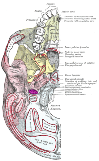
The jugular fossa is a deep depression in the inferior part of the temporal bone at the base of the skull. It lodges the bulb of the internal jugular vein.

A jugular foramen is one of the two large foramina (openings) in the base of the skull, located behind the carotid canal. It is formed by the temporal bone and the occipital bone. It allows many structures to pass, including the inferior petrosal sinus, three cranial nerves, the sigmoid sinus, and meningeal arteries.

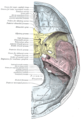
The internal auditory meatus is a canal within the petrous part of the temporal bone of the skull between the posterior cranial fossa and the inner ear.

The petrous part of the temporal bone is pyramid-shaped and is wedged in at the base of the skull between the sphenoid and occipital bones. Directed medially, forward, and a little upward, it presents a base, an apex, three surfaces, and three angles, and houses in its interior the components of the inner ear. The petrous portion is among the most basal elements of the skull and forms part of the endocranium. Petrous comes from the Latin word petrosus, meaning "stone-like, hard". It is one of the densest bones in the body. In other mammals, it is a separate bone, the petrosal bone.

The middle cranial fossa is formed by the sphenoid bones, and the temporal bones. It lodges the temporal lobes, and the pituitary gland. It is deeper than the anterior cranial fossa, is narrow medially and widens laterally to the sides of the skull. It is separated from the posterior cranial fossa by the clivus and the petrous crest.
The subarcuate fossa is a shallow depression upon the internal surface of the petrous part of the temporal bone forming the wall of the posterior cranial fossa. The fossa accommodates the flocculus of the cerebellum. It is situated lateral/posterior to the internal auditory meatus.

Medial to the opening for the carotid canal and close to its posterior border, in front of the jugular fossa, is a triangular depression; at the apex of this is a small opening, the aquaeductus cochleae, which lodges a tubular prolongation of the dura mater establishing a communication between the perilymphatic space and the subarachnoid space, and transmits a vein from the cochlea to join the internal jugular vein. The cochlear aqueduct lies perpendicular to the petrous apex, in contrast with the vestibular aqueduct, which lies parallel to the petrous apex.
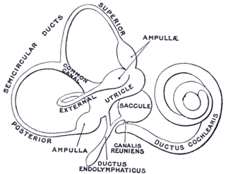
From the posterior wall of the saccule a canal, the endolymphatic duct, is given off; this duct is joined by the ductus utriculosaccularis, and then passes along the aquaeductus vestibuli and ends in a blind pouch on the posterior surface of the petrous portion of the temporal bone, where it is in contact with the dura mater.
From the posterior wall of the saccule a canal, the endolymphatic duct, is given off; this duct is joined by the utriculosaccular duct, and then passes along the vestibular aqueduct and ends in a blind pouch, the endolymphatic sac, on the posterior surface of the petrous portion of the temporal bone, where it is in contact with the dura mater. Studies suggest that the endolymphatic duct and endolymphatic sac perform both absorptive and secretory, as well as phagocytic and immunodefensive, functions.

The vestibule is the central part of the bony labyrinth in the inner ear, and is situated medial to the eardrum, behind the cochlea, and in front of the three semicircular canals.

Large vestibular aqueduct is a structural deformity of the inner ear. Enlargement of this duct is one of the most common inner ear deformities and is commonly associated with hearing loss during childhood. The term was first discovered in 1791 by Mondini when he was completing a temporal bone dissection. It was then defined by Valvassori and Clemis as a vestibular aqueduct that is greater than or equal to 2.0 mm at the operculum and/or greater than or equal to 1.0 mm at the midpoint. Some use the term enlarged vestibular aqueduct syndrome, but this is felt by others to be erroneous as it is a clinical finding which can occur in several syndromes.

An endolymphatic sac tumor (ELST) is a very uncommon papillary epithelial neoplasm arising within the endolymphatic sac or endolymphatic duct. This tumor shows a very high association with Von Hippel–Lindau syndrome (VHL).
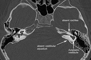
Michel aplasia, also known as complete labyrinthine aplasia (CLA), is a congenital abnormality of the inner ear. It is characterized by the bilateral absence of differentiated inner ear structures and results in complete deafness (anacusis). Michel aplasia should not be confused with michel dysplasia. It may affect one or both ears.