
The inner ear is the innermost part of the vertebrate ear. In vertebrates, the inner ear is mainly responsible for sound detection and balance. In mammals, it consists of the bony labyrinth, a hollow cavity in the temporal bone of the skull with a system of passages comprising two main functional parts:

The cochlea is the part of the inner ear involved in hearing. It is a spiral-shaped cavity in the bony labyrinth, in humans making 2.75 turns around its axis, the modiolus. A core component of the cochlea is the organ of Corti, the sensory organ of hearing, which is distributed along the partition separating the fluid chambers in the coiled tapered tube of the cochlea.
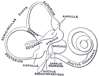
The semicircular canals are three semicircular interconnected tubes located in the innermost part of each ear, the inner ear. The three canals are the lateral, anterior and posterior semicircular canals. They are the part of the bony labyrinth, a periosteum-lined cavity on the petrous part of the temporal bone filled with perilymph.

The basilar membrane is a stiff structural element within the cochlea of the inner ear which separates two liquid-filled tubes that run along the coil of the cochlea, the scala media and the scala tympani. The basilar membrane moves up and down in response to incoming sound waves, which are converted to traveling waves on the basilar membrane.
The organ of Corti, or spiral organ, is the receptor organ for hearing and is located in the mammalian cochlea. This highly varied strip of epithelial cells allows for transduction of auditory signals into nerve impulses' action potential. Transduction occurs through vibrations of structures in the inner ear causing displacement of cochlear fluid and movement of hair cells at the organ of Corti to produce electrochemical signals.

The auditory system is the sensory system for the sense of hearing. It includes both the sensory organs and the auditory parts of the sensory system.

An ear is the organ that enables hearing and body balance using the vestibular system. In mammals the ear is usually described as having three parts: the outer ear, the middle ear and the inner ear. The outer ear consists of the pinna and the ear canal. Since the outer ear is the only visible portion of the ear in most animals, the word "ear" often refers to the external part alone. The middle ear includes the tympanic cavity and the three ossicles. The inner ear sits in the bony labyrinth, and contains structures which are key to several senses: the semicircular canals, which enable balance and eye tracking when moving; the utricle and saccule, which enable balance when stationary; and the cochlea, which enables hearing. The ear is a self cleaning organ through its relationship with earwax and the ear canals. The ears of vertebrates are placed somewhat symmetrically on either side of the head, an arrangement that aids sound localization.
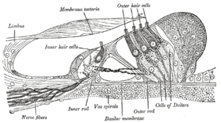
Hair cells are the sensory receptors of both the auditory system and the vestibular system in the ears of all vertebrates, and in the lateral line organ of fishes. Through mechanotransduction, hair cells detect movement in their environment.

In the inner ear, stereocilia are the mechanosensing organelles of hair cells, which respond to fluid motion in numerous types of animals for various functions, including hearing and balance. They are about 10–50 micrometers in length and share some similar features of microvilli. The hair cells turn the fluid pressure and other mechanical stimuli into electric stimuli via the many microvilli that make up stereocilia rods. Stereocilia exist in the auditory and vestibular systems.
Perilymph is an extracellular fluid located within the inner ear. It is found within the scala tympani and scala vestibuli of the cochlea. The ionic composition of perilymph is comparable to that of plasma and cerebrospinal fluid. The major cation in perilymph is sodium, with the values of sodium and potassium concentration in the perilymph being 138 mM and 6.9 mM, respectively. It is also named Cotunnius' liquid and liquor cotunnii for Domenico Cotugno.
The cochlear duct is an endolymph filled cavity inside the cochlea, located between the tympanic duct and the vestibular duct, separated by the basilar membrane and the vestibular membrane respectively. The cochlear duct houses the organ of Corti.
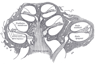
The vestibular membrane, vestibular wall or Reissner's membrane, is a membrane inside the cochlea of the inner ear. It separates the cochlear duct from the vestibular duct. It helps to transmit vibrations from fluid in the vestibular duct to the cochlear duct. Together with the basilar membrane, it creates a compartment in the cochlea filled with endolymph, which is important for the function of the spiral organ of Corti. It allows nutrients to travel from the perilymph to the endolymph of the membranous labyrinth. It may be damaged in Ménière's disease. It is named after the German anatomist Ernst Reissner.
The membranous labyrinth is a collection of fluid filled tubes and chambers which contain the receptors for the senses of equilibrium and hearing. It is lodged within the bony labyrinth in the inner ear and has the same general form; it is, however, considerably smaller and is partly separated from the bony walls by a quantity of fluid, the perilymph.
The stria vascularis of the cochlear duct is a capillary loop in the upper portion of the spiral ligament. It produces endolymph for the scala media in the cochlea.

The crista ampullaris is the sensory organ of rotation. They are found in the ampullae of each of the semicircular canals of the inner ear, meaning that there are three pairs in total. The function of the crista ampullaris is to sense angular acceleration and deceleration.
Endolymphatic hydrops is a disorder of the inner ear. It consists of an excessive build-up of the endolymph fluid, which fills the hearing and balance structures of the inner ear. Endolymph fluid, which is partly regulated by the endolymph sac, flows through the inner ear and is critical to the function of all sensory cells in the inner ear. In addition to water, endolymph fluid contains salts such as sodium, potassium, chloride and other electrolytes. If the inner ear is damaged by disease or injury, the volume and composition of the endolymph fluid can change, causing the symptoms of endolymphatic hydrops.
The vestibular evoked myogenic potential is a neurophysiological assessment technique used to determine the function of the otolithic organs of the inner ear. It complements the information provided by caloric testing and other forms of inner ear testing. There are two different types of VEMPs. One is the oVEMP and another is the cVEMP. The oVEMP measures integrity of the utricule and superior vestibular nerve and the cVemp measures the saccule and the inferior vestibular nerve.
Electrocochleography is a technique of recording electrical potentials generated in the inner ear and auditory nerve in response to sound stimulation, using an electrode placed in the ear canal or tympanic membrane. The test is performed by an otologist or audiologist with specialized training, and is used for detection of elevated inner ear pressure or for the testing and monitoring of inner ear and auditory nerve function during surgery.
Dark cells are specialized nonsensory epithelial cells found on either side of the vestibular organs and lining the endolymphatic space. These dark-cell areas in the vestibular organ are structures involved in the production of endolymph, an inner ear fluid, secreting potassium towards the endolymphatic fluid. Dark cells take part in fluid homeostasis to preserve the unique high-potassium and low-sodium content of the endolymph and also maintain the calcium homeostasis of the inner ear.
Cochlear hydrops is a condition of the inner ear involving a pathological increase of fluid affecting the cochlea. This results in swelling that can lead to hearing loss or changes in hearing perception. It is a form of endolymphatic hydrops and related to Ménière's disease. Cochlear hydrops refers to a case of inner-ear hydrops that only involves auditory symptoms and does not cause vestibular issues.


