Basilar membrane

Last updated
Basilar membrane.
Organ of corti.svg
Section through organ of corti, showing basilar membrane
Cochlea-crosssection.svg
Cross section of the cochlea.
Details
Identifiers
Latin membrana basilaris ductus cochlearis
MeSH D001489
Anatomical terminology

The basilar membrane is a stiff structural element within the cochlea of the inner ear which separates two liquid-filled tubes that run along the coil of the cochlea, the scala media and the scala tympani. The basilar membrane moves up and down in response to incoming sound waves, which are converted to traveling waves on the basilar membrane.

Contents

Structure

The basilar membrane is a pseudo-resonant structure [1] that, like the strings on an instrument, varies in width and stiffness. But unlike the parallel strings of a guitar, the basilar membrane is not a discrete set of resonant structures, but a single structure with varying width, stiffness, mass, damping, and duct dimensions along its length. The motion of the basilar membrane is generally described as a traveling wave. [2] The properties of the membrane at a given point along its length determine its characteristic frequency (CF), the frequency at which it is most sensitive to sound vibrations. The basilar membrane is widest (0.42–0.65 mm) and least stiff at the apex of the cochlea, and narrowest (0.08–0.16 mm) and stiffest at the base (near the round and oval windows). [3] High-frequency sounds localize near the base of the cochlea, while low-frequency sounds localize near the apex.

Function

Sinusoidal drive through the oval window (top) causes a traveling wave of fluid-membrane motion. A modeled snapshot of fluid streamlines is shown. The wavelength is long compared to the duct height near the base, in what is called the long-wave region, and short (0.5 to 1.0 mm in typical observations ) near the place where the displacement and velocity are maximized, just before cutoff, in the short-wave region. Cochlea Traveling Wave.png
Sinusoidal drive through the oval window (top) causes a traveling wave of fluid–membrane motion. A modeled snapshot of fluid streamlines is shown. The wavelength is long compared to the duct height near the base, in what is called the long-wave region, and short (0.5 to 1.0 mm in typical observations ) near the place where the displacement and velocity are maximized, just before cutoff, in the short-wave region.

Endolymph/perilymph separation

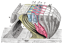
Along with the vestibular membrane, several tissues held by the basilar membrane segregate the fluids of the endolymph and perilymph, such as the inner and outer sulcus cells (shown in yellow) and the reticular lamina of the organ of Corti (shown in magenta). For the organ of Corti, the basilar membrane is permeable to perilymph. Here the border between endolymph and perilymph occurs at the reticular lamina, the endolymph side of the organ of Corti. [6]

A base for the sensory cells

The basilar membrane is also the base for the hair cells. This function is present in all land vertebrates. Due to its location, the basilar membrane places the hair cells adjacent to both the endolymph and the perilymph, which is a precondition of hair cell function.

Frequency dispersion

A third, evolutionarily younger, function of the basilar membrane is strongly developed in the cochlea of most mammalian species and weakly developed in some bird species: [7] the dispersion of incoming sound waves to separate frequencies spatially. In brief, the membrane is tapered and it is stiffer at one end than at the other. Furthermore, sound waves travelling to the "floppier" end of the basilar membrane have to travel through a longer fluid column than sound waves travelling to the nearer, stiffer end. Each part of the basilar membrane, together with the surrounding fluid, can therefore be thought of as a "mass-spring" system with different resonant properties: high stiffness and low mass, hence high resonant frequencies at the near (base) end, and low stiffness and high mass, hence low resonant frequencies, at the far (apex) end. [8] This causes sound input of a certain frequency to vibrate some locations of the membrane more than other locations. The distribution of frequencies to places is called the tonotopic organization of cochlea.

Sound-driven vibrations travel as waves along this membrane, along which, in humans, lie about 3,500 inner hair cells spaced in a single row. Each cell is attached to a tiny triangular frame. The 'hairs' are minute processes on the end of the cell, which are very sensitive to movement. When the vibration of the membrane rocks the triangular frames, the hairs on the cells are repeatedly displaced, and that produces streams of corresponding pulses in the nerve fibers, which are transmitted to the auditory pathway. [9] The outer hair cells feed back energy to amplify the traveling wave, by up to 65 dB at some locations. [10] [11] In the membrane of the outer hair cells there are motor proteins associated with the membrane. Those proteins are activated by sound-induced receptor potentials as the basilar membrane moves up and down. These motor proteins can amplify the movement, causing the basilar membrane to move a little bit more, amplifying the traveling wave. Consequently, the inner hair cells get more displacement of their cilia and move a little bit more and get more information than they would in a passive cochlea.

Generating receptor potential

The movement of the basilar membrane causes hair cell stereocilia movement. The hair cells are attached to the basilar membrane, and with the moving of the basilar membrane, the tectorial membrane and the hair cells are also moving, with the stereocilia bending with the relative motion of the tectorial membrane. This can cause opening and closing of the mechanically gated potassium channels on the cilia of the hair cell. The cilia of the hair cell are in the endolymph. Unlike the normal cellular solution, low concentration of potassium and high of sodium, the endolymph is high concentration of potassium and low of sodium. And it is isolated, which means it does not have a resting potential of −70mV comparing with other normal cells, but rather maintains a potential about +80mV. However, the base of the hair cell is in the perilymph, with a 0 mV potential. This leads to the hair cell have a resting potential of -45 mV. As the basilar membrane moves upward, the cilia move in the direction causing opening of the mechanically gated potassium channel. The influx of potassium ions leads to depolarization. On the contrary, the cilia move the other way as the basilar membrane moves down, closing more mechanically gated potassium channels and leading to hyperpolarization. Depolarization will open the voltage gated calcium channel, releasing neurotransmitter (glutamate) at the nerve ending, acting on the spiral ganglion cell, the primary auditory neurons, making them more likely to spike. Hyperpolarization causes less calcium influx, thus less neurotransmitter release, and a reduced probability of spiral ganglion cell spiking.

Additional images

See also

Deiters cells

Related Research Articles

<span class="mw-page-title-main">Inner ear</span> Innermost part of the vertebrate ear

The inner ear is the innermost part of the vertebrate ear. In vertebrates, the inner ear is mainly responsible for sound detection and balance. In mammals, it consists of the bony labyrinth, a hollow cavity in the temporal bone of the skull with a system of passages comprising two main functional parts:

<span class="mw-page-title-main">Cochlea</span> Snail-shaped part of inner ear involved in hearing

The cochlea is the part of the inner ear involved in hearing. It is a spiral-shaped cavity in the bony labyrinth, in humans making 2.75 turns around its axis, the modiolus. A core component of the cochlea is the Organ of Corti, the sensory organ of hearing, which is distributed along the partition separating the fluid chambers in the coiled tapered tube of the cochlea.

<span class="mw-page-title-main">Organ of Corti</span> Receptor organ for hearing

The organ of Corti, or spiral organ, is the receptor organ for hearing and is located in the mammalian cochlea. This highly varied strip of epithelial cells allows for transduction of auditory signals into nerve impulses' action potential. Transduction occurs through vibrations of structures in the inner ear causing displacement of cochlear fluid and movement of hair cells at the organ of Corti to produce electrochemical signals.

<span class="mw-page-title-main">Auditory system</span> Sensory system used for hearing

The auditory system is the sensory system for the sense of hearing. It includes both the sensory organs and the auditory parts of the sensory system.

<span class="mw-page-title-main">Hair cell</span> Auditory sensory receptor nerve cells

Hair cells are the sensory receptors of both the auditory system and the vestibular system in the ears of all vertebrates, and in the lateral line organ of fishes. Through mechanotransduction, hair cells detect movement in their environment.

<span class="mw-page-title-main">Endolymph</span> Inner ear fluid

Endolymph is the fluid contained in the membranous labyrinth of the inner ear. The major cation in endolymph is potassium, with the values of sodium and potassium concentration in the endolymph being 0.91 mM and 154 mM, respectively. It is also called Scarpa's fluid, after Antonio Scarpa.

<span class="mw-page-title-main">Sensorineural hearing loss</span> Hearing loss caused by an inner ear or vestibulocochlear nerve defect

Sensorineural hearing loss (SNHL) is a type of hearing loss in which the root cause lies in the inner ear or sensory organ or the vestibulocochlear nerve. SNHL accounts for about 90% of reported hearing loss. SNHL is usually permanent and can be mild, moderate, severe, profound, or total. Various other descriptors can be used depending on the shape of the audiogram, such as high frequency, low frequency, U-shaped, notched, peaked, or flat.

In physiology, tonotopy is the spatial arrangement of where sounds of different frequency are processed in the brain. Tones close to each other in terms of frequency are represented in topologically neighbouring regions in the brain. Tonotopic maps are a particular case of topographic organization, similar to retinotopy in the visual system.

In audiology and psychoacoustics the concept of critical bands, introduced by Harvey Fletcher in 1933 and refined in 1940, describes the frequency bandwidth of the "auditory filter" created by the cochlea, the sense organ of hearing within the inner ear. Roughly, the critical band is the band of audio frequencies within which a second tone will interfere with the perception of the first tone by auditory masking.

<span class="mw-page-title-main">Stereocilia (inner ear)</span> Mechanosensing organelles of hair cells

In the inner ear, stereocilia are the mechanosensing organelles of hair cells, which respond to fluid motion in numerous types of animals for various functions, including hearing and balance. They are about 10–50 micrometers in length and share some similar features of microvilli. The hair cells turn the fluid pressure and other mechanical stimuli into electric stimuli via the many microvilli that make up stereocilia rods. Stereocilia exist in the auditory and vestibular systems.

<span class="mw-page-title-main">Tympanic duct</span>

The tympanic duct or scala tympani is one of the perilymph-filled cavities in the inner ear of humans. It is separated from the cochlear duct by the basilar membrane, and it extends from the round window to the helicotrema, where it continues as vestibular duct.

<span class="mw-page-title-main">Cochlear duct</span> Cavity in the cochlea of the inner ear

The cochlear duct is an endolymph filled cavity inside the cochlea, located between the tympanic duct and the vestibular duct, separated by the basilar membrane and the vestibular membrane respectively. The cochlear duct houses the organ of Corti.

<span class="mw-page-title-main">Vestibular membrane</span> Membrane in the cochlea in the inner ear

The vestibular membrane, vestibular wall or Reissner's membrane, is a membrane inside the cochlea of the inner ear. It separates the cochlear duct from the vestibular duct. It helps to transmit vibrations from fluid in the vestibular duct to the cochlear duct. Together with the basilar membrane, it creates a compartment in the cochlea filled with endolymph, which is important for the function of the spiral organ of Corti. It allows nutrients to travel from the perilymph to the endolymph of the membranous labyrinth. It may be damaged in Ménière's disease. It is named after the German anatomist Ernst Reissner.

<span class="mw-page-title-main">Tectorial membrane</span>

The tectoria membrane (TM) is one of two acellular membranes in the cochlea of the inner ear, the other being the basilar membrane (BM). "Tectorial" in anatomy means forming a cover. The TM is located above the spiral limbus and the spiral organ of Corti and extends along the longitudinal length of the cochlea parallel to the BM. Radially the TM is divided into three zones, the limbal, middle and marginal zones. Of these the limbal zone is the thinnest (transversally) and overlies the auditory teeth of Huschke with its inside edge attached to the spiral limbus. The marginal zone is the thickest (transversally) and is divided from the middle zone by Hensen's Stripe. It overlies the sensory inner hair cells and electrically-motile outer hair cells of the organ of Corti and during acoustic stimulation stimulates the inner hair cells through fluid coupling, and the outer hair cells via direct connection to their tallest stereocilia.

<span class="mw-page-title-main">Hearing</span> Sensory perception of sound by living organisms

Hearing, or auditory perception, is the ability to perceive sounds through an organ, such as an ear, by detecting vibrations as periodic changes in the pressure of a surrounding medium. The academic field concerned with hearing is auditory science.

The cochlear amplifier is a positive feedback mechanism within the cochlea that provides acute sensitivity in the mammalian auditory system. The main component of the cochlear amplifier is the outer hair cell (OHC) which increases the amplitude and frequency selectivity of sound vibrations using electromechanical feedback.

The neural encoding of sound is the representation of auditory sensation and perception in the nervous system. The complexities of contemporary neuroscience are continually redefined. Thus what is known of the auditory system has been continually changing. The encoding of sounds includes the transduction of sound waves into electrical impulses along auditory nerve fibers, and further processing in the brain.

Electrocochleography is a technique of recording electrical potentials generated in the inner ear and auditory nerve in response to sound stimulation, using an electrode placed in the ear canal or tympanic membrane. The test is performed by an otologist or audiologist with specialized training, and is used for detection of elevated inner ear pressure or for the testing and monitoring of inner ear and auditory nerve function during surgery.

<span class="mw-page-title-main">Reticular membrane</span> Thin, stiff lamina that extends from the outer hair cells to the Hensens cells

The reticular membrane is a thin, stiff lamina that extends from the outer hair cells to the Hensen's cells. The RM is composed of "minute-fiddle-shaped cuticular structures" called the phalangeal extensions of the outer hair cells, interspaced with extensions coming from the outer phalangeal cells. The RM separates endolymph in the cochlear duct from underlying corticolymph and perilymph of the scala tympani.

Cochlea is Latin for “snail, shell or screw” and originates from the Greek word κοχλίας kokhlias. The modern definition, the auditory portion of the inner ear, originated in the late 17th century. Within the mammalian cochlea exists the organ of Corti, which contains hair cells that are responsible for translating the vibrations it receives from surrounding fluid-filled ducts into electrical impulses that are sent to the brain to process sound.

References

  1. Holmes M, Cole JS (1983). "Pseudoresonance in the cochlea". In de Boer E, Viergever MA (eds.). Mechanics of Hearing. Delft: Proceedings of the IUTAM/ICA Symposium. pp. 45–52.
  2. Fay RR, Popper AN, Bacon SP (2004). Compression: From Cochlea to Cochlear Implants. Springer. ISBN   0-387-00496-3.
  3. Oghalai JS (October 2004). "The cochlear amplifier: augmentation of the traveling wave within the inner ear". Current Opinion in Otolaryngology & Head and Neck Surgery. 12 (5): 431–8. doi:10.1097/01.moo.0000134449.05454.82. PMC   1315292 . PMID   15377957.
  4. Shera CA (November 2007). "Laser amplification with a twist: traveling-wave propagation and gain functions from throughout the cochlea". The Journal of the Acoustical Society of America. 122 (5): 2738–58. Bibcode:2007ASAJ..122.2738S. doi:10.1121/1.2783205. PMID   18189566. Archived from the original on 3 July 2013.
  5. Robles L, Ruggero MA (July 2001). "Mechanics of the mammalian cochlea". Physiological Reviews. 81 (3): 1305–52. doi:10.1152/physrev.2001.81.3.1305. PMC   3590856 . PMID   11427697.
  6. Salt AN, Konishi T (1986). "The cochlear fluids: Perilymph and endolymph.". In Altschuler RA, Hoffman DW, Bobbin RP (eds.). Neurobiology of Hearing: The Cochlea. New York: Raven Press. pp. 109–122.
  7. Fritzsch B: The water-to-land transition: Evolution of the tetrapod basilar papilla; middle ear, and auditory nuclei. In: Webster DB, Fay RA, Popper AN, eds. (1992). The Evolutionary biology of hearing. Berlin: Springer-Verlag. pp. 351–375. ISBN   0-387-97588-8.
  8. Schnupp J, Nelken I, King A (2011). Auditory Neuroscience. Cambridge MA: MIT Press. ISBN   978-0-262-11318-2.
  9. Beament J (2001). "How We Hear Music: the Relationship Between Music and the Hearing Mechanism". Woodbridge: Boydell Press: 97.{{cite journal}}: Cite journal requires |journal= (help)
  10. Nilsen KE, Russell IJ (July 1999). "Timing of cochlear feedback: spatial and temporal representation of a tone across the basilar membrane". Nature Neuroscience. 2 (7): 642–8. doi:10.1038/10197. PMID   10404197. S2CID   2380374.
  11. Nilsen KE, Russell IJ (October 2000). "The spatial and temporal representation of a tone on the guinea pig basilar membrane". Proceedings of the National Academy of Sciences of the United States of America. 97 (22): 11751–8. Bibcode:2000PNAS...9711751N. doi: 10.1073/pnas.97.22.11751 . PMC   34345 . PMID   11050205.