
The outer ear, external ear, or auris externa is the external part of the ear, which consists of the auricle and the ear canal. It gathers sound energy and focuses it on the eardrum.

Anotia describes a rare congenital deformity that involves the complete absence of the pinna, the outer projected portion of the ear, and narrowing or absence of the ear canal. This contrasts with microtia, in which a small part of the pinna is present. Anotia and microtia may occur unilaterally or bilaterally. This deformity results in conductive hearing loss, deafness.

Auriculotherapy is a form of alternative medicine based on the idea that the ear is a micro system and an external organ, which reflects the entire body, represented on the auricle, the outer portion of the ear. Conditions affecting the physical, mental or emotional health of the patient are assumed to be treatable by stimulation of the surface of the ear exclusively. Similar mappings are used by several modalities, including the practices of reflexology and iridology. These mappings are not based on or supported by any medical or scientific evidence, and are therefore considered to be pseudoscience.

An ear is the organ that enables hearing and body balance using the vestibular system. In mammals, the ear is usually described as having three parts: the outer ear, the middle ear and the inner ear. The outer ear consists of the pinna and the ear canal. Since the outer ear is the only visible portion of the ear in most animals, the word "ear" often refers to the external part alone. The middle ear includes the tympanic cavity and the three ossicles. The inner ear sits in the bony labyrinth, and contains structures which are key to several senses: the semicircular canals, which enable balance and eye tracking when moving; the utricle and saccule, which enable balance when stationary; and the cochlea, which enables hearing. The ear is a self cleaning organ through its relationship with earwax and the ear canals. The ears of vertebrates are placed somewhat symmetrically on either side of the head, an arrangement that aids sound localization.

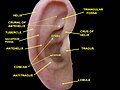
The auricle or auricula is the visible part of the ear that is outside the head. It is also called the pinna, a term that is used more in zoology.
Otoplasty is a procedure for correcting the deformities and defects of the pinna, whether these defects are congenital conditions or caused by trauma. Otoplastic surgeons may reshape, move, or augment the cartilaginous support framework of the pinna to correct these defects.

The ear canal is a pathway running from the outer ear to the middle ear. The adult human ear canal extends from the auricle to the eardrum and is about 2.5 centimetres (1 in) in length and 0.7 centimetres (0.3 in) in diameter.
The helix is the prominent rim of the auricle. Where the helix turns downwards posteriorly, a small tubercle is sometimes seen, namely the auricular tubercle of Darwin.
The tragus is a small pointed eminence of the external ear, situated in front of the concha, and projecting backward over the meatus. It also is the name of hair growing at the entrance of the ear. Its name comes the Ancient Greek tragos, meaning 'goat', and is descriptive of its general covering on its under surface with a tuft of hair, resembling a goat's beard. The nearby antitragus projects forwards and upwards.
The antitragus is a feature of mammalian ear anatomy.

The antitragicus is an intrinsic muscle of the outer ear.

The oblique muscle of auricle is an intrinsic muscle of the outer ear.
The transverse muscle of auricle is an intrinsic muscle of the outer ear.
The Helicis minor is a small skeletal muscle. The helicis minor is an intrinsic muscle of the outer ear. The muscle runs obliques and covers the helical crus, part of the helix located just above the tragus.

The anterior auricular branches of the superficial temporal artery are distributed to the anterior portion of the auricula, the lobule, and part of the external meatus, anastomosing with the posterior auricular. They supply the external acoustic meatus and the visible part of the ear.

Macrotia refers to an ear that is larger than would be expected. The normal auricular axis length is 58–62 mm (2.3–2.4 in) among females and 62–66 mm (2.4–2.6 in) among males. The average width of an adult ear, specifically the distance between the helix root and the posterior auricle, is between 30 and 40 mm.
The posterior auricular muscle is a muscle behind the auricle of the outer ear. It arises from the mastoid part of the temporal bone, and inserts into the lower part of the cranial surface of the auricle of the outer ear. It draws the auricle backwards, usually a very slight effect.

Cryptotia is the condition where an ear appears to have its upper portion buried underneath the side of the head. The condition also involves underdeveloped scapha and antihelical crura. Cryptotia is also known as buried ear or hidden ear.
The Converse technique, together with the Mustardé technique and Stenström technique, belongs to the standard methods of traditional otoplasty. Numerous variations for pinning ears have been derived from them.
The Incisionless Fritsch otoplasty is a minimally invasive procedure for pinning protruding ears.


