The outer ear, external ear, or auris externa is the external part of the ear, which consists of the auricle and the ear canal. It gathers sound energy and focuses it on the eardrum.

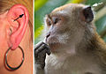
Auriculotherapy is a form of alternative medicine based on the idea that the ear is a micro system and an external organ, which reflects the entire body, represented on the auricle, the outer portion of the ear. Conditions affecting the physical, mental or emotional health of the patient are assumed to be treatable by stimulation of the surface of the ear exclusively. Similar mappings are used by several modalities, including the practices of reflexology and iridology. These mappings are not based on or supported by any medical or scientific evidence, and are therefore considered to be pseudoscience.

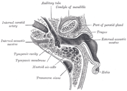
An ear is the organ that enables hearing and body balance using the vestibular system. In mammals, the ear is usually described as having three parts: the outer ear, the middle ear and the inner ear. The outer ear consists of the pinna and the ear canal. Since the outer ear is the only visible portion of the ear in most animals, the word "ear" often refers to the external part alone. The middle ear includes the tympanic cavity and the three ossicles. The inner ear sits in the bony labyrinth, and contains structures which are key to several senses: the semicircular canals, which enable balance and eye tracking when moving; the utricle and saccule, which enable balance when stationary; and the cochlea, which enables hearing. The ear canal is cleaned via earwax, which naturally migrates to the auricle. The ears of vertebrates are placed somewhat symmetrically on either side of the head, an arrangement that aids sound localization.

In anatomy, a tubercle is any round nodule, small eminence, or warty outgrowth found on external or internal organs of a plant or an animal.

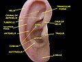
The auricle or auricula is the visible part of the ear that is outside the head. It is also called the pinna, a term that is used more in zoology.

The cervical plexus is a nerve plexus of the anterior rami of the first four cervical spinal nerves C1-C4. The cervical plexus provides motor innervation to some muscles of the neck, and the diaphragm; it provides sensory innervation to parts of the head, neck, and chest.
Otoplasty is a procedure for correcting the deformities and defects of the auricle, whether these defects are congenital conditions or caused by trauma. Otoplastic surgeons may reshape, move, or augment the cartilaginous support framework of the auricle to correct these defects.

The ear canal is a pathway running from the outer ear to the middle ear. The adult human ear canal extends from the auricle to the eardrum and is about 2.5 centimetres (1 in) in length and 0.7 centimetres (0.3 in) in diameter.

Darwin's tubercle is a congenital ear condition which often presents as a thickening on the helix at the junction of the upper and middle thirds.

The inguinal ligament, also known as Poupart's ligament or groin ligament, is a band running from the pubic tubercle to the anterior superior iliac spine. It forms the base of the inguinal canal through which an indirect inguinal hernia may develop.

The auriculotemporal nerve is a sensory branch of the mandibular nerve (CN V3) that runs with the superficial temporal artery and vein, and provides sensory innervation to parts of the external ear, scalp, and temporomandibular joint. The nerve also conveys post-ganglionic parasympathetic fibres from the otic ganglion to the parotid gland.

The external iliac arteries are two major arteries which bifurcate off the common iliac arteries anterior to the sacroiliac joint of the pelvis.
The tragus is a small pointed eminence of the external ear, situated in front of the concha, and projecting backward over the meatus. It also is the name of hair growing at the entrance of the ear. Its name comes the Ancient Greek tragos, meaning 'goat', and is descriptive of its general covering on its under surface with a tuft of hair, resembling a goat's beard. The nearby antitragus projects forwards and upwards.

The condyloid process or condylar process is the process on the human and other mammalian species' mandibles that ends in a condyle, the mandibular condyle. It is thicker than the coronoid process of the mandible and consists of two portions: the condyle and the constricted portion which supports it, the neck.

The antihelix (anthelix) is a part of the visible ear; the pinna. The antihelix is a curved prominence of cartilage parallel with and in front of the helix on the pinna.
The antitragus is a feature of mammalian ear anatomy.

The Helicis minor is a small skeletal muscle. The helicis minor is an intrinsic muscle of the outer ear. The muscle runs obliques and covers the helical crus, part of the helix located just above the tragus.
A vestigial response or vestigial reflex in a species is a response that has lost its original function. In humans, vestigial responses include ear perking, goose bumps and the hypnic jerk.

The following outline is provided as an overview of and topical guide to human anatomy:

The anterior auricular muscle, the smallest of the three auricular muscles, is thin and fan-shaped, and its fibers are pale and indistinct. It arises from the lateral edge of the epicranial aponeurosis, and its fibers converge to be inserted into a projection on the front of the helix.