Outer ear

Last updated
Outer ear
Anatomy of the Human Ear.svg
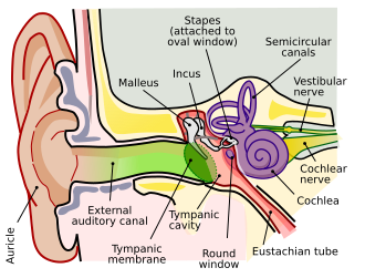
A diagram of the anatomy of the human ear:
  Brown is outer ear.
  Red is middle ear.
  Purple is inner ear.
Gray904.png
The auricula. Lateral surface.
Details
Identifiers
Latin auris externa
MeSH D004431
NeuroLex ID birnlex_1705
TA98 A15.3.01.001
TA2 6862
FMA 52781
Anatomical terminology

The outer ear, external ear, or auris externa is the external part of the ear, which consists of the auricle (also pinna) and the ear canal. [1] It gathers sound energy and focuses it on the eardrum (tympanic membrane).

Contents

Structure

Auricle

The visible part is called the auricle, also known as the pinna, especially in other animals. It is composed of a thin plate of yellow elastic cartilage, covered with integument, and connected to the surrounding parts by ligaments and muscles; and to the commencement of the ear canal by fibrous tissue. Many mammals can move the pinna (with the auriculares muscles) in order to focus their hearing in a certain direction in much the same way that they can turn their eyes. Most humans do not have this ability. [2]

Ear canal

From the pinna, the sound waves move into the ear canal (also known as the external acoustic meatus) a simple tube running through to the middle ear. This tube leads inward from the bottom of the auricula and conducts the vibrations to the tympanic cavity and amplifies frequencies in the range 2  kHz to 5 kHz. [3]

Auricular muscles

Intrinsic muscles

Intrinsic muscles of external ear
Gray906.png
The muscles of the auricula
Details
Nerve Facial nerve
Actions Undeveloped in humans
Identifiers
MeSH D004431
NeuroLex ID birnlex_1705
TA98 A15.3.01.001
TA2 6862
FMA 52781
Anatomical terms of muscle

The intrinsic auricular muscles are:

  • The helicis major is a narrow vertical band situated upon the anterior margin of the helix. It arises below, from the spina helicis, and is inserted into the anterior border of the helix, just where it is about to curve backward.
  • The helicis minor is an oblique fasciculus, covering the crus helicis.
  • The tragicus is a short, flattened vertical band on the lateral surface of the tragus. Also known as the mini lobe.
  • The antitragicus arises from the outer part of the antitragus, and is inserted into the cauda helicis and antihelix.
  • The transverse muscle is placed on the cranial surface of the pinna. It consists of scattered fibers, partly tendinous and partly muscular, extending from the eminentia conchae to the prominence corresponding with the scapha.
  • The oblique muscle also on the cranial surface, consists of a few fibers extending from the upper and back part of the concha to the convexity immediately above it.

The intrinsic muscles contribute to the topography of the auricle, while also function as a sphincter of the external auditory meatus. It has been suggested that during prenatal development in the womb, these muscles exert forces on the cartilage which in turn affects the shaping of the ear. [4]

Extrinsic muscles

Auricular muscles
Gray906.png
The muscles of the pinna
Gray378.png
Auricular muscles in context with the other facial muscles
Details
Origin Galeal aponeurosis
Insertion Front of the helix, cranial surface of the pinna
Artery Posterior auricular artery
Nerve Facial nerve
Actions Subtle auricle movements (forwards, backwards and upwards)
Identifiers
Latin musculi auriculares
MeSH D004431
NeuroLex ID birnlex_1705
TA98 A15.3.01.001
TA2 6862
FMA 52781
Anatomical terms of muscle

The extrinsic auricular muscles are the three muscles surrounding the auricula or outer ear:

The superior muscle is the largest of the three, followed by the posterior and the anterior.

In some mammals these muscles can adjust the direction of the pinna. In humans these muscles possess very little action. The auricularis anterior draws the auricula forward and upward, the auricularis superior slightly raises it, and the auricularis posterior draws it backward. The superior auricular muscle also acts as a stabilizer of the occipitofrontalis muscle and as a weak brow lifter. [5] The presence of auriculomotor activity in the posterior auricular muscle causes the muscle to contract and cause the pinna to be pulled backwards and flatten when exposed to sudden, surprising sounds. [6]

Function

One consequence of the configuration of the outer ear is selectively to boost the sound pressure 30- to 100-fold for frequencies around 3 kHz. This amplification makes humans most sensitive to frequencies in this range—and also explains why they are particularly prone to acoustical injury and hearing loss near this frequency. Most human speech sounds are also distributed in the bandwidth around 3 kHz. [7]

Clinical significance

Malformations of the external ear can be a consequence of hereditary disease, or exposure to environmental factors such as radiation, infection. Such defects include:

Surgery

Usually, malformations are treated with surgery, although artificial prostheses are also sometimes used. [10]

If malformations are accompanied by hearing loss amenable to correction, then the early use of hearing aids may prevent complete hearing loss. [18]

Evolution

The outer ear's cartilage is homologous to the cartilage in gills of amphibians, fishes, and invertebrates such as the horseshoe crab. The extracolumella cartilage of reptiles is likely also homologous. [19]

Additional images

References

PD-icon.svgThis article incorporates text in the public domain from page 1033 of the 20th edition of Gray's Anatomy (1918)

  1. nyu.edu/classes/bello/FMT_files/2_hearing.pdf "Hearing" by Juan P Bello
  2. "Why Can Some People Wiggle Their Ears?". Live Science . 30 March 2012.
  3. "Acoustics Chapter One: The ear". cmtext.indiana.edu. Retrieved 2024-10-21.
  4. Liugan, Mikee; Zhang, Ming; Cakmak, Yusuf Ozgur (2018). "Neuroprosthetics for Auricular Muscles: Neural Networks and Clinical Aspects". Frontiers in Neurology. 8: 752. doi: 10.3389/fneur.2017.00752 . ISSN   1664-2295. PMC   5775970 . PMID   29387041.
  5. Chon, Brian H.; Blandford, Alex D.; Hwang, Catherine J.; Petkovsek, Daniel; Zheng, Andrew; Zhao, Carrie; Cao, Jessica; Grissom, Nick; Perry, Julian D. (February 2021). "Dimensions, Function and Applications of the Auricular Muscle in Facial Plastic Surgery". Aesthetic Plastic Surgery. 45 (1): 309–314. doi:10.1007/s00266-020-02045-x. ISSN   1432-5241. PMID   33258010. S2CID   227236615.
  6. Strauss, Daniel J; Corona-Strauss, Farah I; Schroeer, Andreas; Flotho, Philipp; Hannemann, Ronny; Hackley, Steven A (2020-07-03). Groh, Jennifer M; Shinn-Cunningham, Barbara G; Verhulst, Sarah; Shera, Christopher; Corneil, Brian D (eds.). "Vestigial auriculomotor activity indicates the direction of auditory attention in humans". eLife. 9: e54536. doi: 10.7554/eLife.54536 . ISSN   2050-084X. PMC   7334025 . PMID   32618268.
  7. Purves, Dale, George J. Augustine, David Fitzpatrick, William C. Hall, Anthony-Samuel LaMantia, James O. McNamara, and Leonard E. White (2008). "Chapter 13". Neuroscience. 4th ed. Sinauer Associates. p. 317. ISBN   978-0-87893-697-7.{{cite book}}: CS1 maint: multiple names: authors list (link)
  8. Богомильский, Чистякова 2002, pp. 68–69.
  9. Богомильский, Чистякова 2002, pp. 65–66.
  10. 1 2 3 Пальчун, Крюков 2001, p. 489.
  11. СЭС 1986, p. 89.
  12. СЭС 1986, p. 68.
  13. Богомильский, Чистякова 2002, pp. 66–67.
  14. Богомильский, Чистякова 2002, p. 67.
  15. Богомильский, Чистякова 2002, pp. 67–68.
  16. Асанов и др. 2003, pp. 198–199.
  17. Асанов и др. 2003, p. 198.
  18. 1 2 Богомильский, Чистякова 2002, p. 65.
  19. Thiruppathy, Mathi; Teubner, Lauren; Roberts, Ryan R.; Lasser, Micaela; Moscatello, Alessandra; Chen, Ya-Wen; Hochstim, Christian; Ruffins, Seth; Sarkar, Arijita; Tassey, Jade; Evseenko, Denis; Lozito, Thomas P.; Willsey, Helen Rankin; Gillis, J. Andrew; Crump, J. Gage (9 January 2025). "Repurposing of a gill gene regulatory program for outer ear evolution". Nature. doi:10.1038/s41586-024-08577-5.

Commons-logo.svg Media related to Outer ear at Wikimedia Commons