Muscle fascicle

Last updated
Muscle fascicle
Illu muscle structure.jpg
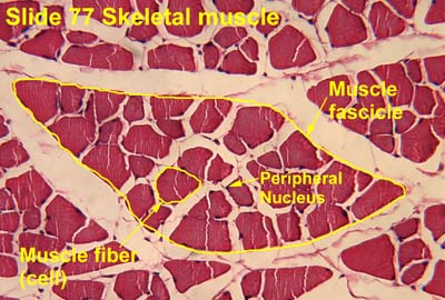
Structure of a skeletal muscle. (Fascicle labeled at bottom right.)
Details
Part of Skeletal muscle
Identifiers
Latin fasiculus muscularis
TA2 2006
TH H3.03.00.0.00003
Anatomical terminology

A muscle fascicle is a bundle of skeletal muscle fibers surrounded by perimysium, a type of connective tissue. [1]

Contents

Structure

Muscle cells are grouped into muscle fascicles by enveloping perimysium connective tissue. [1] Fascicles are bundled together by epimysium connective tissue. [1] Muscle fascicles typically only contain one type of muscle cell (either type I fibres or type II fibres), but can contain a mixture of both types. [2]

Function

In the heart, specialized cardiac muscle cells transmit electrical impulses from the atrioventricular node (AV node) to the Purkinje fibers – fascicles, also referred to as bundle branches.[ citation needed ] These start as a single fascicle of fibers at the AV node called the bundle of His that then splits into three bundle branches: the right fascicular branch, left anterior fascicular branch, and left posterior fascicular branch.

Clinical significance

Myositis may cause thickening of the muscle fascicles. [3] This may be detected with ultrasound scans. [3]

Muscle fascicle structure is a useful diagnostic tool for dermatomyositis. Myocytes towards the edges of the muscle fascicle are typically narrower, while those at the centre of the muscle fascicle are a normal thickness. [4]

Muscle fascicles may be involved in myokymia, although commonly only individual myocytes are involved. [5]

See also

Related Research Articles

<span class="mw-page-title-main">Motor nerve</span> Nerve located in the central nervous system

A motor nerve is a nerve that transmits motor signals from the central nervous system (CNS) to the muscles of the body. This is different from the motor neuron, which includes a cell body and branching of dendrites, while the nerve is made up of a bundle of axons. Motor nerves act as efferent nerves which carry information out from the CNS to muscles, as opposed to afferent nerves, which transfer signals from sensory receptors in the periphery to the CNS. Efferent nerves can also connect to glands or other organs/issues instead of muscles. In addition, there are nerves that serve as both sensory and motor nerves called mixed nerves.

<span class="mw-page-title-main">Purkinje fibers</span> Fibers in the wall of the heart

The Purkinje fibers are located in the inner ventricular walls of the heart, just beneath the endocardium in a space called the subendocardium. The Purkinje fibers are specialized conducting fibers composed of electrically excitable cells. They are larger than cardiomyocytes with fewer myofibrils and many mitochondria. They conduct cardiac action potentials more quickly and efficiently than any of the other cells in the heart's electrical conduction system. Purkinje fibers allow the heart's conduction system to create synchronized contractions of its ventricles, and are essential for maintaining a consistent heart rhythm.

<span class="mw-page-title-main">Muscle cell</span> Type of cell found in muscle tissue

A muscle cell, also known as a myocyte, is a mature contractile cell in the muscle of an animal. In humans and other vertebrates there are three types: skeletal, smooth, and cardiac (cardiomyocytes). A skeletal muscle cell is long and threadlike with many nuclei and is called a muscle fiber. Muscle cells develop from embryonic precursor cells called myoblasts.

<span class="mw-page-title-main">Bundle of His</span> Collection of heart muscle cells

The bundle of His (BH) or His bundle (HB) ( "hiss") is a collection of heart muscle cells specialized for electrical conduction. As part of the electrical conduction system of the heart, it transmits the electrical impulses from the atrioventricular node to the point of the apex of the fascicular branches via the bundle branches. The fascicular branches then lead to the Purkinje fibers, which provide electrical conduction to the ventricles, causing the cardiac muscle of the ventricles to contract at a paced interval.

<span class="mw-page-title-main">Striated muscle tissue</span> Muscle tissue with repeating functional units called sarcomeres

Striated muscle tissue is a muscle tissue that features repeating functional units called sarcomeres. The presence of sarcomeres manifests as a series of bands visible along the muscle fibers, which is responsible for the striated appearance observed in microscopic images of this tissue. There are two types of striated muscle:

<span class="mw-page-title-main">Cardiac conduction system</span> Aspect of heart function

The cardiac conduction system transmits the signals generated by the sinoatrial node – the heart's pacemaker, to cause the heart muscle to contract, and pump blood through the body's circulatory system. The pacemaking signal travels through the right atrium to the atrioventricular node, along the bundle of His, and through the bundle branches to Purkinje fibers in the walls of the ventricles. The Purkinje fibers transmit the signals more rapidly to stimulate contraction of the ventricles.

<span class="mw-page-title-main">Endomysium</span> Connective tissue ensheathing individual muscle fibres

The endomysium, meaning within the muscle, is a wispy layer of areolar connective tissue that ensheaths each individual muscle fiber, or muscle cell. It also contains capillaries and nerves. It overlies the muscle fiber's cell membrane: the sarcolemma. Endomysium is the deepest and smallest component of muscle connective tissue. This thin layer helps provide an appropriate chemical environment for the exchange of calcium, sodium, and potassium, which is essential for the excitation and subsequent contraction of a muscle fiber.

<span class="mw-page-title-main">Perimysium</span> Layer of connective tissue that groups muscle fibers into bundles

Perimysium is a sheath of dense irregular connective tissue that groups muscle fibers into bundles or fascicles.

<span class="mw-page-title-main">Bundle branch block</span> Medical condition

A bundle branch block is a defect in one of the bundle branches in the electrical conduction system of the heart.

<span class="mw-page-title-main">Epineurium</span> Outermost layer of a nerve

The epineurium is the outermost layer of dense irregular connective tissue surrounding a peripheral nerve. It usually surrounds multiple nerve fascicles as well as blood vessels which supply the nerve. Smaller branches of these blood vessels penetrate into the perineurium. In addition to blood vessels which supply the nerve, lymphocytes and fibroblasts are also present and contribute to the production of collagen fibers that form the backbone of the epineurium. In addition to providing structural support, lymphocytes and fibroblasts also play a vital role in maintenance and repair of the surrounding tissues.

<span class="mw-page-title-main">Platysma muscle</span> Human neck muscle

The platysma muscle is a superficial muscle of the human neck that overlaps the sternocleidomastoid. It covers the anterior surface of the neck superficially. When it contracts, it produces a slight wrinkling of the neck, and a "bowstring" effect on either side of the neck.

<span class="mw-page-title-main">Intrafusal muscle fiber</span> Skeletal muscle fibers

Intrafusal muscle fibers are skeletal muscle fibers that serve as specialized sensory organs (proprioceptors). They detect the amount and rate of change in length of a muscle. They constitute the muscle spindle, and are innervated by both sensory (afferent) and motor (efferent) fibers.

<span class="mw-page-title-main">Lingual artery</span>

The lingual artery arises from the external carotid artery between the superior thyroid artery and facial artery. It can be located easily in the tongue.

<span class="mw-page-title-main">Lower subscapular nerve</span>

The lower subscapular nerve, also known as the inferior subscapular nerve, is the third branch of the posterior cord of the brachial plexus. It innervates the inferior portion of the subscapularis muscle and the teres major muscle.

<span class="mw-page-title-main">Perineurium</span> Sheath around nerve fasciculi

The perineurium is a protective sheath that surrounds a nerve fascicle. This bundles together axons targeting the same anatomical location. The perineurium is composed from fibroblasts.

<span class="mw-page-title-main">Endoneurium</span> Connective tissue layer around myelinated nerve fibers in peripheral nervous system

The endoneurium is a layer of delicate connective tissue around the myelin sheath of each myelinated nerve fiber in the peripheral nervous system. Its component cells are called endoneurial cells. The endoneuria with their enclosed nerve fibers are bundled into groups called nerve fascicles, each fascicle within its own protective sheath called a perineurium. In sufficiently large nerves multiple fascicles, each with its blood supply and fatty tissue, may be bundled within yet another sheath, the epineurium.

A pennate or pinnate muscle is a type of skeletal muscle with fascicles that attach obliquely to its tendon. This type of muscle generally allows higher force production but a smaller range of motion. When a muscle contracts and shortens, the pennation angle increases.

<span class="mw-page-title-main">Muscle</span> Basic biological tissue present in animals

Muscle is a soft tissue, one of the four basic types of animal tissue. Muscle tissue gives skeletal muscles the ability to contract. Muscle is formed during embryonic development, in a process known as myogenesis. Muscle tissue contains special contractile proteins called actin and myosin which interact to cause movement. Among many other muscle proteins present are two regulatory proteins, troponin and tropomyosin.

Fascicle or fasciculus may refer to:

<span class="mw-page-title-main">Lateral force transmission in skeletal muscle</span>

A key component in lateral force transmission in skeletal muscle is the extracellular matrix (ECM). Skeletal muscle is a complex biological material that is composed of muscle fibers and an ECM consisting of the epimysium, perimysium, and endomysium. It can be described as a collagen fiber-reinforced composite. The ECM has at least three functions: (1) to provide a framework binding muscle fibers together and ensure their proper alignment, (2) to transmit the forces, either from active muscle contraction or ones passively imposed on it, and (3) providing lubricated surfaces between muscle fibers and bundles enabling the muscle to change shape. The mechanical properties of skeletal muscle depend on both the properties of muscle fibers and the ECM, and the interaction between the two. Contractile forces are transmitted laterally within intramuscular connective tissue to the epimysium and then to the tendon. Due to the nature of skeletal muscle, direct measurements are not possible, but many indirect studies and analyses have shown that the ECM is an important part of force transmission during muscle contraction.

References

  1. 1 2 3 Damjanov, Ivan (2009-01-01), Damjanov, Ivan (ed.), "Chapter 21 - Skeletal Muscles", Pathology Secrets (Third Edition), Philadelphia: Mosby, pp. 434–447, doi:10.1016/b978-0-323-05594-9.00021-0, ISBN   978-0-323-05594-9 , retrieved 2020-11-04
  2. Gandevia, SIMON C.; Burke, DAVID (2004-01-01), Paxinos, GEORGE; Mai, JÜRGEN K. (eds.), "CHAPTER 5 - Peripheral Motor System", The Human Nervous System (Second Edition), San Diego: Academic Press, pp. 113–133, doi:10.1016/b978-012547626-3/50006-5, ISBN   978-0-12-547626-3 , retrieved 2020-11-04
  3. 1 2 Möller, Ingrid; Bong, David; Mendieta, Eugenio de Miguel (2010-01-01), Wakefield, Richard J.; D'Agostino, Maria Antonietta (eds.), "Chapter 19 - Soft Tissue Rheumatism", Essential Applications of Musculoskeletal Ultrasound in Rheumatology, Philadelphia: W.B. Saunders, pp. 219–235, doi:10.1016/b978-1-4377-0127-2.10019-x, ISBN   978-1-4377-0127-2 , retrieved 2020-11-04
  4. Harati, Yadollah; Biliciler, Suur (2010-01-01), Rolak, Loren A. (ed.), "CHAPTER 4 - Myopathies", Neurology Secrets (Fifth Edition), Philadelphia: Mosby, pp. 63–82, doi:10.1016/b978-0-323-05712-7.00004-0, ISBN   978-0-323-05712-7 , retrieved 2020-11-13
  5. Ha, Ainhi D.; Jankovic, Joseph (2011-01-01), Brotchie, Jonathan; Bezard, Erwan; Jenner, Peter (eds.), "An Introduction to Dyskinesia—The Clinical Spectrum", International Review of Neurobiology, Pathophysiology, Pharmacology, and Biochemistry of Dyskinesia, Academic Press, 98: 1–29, doi:10.1016/b978-0-12-381328-2.00001-8, ISBN   9780123813282, PMID   21907081 , retrieved 2020-11-13