Dysferlin also known as dystrophy-associated fer-1-like protein is a protein that in humans is encoded by the DYSFgene.[5] Dysferlin is linked with plasma membrane repair.,[6] stabilization of calcium signaling[7][8][9] and the development of the T-tubule system of the muscle[10] A defect in the DYSF gene, located on chromosome 2p12-14, results in several types of muscular dystrophy; including Miyoshi myopathy (MM), Limb-girdle muscular dystrophy type 2B (LGMD2B) and Distal Myopathy (DM). A reduction or absence of dysferlin, termed dysferlinopathy, usually becomes apparent in the third or fourth decade of life and is characterised by weakness and wasting of various voluntary skeletal muscles.[11] Pathogenic mutations leading to dysferlinopathy can occur throughout the DYSF gene.
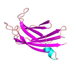
The human dysferlin protein is a 237 kilodalton type-II transmembrane protein.[12][13][14][15][16] It contains a large intracellular cytoplasmic N-terminal domain, an extreme C-terminal transmembrane domain, and a short C-terminal extracellular domain. The cytosolic domain of dysferlin is composed of seven highly conserved C2 domains (C2A-G) which are conserved across several proteins within the ferlin family, including dysferlin homolog myoferlin.[17][18][13] In fact, the C2 domain at any given position is more similar to the C2 domain at the corresponding position within other ferlin family members than the adjacent C2 domain within the same protein. This suggests that each individual C2 domain may in fact play a specific role in dysferlin function and each has in fact been shown to be required for two of dysferlin's roles stabilization of calcium signaling and membrane repair.[19] Mutations in each of these domains can cause dysferlinopathy. A crystal structure of the C2A domain of human dysferlin has been solved, and reveals that the C2A domain changes conformation when interacting with calcium ions,[13] which is consistent with a growing body of evidence suggesting that the C2A domain plays a role in calcium-dependent lipid binding.[20] Its ability to stabilize calcium signaling in the intact dysferlin protein depends on its calcium binding activity.[21] In addition to the C2 domains, dysferlin also contains "FerA" and "DysF" domains. Mutations in both FerA[22] and DysF[23] can cause muscular dystrophies. DysF domain has an interesting structure as in contains one DysF domain within another DysF domain, a result of gene duplication; however, the function of this domain is currently unknown.[23] FerA domain is conserved among all members of ferlin protein family. FerA domain is a four helix bundle and it can interact with membrane, usually in a calcium-dependent manner.[22]
Function
The most intensively studied role for dysferlin is in a cellular process called membrane repair. Membrane repair is a critical mechanism by which cells are able to seal dramatic wounds to the plasma membrane. Muscle is thought to be particularly prone to membrane wounds given that muscle cells transmit high force and undergo cycles of contraction. Dysferlin is highly expressed in muscle, and is homologous to the ferlin family of proteins, which are thought to regulate membrane fusion across a wide variety of species and cell types.[24] Several lines of evidence suggest that dysferlin may be involved in membrane repair in muscle. First, dysferlin-deficient muscle fibers show accumulation of vesicles (which are critical for membrane repair in non-muscle cell types) near membrane lesions, indicating that dysferlin may be required for fusion of repair vesicles with the plasma membrane. Further, dysferlin-deficient muscle fibers take up extracellular dyes to a greater extent than wild-type muscle fibers following laser-induced wounding in-vitro.[25] Dysferlin is also markedly enriched at membrane lesions with several additional proteins thought to be involved in membrane resealing, including annexin and MG53.[26] Exactly how dysferlin contributes to membrane resealing is not clear, but biochemical evidence indicates that dysferlin may bind lipids in a calcium-dependent manner, consistent with a role for dysferlin in regulating fusion of repair vesicles with the sarcolemma during membrane repair.[27] Furthermore, live-cell imaging of dysferlin-eGFP expressing myotubes indicates that dysferlin localizes to a cellular compartment that responds to injury by forming large dysferlin-containing vesicles, and formation of these vesicles may contribute to wound repair.[28] Dysferlin may also be involved in Alzheimer's disease pathogenesis.[29]
Another well studied role for dysferlin is in stabilization of calcium signaling, especially following a mild injury. This approach was based on two observations: that muscle lacking dysferlin that is injured by eccentric contractions can repair its plasma membrane, or sarcolemma, as efficiently as healthy muscle can,[30] and that most of the dysferlin in healthy muscle is concentrated in the transverse tubules at triad junctions,[31][32] where calcium release is regulated. Destabilization of signaling in dysferlinopathic muscle can result in the generation of calcium waves,[33] which can contribute to the disease pathology. Nearly every change in dysferlin that affects membrane repair also destabilizes calcium signaling,[34] suggesting that these two activities are closely linked. Remarkably, however, membrane repair requires calcium ions, whereas calcium ions contribute to the destabilization of signaling when dysferlin is absent or mutated.[35] These paradoxical results have yet to be reconciled.
Interactions
Dysferlin has been shown to bind to itself, to form dimers and perhaps larger oligomers.[36] It can also has been shown to interact with Caveolin 3 in skeletal muscle,[37] and this interaction is thought to retain dysferlin within the plasma membrane.[38] Dysferlin also interacts with MG53, and a functional interaction between dysferlin, caveolin-3 and MG53 is thought to be critical for membrane repair in skeletal muscle.[39]
↑ Vafiadaki E, Reis A, Keers S, Harrison R, Anderson LV, Raffelsberger T, etal. (March 2001). "Cloning of the mouse dysferlin gene and genomic characterization of the SJL-Dysf mutation". NeuroReport. 12 (3): 625–629. doi:10.1097/00001756-200103050-00039. PMID11234777. S2CID22800606.
↑ Bashir R, Britton S, Strachan T, Keers S, Vafiadaki E, Lako M, Richard I, Marchand S, Bourg N, Argov Z, Sadeh M, Mahjneh I, Marconi G, Passos-Bueno MR, Moreira Ede S, Zatz M, Beckmann JS, Bushby K (1998). "A gene related to Caenorhabditis elegans spermatogenesis factor fer-1 is mutated in limb-girdle muscular dystrophy type 2B". Nat. Genet. 20 (1): 37–42. doi:10.1038/1689. PMID9731527. S2CID24234676.
Bejaoui K, Hirabayashi K, Hentati F, Haines JL, Ben Hamida C, Belal S, Miller RG, McKenna-Yasek D, Weissenbach J, Rowland LP (1995). "Linkage of Miyoshi myopathy (distal autosomal recessive muscular dystrophy) locus to chromosome 2p12-14". Neurology. 45 (4): 768–72. doi:10.1212/wnl.45.4.768. PMID7723968. S2CID31029040.
Bashir R, Strachan T, Keers S, Stephenson A, Mahjneh I, Marconi G, Nashef L, Bushby KM (1994). "A gene for autosomal recessive limb-girdle muscular dystrophy maps to chromosome 2p". Hum. Mol. Genet. 3 (3): 455–7. doi:10.1093/hmg/3.3.455. PMID8012357.
Liu J, Aoki M, Illa I, Wu C, Fardeau M, Angelini C, Serrano C, Urtizberea JA, Hentati F, Hamida MB, Bohlega S, Culper EJ, Amato AA, Bossie K, Oeltjen J, Bejaoui K, McKenna-Yasek D, Hosler BA, Schurr E, Arahata K, de Jong PJ, Brown RH (1998). "Dysferlin, a novel skeletal muscle gene, is mutated in Miyoshi myopathy and limb girdle muscular dystrophy". Nat. Genet. 20 (1): 31–6. doi:10.1038/1682. PMID9731526. S2CID12018395.
Bashir R, Britton S, Strachan T, Keers S, Vafiadaki E, Lako M, Richard I, Marchand S, Bourg N, Argov Z, Sadeh M, Mahjneh I, Marconi G, Passos-Bueno MR, Moreira Ede S, Zatz M, Beckmann JS, Bushby K (1998). "A gene related to Caenorhabditis elegans spermatogenesis factor fer-1 is mutated in limb-girdle muscular dystrophy type 2B". Nat. Genet. 20 (1): 37–42. doi:10.1038/1689. PMID9731527. S2CID24234676.
Anderson LV, Davison K, Moss JA, Young C, Cullen MJ, Walsh J, Johnson MA, Bashir R, Britton S, Keers S, Argov Z, Mahjneh I, Fougerousse F, Beckmann JS, Bushby KM (1999). "Dysferlin is a plasma membrane protein and is expressed early in human development". Hum. Mol. Genet. 8 (5): 855–61. doi:10.1093/hmg/8.5.855. PMID10196375.
Weiler T, Bashir R, Anderson LV, Davison K, Moss JA, Britton S, Nylen E, Keers S, Vafiadaki E, Greenberg CR, Bushby CR, Wrogemann K (1999). "Identical mutation in patients with limb girdle muscular dystrophy type 2B or Miyoshi myopathy suggests a role for modifier gene(s)". Hum. Mol. Genet. 8 (5): 871–7. doi:10.1093/hmg/8.5.871. PMID10196377.
Matsuda C, Aoki M, Hayashi YK, Ho MF, Arahata K, Brown RH (1999). "Dysferlin is a surface membrane-associated protein that is absent in Miyoshi myopathy". Neurology. 53 (5): 1119–22. doi:10.1212/wnl.53.5.1119. PMID10496277. S2CID6068681.
Illa I, Serrano-Munuera C, Gallardo E, Lasa A, Rojas-García R, Palmer J, Gallano P, Baiget M, Matsuda C, Brown RH (2001). "Distal anterior compartment myopathy: a dysferlin mutation causing a new muscular dystrophy phenotype". Ann. Neurol. 49 (1): 130–4. doi:10.1002/1531-8249(200101)49:1<130::AID-ANA22>3.0.CO;2-0. PMID11198284. S2CID9278818.
Aoki M, Liu J, Richard I, Bashir R, Britton S, Keers SM, Oeltjen J, Brown HE, Marchand S, Bourg N, Beley C, McKenna-Yasek D, Arahata K, Bohlega S, Cupler E, Illa I, Majneh I, Barohn RJ, Urtizberea JA, Fardeau M, Amato A, Angelini C, Bushby K, Beckmann JS, Brown RH (2001). "Genomic organization of the dysferlin gene and novel mutations in Miyoshi myopathy". Neurology. 57 (2): 271–8. doi:10.1212/wnl.57.2.271. PMID11468312. S2CID31959549.
Ikezoe K, Furuya H, Ohyagi Y, Osoegawa M, Nishino I, Nonaka I, Kira J (2003). "Dysferlin expression in tubular aggregates: their possible relationship to endoplasmic reticulum stress". Acta Neuropathol. 105 (6): 603–9. doi:10.1007/s00401-003-0686-1. PMID12664320. S2CID7734282.
von Tell D, Bruder CE, Anderson LV, Anvret M, Ahlberg G (2003). "Refined mapping of the Welander distal myopathy region on chromosome 2p13 positions the new candidate region telomeric of the DYSF locus". Neurogenetics. 4 (4): 173–7. doi:10.1007/s10048-003-0154-z. PMID12836053. S2CID27539044.
Katz JS, Rando TA, Barohn RJ, Saperstein DS, Jackson CE, Wicklund M, Amato AA (2003). "Late-onset distal muscular dystrophy affecting the posterior calves". Muscle Nerve. 28 (4): 443–8. doi:10.1002/mus.10458. PMID14506716. S2CID29825709.
Confalonieri P, Oliva L, Andreetta F, Lorenzoni R, Dassi P, Mariani E, Morandi L, Mora M, Cornelio F, Mantegazza R (2003). "Muscle inflammation and MHC class I up-regulation in muscular dystrophy with lack of dysferlin: an immunopathological study". J. Neuroimmunol. 142 (1–2): 130–6. doi:10.1016/S0165-5728(03)00255-8. PMID14512171. S2CID37727809.
Cagliani R, Fortunato F, Giorda R, Rodolico C, Bonaglia MC, Sironi M, D'Angelo MG, Prelle A, Locatelli F, Toscano A, Bresolin N, Comi GP (2003). "Molecular analysis of LGMD-2B and MM patients: identification of novel DYSF mutations and possible founder effect in the Italian population". Neuromuscul. Disord. 13 (10): 788–95. doi:10.1016/S0960-8966(03)00133-0. PMID14678801. S2CID23179310.
Brüss M, Homann J, Molderings GJ (2004). "[Dysferlinopathy as an extrahepatic cause for the elevation of serum transaminases]". Med. Klin. (Munich). 99 (6): 326–9. doi:10.1007/s00063-004-1046-1. PMID15221058. S2CID30657667.
This page is based on this Wikipedia article Text is available under the CC BY-SA 4.0 license; additional terms may apply. Images, videos and audio are available under their respective licenses.