Costamere

Last updated
Costamere
Costamere structure in mouse quadriceps - journal.pone.0002604.g003-cropped.png
Costamere structure in mouse quadriceps
Details
Part of Striated muscle
Identifiers
Latin costamerum
MeSH D054974
TH H2.00.05.2.01013
Anatomical terminology

The costamere is a structural-functional component of striated muscle cells [1] which connects the sarcomere of the muscle to the cell membrane (i.e. the sarcolemma). [2]

Contents

Costameres are sub-sarcolemmal protein assemblies circumferentially aligned in register with the Z-disk of peripheral myofibrils. [3] [4] [5] They physically couple force-generating sarcomeres with the sarcolemma in striated muscle cells and are thus considered one of several "Achilles' heels" of skeletal muscle, a critical component of striated muscle morphology which, when compromised, is thought to directly contribute to the development of several distinct myopathies. [6]

The dystrophin-associated protein complex, also referred to as the dystrophin-associated glycoprotein complex (DGC or DAGC), [2] contains various integral and peripheral membrane proteins such as dystroglycans and sarcoglycans, which are thought to be responsible for linking the internal cytoskeletal system of individual myofibers to structural proteins within the extracellular matrix (such as collagen and laminin). Therefore, it is one of the features of the sarcolemma which helps to couple the sarcomere to the extracellular connective tissue as some experiments have shown. [7] Desmin protein may also bind to the DAG complex, and regions of it are known to be involved in signaling.

Structure

Costameres are highly complex networks of proteins and glycoproteins, [8] and can be considered as consisting of two major protein complexes: the dystrophin-glycoprotein complex (DGC) and the integrin-vinculin-talin complex. [9] The sarcoglycans of the DGC and the integrins of the integrin-vinculin-talin complex attach directly to filamin C, a component of the Z-disk, linking these protein complexes of costameres to complexes of the Z-disk. [9] Restated, filamin C physically links the two complexes that constitute the costamere to sarcomeres by interacting with the sarcoglycans in the DGC and the integrins of the integrin-vinculin-talin complex. [9]

The DGC consists of peripheral and integral proteins that physically traverse the sarcolemma and connect the ECM to the F-actin based cytoskeleton. [9] The core proteins of DGC are dystrophin, the sarcoglycans (including alpha, beta, gamma, and lambda sarcoglycan), sarcospan, dystroglycan (alpha and beta), and syntrophin. [9] These proteins are thought to play an important role in maintaining the structural integrity of sarcolemma during contraction and stretching, and loss of these core proteins results in progressive contraction induced damage. [9]

The vinculin and talin components of the integrin-vinculin-talin complex are cytoskeletal proteins physically anchored to the costamere as a whole via the integrin components, which are transmembrane proteins that interact directly with filamin C of the Z disk. [9]

Function

Costameres have several primary functions. [8] [9] [10] First, they keep the sarcolemma in line with the sarcomere during contraction and subsequent relaxation. [10] They are also responsible for the lateral transmission of the sarcomere-generated contractile force to the sarcolemma and the extracellular matrix. [9] [10] Only 20-30% of the total force generated by sarcomere contraction is transmitted longitudinally, suggesting that the majority of the force generated by sarcomeres is transduced in the lateral direction, perpendicular to the contracting myofibril fibers. [9] Most of the force generated by the sarcomeres deep inside the muscle fiber is transmitted perpendicularly to adjacent myofibrils until it reaches the peripheral myofibrils. At that point, the costameric complex channels the force through the sarcolemma to the extracellular matrix. The lateral transmission of force by costameres helps maintain uniform sarcomere lengths in adjacent muscle cells that are under the control of different motor units and are therefore not synchronized in their active contractions; restated, if one muscle fiber is actively contracting and an adjacent one is not, the lateral force transmission helps this second fiber to shorten as well. [8] Costameres also transmit forces in the opposite direction, transmitting the forces of external mechanical stress from the sarcolemma to the Z-disk. [9] Costameres are also involved in protecting the relatively weak and labile sarcolemma from the mechanical stresses of contraction and stretching. [8] [10] The proteins mechanically support the lipid bilayer, and also may facilitate an organized folding of the plasma membrane ("festooning") that minimizes stress on the bilayer during contraction and stretching. [8] Finally, costameres are also involved in the orchestration of mechanically related signaling. [9]

Pathology

The dysfunction of the proteins involved in costameres contributes to some muscular diseases, including muscular dystrophies and cardiomyopathies. [8] [10]

Dynamics

Costameres are dynamic structures. [8] Several studies have suggested that costameres are responsive to mechanical, electrical, and chemical stimuli. [8] For instance, mechanical tension is critical in regulating costameric protein expression, stability, and organization, and dystrophin deficient costameres may sense increased mechanical stress and attempt to compensate with filament recruitment. [8]

Related Research Articles

<span class="mw-page-title-main">Myofibril</span> Contractile element of muscle

A myofibril is a basic rod-like organelle of a muscle cell. Skeletal muscles are composed of long, tubular cells known as muscle fibers, and these cells contain many chains of myofibrils. Each myofibril has a diameter of 1–2 micrometres. They are created during embryonic development in a process known as myogenesis.

<span class="mw-page-title-main">Sarcomere</span> Repeating unit of a myofibril in a muscle cell

A sarcomere is the smallest functional unit of striated muscle tissue. It is the repeating unit between two Z-lines. Skeletal muscles are composed of tubular muscle cells which are formed during embryonic myogenesis. Muscle fibers contain numerous tubular myofibrils. Myofibrils are composed of repeating sections of sarcomeres, which appear under the microscope as alternating dark and light bands. Sarcomeres are composed of long, fibrous proteins as filaments that slide past each other when a muscle contracts or relaxes. The costamere is a different component that connects the sarcomere to the sarcolemma.

<span class="mw-page-title-main">Muscle cell</span> Type of cell found in muscle tissue

A muscle cell is also known as a myocyte when referring to either a cardiac muscle cell (cardiomyocyte) or a smooth muscle cell, as these are both small cells. A skeletal muscle cell is long and threadlike with many nuclei and is called a muscle fiber. Muscle cells develop from embryonic precursor cells called myoblasts.

<span class="mw-page-title-main">Striated muscle tissue</span> Muscle tissue with repeating functional units called sarcomeres

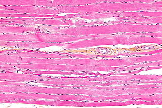
Striated muscle tissue is a muscle tissue that features repeating functional units called sarcomeres. The presence of sarcomeres manifests as a series of bands visible along the muscle fibers, which is responsible for the striated appearance observed in microscopic images of this tissue. There are two types of striated muscle:

<span class="mw-page-title-main">Dystrophin</span> Rod-shaped cytoplasmic protein

Dystrophin is a rod-shaped cytoplasmic protein, and a vital part of a protein complex that connects the cytoskeleton of a muscle fiber to the surrounding extracellular matrix through the cell membrane. This complex is variously known as the costamere or the dystrophin-associated protein complex (DAPC). Many muscle proteins, such as α-dystrobrevin, syncoilin, synemin, sarcoglycan, dystroglycan, and sarcospan, colocalize with dystrophin at the costamere. It has a molecular weight of 427 kDa

<span class="mw-page-title-main">Muscle contraction</span> Activation of tension-generating sites in muscle

Muscle contraction is the activation of tension-generating sites within muscle cells. In physiology, muscle contraction does not necessarily mean muscle shortening because muscle tension can be produced without changes in muscle length, such as when holding something heavy in the same position. The termination of muscle contraction is followed by muscle relaxation, which is a return of the muscle fibers to their low tension-generating state.

<span class="mw-page-title-main">Vinculin</span> Mammalian protein found in Homo sapiens

In mammalian cells, vinculin is a membrane-cytoskeletal protein in focal adhesion plaques that is involved in linkage of integrin adhesion molecules to the actin cytoskeleton. Vinculin is a cytoskeletal protein associated with cell-cell and cell-matrix junctions, where it is thought to function as one of several interacting proteins involved in anchoring F-actin to the membrane.

<span class="mw-page-title-main">Myofilament</span> The two protein filaments of myofibrils in muscle cells

Myofilaments are the three protein filaments of myofibrils in muscle cells. The main proteins involved are myosin, actin, and titin. Myosin and actin are the contractile proteins and titin is an elastic protein. The myofilaments act together in muscle contraction, and in order of size are a thick one of mostly myosin, a thin one of mostly actin, and a very thin one of mostly titin.

The sarcoglycans are a family of transmembrane proteins involved in the protein complex responsible for connecting the muscle fibre cytoskeleton to the extracellular matrix, preventing damage to the muscle fibre sarcolemma through shearing forces.

The dystrophin-associated protein complex, also known as the dystrophin-associated glycoprotein complex is a multiprotein complex that includes dystrophin and the dystrophin-associated proteins. It is one of the two protein complexes that make up the costamere in striated muscle cells. The other complex is the integrin-vinculin-talin complex.

<span class="mw-page-title-main">Paxillin</span> Protein-coding gene in the species Homo sapiens

Paxillin is a protein that in humans is encoded by the PXN gene. Paxillin is expressed at focal adhesions of non-striated cells and at costameres of striated muscle cells, and it functions to adhere cells to the extracellular matrix. Mutations in PXN as well as abnormal expression of paxillin protein has been implicated in the progression of various cancers.

<span class="mw-page-title-main">Integrin beta 1</span> Mammalian protein found in Homo sapiens

Integrin beta-1 (ITGB1), also known as CD29, is a cell surface receptor that in humans is encoded by the ITGB1 gene. This integrin associates with integrin alpha 1 and integrin alpha 2 to form integrin complexes which function as collagen receptors. It also forms dimers with integrin alpha 3 to form integrin receptors for netrin 1 and reelin. These and other integrin beta 1 complexes have been historically known as very late activation (VLA) antigens.

Talin is a high-molecular-weight cytoskeletal protein concentrated at regions of cell–substratum contact and, in lymphocytes, at cell–cell contacts. Discovered in 1983 by Keith Burridge and colleagues, talin is a ubiquitous cytosolic protein that is found in high concentrations in focal adhesions. It is capable of linking integrins to the actin cytoskeleton either directly or indirectly by interacting with vinculin and α-actinin.

<span class="mw-page-title-main">Sarcospan</span>

Originally identified as Kirsten ras associated gene (KRAG), sarcospan (SSPN) is a 25-kDa transmembrane protein located in the dystrophin-associated protein complex of skeletal muscle cells, where it is most abundant. It contains four transmembrane spanning helices with both N- and C-terminal domains located intracellularly. Loss of SSPN expression occurs in patients with Duchenne muscular dystrophy. Dystrophin is required for proper localization of SSPN. SSPN is also an essential regulator of Akt signaling pathways. Without SSPN, Akt signaling pathways will be hindered and muscle regeneration will not occur.

Dystrobrevin is a protein that binds to dystrophin in the costamere of skeletal muscle cells. In humans, there are at least two isoforms of dystrobrevin, dystrobrevin alpha and dystrobrevin beta.

<span class="mw-page-title-main">Actin, cytoplasmic 2</span> Protein-coding gene in the species Homo sapiens

Actin, cytoplasmic 2, or gamma-actin is a protein that in humans is encoded by the ACTG1 gene. Gamma-actin is widely expressed in cellular cytoskeletons of many tissues; in adult striated muscle cells, gamma-actin is localized to Z-discs and costamere structures, which are responsible for force transduction and transmission in muscle cells. Mutations in ACTG1 have been associated with nonsyndromic hearing loss and Baraitser-Winter syndrome, as well as susceptibility of adolescent patients to vincristine toxicity.

<span class="mw-page-title-main">Integrin alpha 7</span>

Alpha-7 integrin is a protein that in humans is encoded by the ITGA7 gene. Alpha-7 integrin is critical for modulating cell-matrix interactions. Alpha-7 integrin is highly expressed in cardiac muscle, skeletal muscle and smooth muscle cells, and localizes to Z-disc and costamere structures. Mutations in ITGA7 have been associated with congenital myopathies and noncompaction cardiomyopathy, and altered expression levels of alpha-7 integrin have been identified in various forms of muscular dystrophy.

<span class="mw-page-title-main">SGCB</span> Protein-coding gene in the species Homo sapiens

Beta-sarcoglycan is a protein that in humans is encoded by the SGCB gene.

<span class="mw-page-title-main">TLN1</span> Protein-coding gene in the species Homo sapiens

Talin-1 is a protein that in humans is encoded by the TLN1 gene. Talin-1 is ubiquitously expressed, and is localized to costamere structures in cardiac and skeletal muscle cells, and to focal adhesions in smooth muscle and non-muscle cells. Talin-1 functions to mediate cell-cell adhesion via the linkage of integrins to the actin cytoskeleton and in the activation of integrins. Altered expression of talin-1 has been observed in patients with heart failure, however no mutations in TLN1 have been linked with specific diseases.

<span class="mw-page-title-main">TLN2</span> Protein-coding gene in the species Homo sapiens

Talin 2 is a protein in humans that is encoded by the TLN2 gene. It belongs to the talin protein family. This gene encodes a protein related to talin 1, a cytoskeletal protein that plays a significant role in the assembly of actin filaments. Talin-2 is expressed at high levels in cardiac muscle and functions to provide linkages between the extracellular matrix and actin cytoskeleton at costamere structures to transduce force laterally.

References

  1. Costameres at the U.S. National Library of Medicine Medical Subject Headings (MeSH)
  2. 1 2 Srivastava, D.; Yu, S (2006). "Stretching to meet needs: integrin-linked kinase and the cardiac pump". Genes Dev. 20 (17): 2327–2331. doi: 10.1101/gad.1472506 . PMID   16951248.20: 2327-2331
  3. Pardo, Jose V; Siliciano, Janet D'Angelo; Craig, Susan W (February 1983). "A vinculin-containing cortical lattice in skeletal muscle: Transverse lattice elements ("costameres") mark sites of attachment between myofibrils and sarcolemma" (PDF). Proceedings of the National Academy of Sciences. 80 (4): 1008–1012. Bibcode:1983PNAS...80.1008P. doi: 10.1073/pnas.80.4.1008 . PMC   393517 . PMID   6405378.
  4. Pardo, Jose V; Siliciano, Janet D'Angelo; Craig, Susan W (1 October 1983). "Vinculin is a component of an extensive network of myofibril-sarcolemma attachment regions in cardiac muscle fibers". Journal of Cell Biology. 97 (4): 1081–1088. doi:10.1083/jcb.97.4.1081. PMC   2112590 . PMID   6413511.
  5. Craig, Susan W; Pardo, Jose V (1983). "Gamma actin, spectrin, and intermediate filament proteins colocalize with vinculin at costameres, myofibril-to-sarcolemma attachment sites". Cell Motility. 3 (5): 449–462. doi:10.1002/cm.970030513. PMID   6420066.
  6. James M. Ervasti (2003). "Costameres: the Achilles' Heel of Herculean Muscle". J. Biol. Chem. 278 (13591–13594): 13591–4. doi: 10.1074/jbc.R200021200 . PMID   12556452.
  7. García-Pelagio Karla; Bloch Robert; Ortega A; Gonzáles-Serratos Hugo (2011). "Biomechanics of the sarcolemma and costameres in single skeletal muscle fibers from normal and dystrophin- null mice". J Muscle Res Cell Motil. 31 (5–6): 323–336. doi:10.1007/s10974-011-9238-9. PMC   4326082 . PMID   21312057.
  8. 1 2 3 4 5 6 7 8 9 Ervasti, James M. (2003-04-18). "Costameres: the Achilles' Heel of Herculean Muscle". Journal of Biological Chemistry. 278 (16): 13591–13594. doi: 10.1074/jbc.R200021200 . ISSN   0021-9258. PMID   12556452.
  9. 1 2 3 4 5 6 7 8 9 10 11 12 Peter, Angela K.; Cheng, Hongqiang; Ross, Robert S.; Knowlton, Kirk U.; Chen, Ju (May 2011). "The costamere bridges sarcomeres to the sarcolemma in striated muscle". Progress in Pediatric Cardiology. 31 (2): 83–88. doi:10.1016/j.ppedcard.2011.02.003. ISSN   1058-9813. PMC   3770312 . PMID   24039381.
  10. 1 2 3 4 5 Bloch, Robert J.; Capetanaki, Yassemi; O'Neill, Andrea; Reed, Patrick; Williams, McRae W.; Resneck, Wendy G.; Porter, Neil C.; Ursitti, Jeanine A. (October 2002). "Costameres: Repeating Structures at the Sarcolemma of Skeletal Muscle". Clinical Orthopaedics and Related Research. 403 (403 Suppl): S203-10. doi:10.1097/00003086-200210001-00024. PMID   12394470.