Crouzon syndrome

Last updated
Crouzon syndrome
Other namesBrachial arch syndrome
Specialty Medical genetics

Crouzon syndrome is an autosomal dominant genetic disorder known as a branchial arch syndrome. Specifically, this syndrome affects the first branchial (or pharyngeal) arch, which is the precursor of the maxilla and mandible. Because the branchial arches are important developmental features in a growing embryo, disturbances in their development create lasting and widespread effects. The syndrome is caused by a mutation in a gene on chromosome 10 that controls the body's production of fibroblast growth factor receptor 2 (FGFR2).

Contents

Crouzon syndrome is named for Octave Crouzon, [1] [2] a French physician who first described this disorder. First called "craniofacial dysostosis" ("craniofacial" refers to the skull and face, and "dysostosis" refers to malformation of bone), the disorder was characterized by a number of clinical features which can be described by the rudimentary meanings of its former name. The developing fetus's skull and facial bones fuse early or are unable to expand. Thus, normal bone growth cannot occur. Fusion of different sutures leads to abnormal patterns of growth of the skull.

Signs and symptoms

Child with Crouzon syndrome showing characteristic facial features. Patient with Crouzon syndrome (1912).jpg
Child with Crouzon syndrome showing characteristic facial features.
Bedell Oxycephalus 3.jpg
Bedell Oxycephalus 4.jpg
Boy with symptoms of Crouzon syndrome at age 2 (left) and 8 (right)
Cranial sutures Sutures from top.png
Cranial sutures

A defining characteristic of Crouzon syndrome is craniosynostosis, which results in an abnormal head shape. This is present in combinations of: frontal bossing, trigonocephaly (fusion of the metopic suture), brachycephaly (fusion of the coronal suture), dolichocephaly (fusion of the sagittal suture), plagiocephaly (unilateral premature closure of lambdoid and coronal sutures), oxycephaly (fusion of coronal and lambdoidal sutures), and complex craniosynostosis (premature closure of some or all sutures).[ citation needed ]

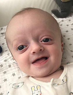
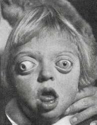
Exophthalmos (bulging eyes due to shallow eye sockets after early fusion of surrounding bones), hypertelorism (greater than normal distance between the eyes), and psittichorhina (beak-like nose) are also very common features. Other facial characteristics that are present in many cases include external strabismus and hypoplastic maxilla (insufficient growth of the midface), which results in relative mandibular prognathism (protruding chin) and gives the effect of the patient having a concave face. [3]

Most symptoms are secondary to the abnormal skull structure. Approximately 30% of people with Crouzon syndrome develop hydrocephalus. Sensorineural hearing loss is present in some cases. The abnormalities in the manner in which the eyes fit in the eye sockets can cause vision problems, the most common of which is corneal exposure that can lead to visual impairment. [4] Some people with the condition have a restricted airway and can experience severe problems breathing. [5]

Common features are a narrow/high-arched palate, posterior bilateral crossbite, hypodontia (missing some teeth), and crowding of teeth. Due to maxillary hypoplasia, people with Crouzon syndrome generally have a considerable permanent underbite. [6]

Causes

The current research indicates fibroblast growth factor receptors (FGFR) FGFR2 and FGFR3 as the leading factors in causing the autosomal dominant Crouzon syndrome. [7] [8] These two transmembrane proteins are two of four fibroblast growth factor receptors involved in osteoblast differentiation during embryonic development; mutations amongst these receptors are involved in several genetic disorders. [7]

There are 40 known mutations, most of which are caused by a missense mutation. [9] FGFR2 is the most commonly mutated gene, a missense at cysteine 342 in exon 9, which creates a gain-of-function. [9] The FGFR2lllc isoform, created via alternative splicing of exon 3 of the FGFR2 gene, uses exon 9 and is used in mesenchymal stem cells to control ossification. However, the mutation constitutively activates the transmembrane protein via a disulfide bond formed incorrectly due to the loss of cysteine 342. [9] FGFR3 is expressed more in the frontal bones during embryonic development, guiding cranial bone development. A point mutation causes constitutive activation of tyrosine in the activation loop, located in the cytosolic region of the protein, leading to accelerated differentiation of frontal osteoblasts, [10] resulting in premature fusion of frontal cranial bones. [10]

Diagnosis

Diagnosis of Crouzon syndrome usually can occur at birth by assessing the physical appearance of the infant. Further analysis, including radiographs, magnetic resonance imaging (MRI) scans, genetic testing and CT scans can be used to confirm the diagnosis of Crouzon syndrome.[ citation needed ]

Treatment

Abnormal fusion of the skull bones is characteristic of Crouzon syndrome. Crouzon4.jpg
Abnormal fusion of the skull bones is characteristic of Crouzon syndrome.

Surgery is typically used to prevent the closure of sutures of the skull from damaging the brain's development. Without surgery, blindness and intellectual disability are typical outcomes. Without treatment, Crouzon syndrome can cause hearing and vision loss, exposure keratitis or conjunctivitis, drying of the cornea, hydrocephalus, sleep apnea, and breathing problems.[ medical citation needed ] To move the orbits forward, surgeons expose the skull and orbits and reshape the bone. To treat the midface deficiency, surgeons can move the lower orbit and midface bones forward.[ medical citation needed ] Additionally, surgery can be performed to relieve pressure inside the skull, fix a cleft lip or palate, correct a malformed jaw, straighten crooked teeth, or correct eye problems.[ medical citation needed ]

People with Crouzon syndrome tend to have multiple sutures involved, most specifically bilateral coronal craniosynostoses, and either open vault surgery or strip craniectomy (if the child is under 6 months) can be performed. In the latter scenario, a helmet is worn for several months following surgery.[ citation needed ]

Once treated for the cranial vault abnormalities, Crouzon patients generally go on to live a normal lifespan.[ citation needed ]

Epidemiology

Incidence of Crouzon syndrome is currently estimated at 1.6 out of every 100,000 people. [11] It is the most common craniostenosis syndrome. [8]

History

Crouzon syndrome was first described by Octave Crouzon in 1912. [12] He noted the affected patients were a mother and her daughter, implying a genetic basis.[ citation needed ]

See also

Related Research Articles

Achondroplasia is a genetic disorder with an autosomal dominant pattern of inheritance whose primary feature is dwarfism. It is the most common cause of dwarfism and affects about 1 in 27,500 people. In those with the condition, the arms and legs are short, while the torso is typically of normal length. Those affected have an average adult height of 131 centimetres for males and 123 centimetres (4 ft) for females. Other features can include an enlarged head with prominent forehead and underdevelopment of the midface. Complications can include sleep apnea or recurrent ear infections. Achondroplasia includes the extremely rare short-limb skeletal dysplasia with severe combined immunodeficiency.

<span class="mw-page-title-main">Trigonocephaly</span> Congenital condition of premature fusion of the metopic suture

Trigonocephaly is a congenital condition due to premature fusion of the metopic suture, leading to a triangular forehead. The premature merging of the two frontal bones leads to transverse growth restriction and parallel growth expansion. It may occur as one component of a syndrome together with other abnormalities, or in isolated form. The term is from the Greek trigonon, "triangle", and kephale, "head".

<span class="mw-page-title-main">Apert syndrome</span> Congenital disorder of the skull and digits

Apert syndrome is a form of acrocephalosyndactyly, a congenital disorder characterized by malformations of the skull, face, hands and feet. It is classified as a branchial arch syndrome, affecting the first branchial arch, the precursor of the maxilla and mandible. Disturbances in the development of the branchial arches in fetal development create lasting and widespread effects.

<span class="mw-page-title-main">Craniosynostosis</span> Premature fusion of bones in the skull

Craniosynostosis is a condition in which one or more of the fibrous sutures in a young infant's skull prematurely fuses by turning into bone (ossification), thereby changing the growth pattern of the skull. Because the skull cannot expand perpendicular to the fused suture, it compensates by growing more in the direction parallel to the closed sutures. Sometimes the resulting growth pattern provides the necessary space for the growing brain, but results in an abnormal head shape and abnormal facial features. In cases in which the compensation does not effectively provide enough space for the growing brain, craniosynostosis results in increased intracranial pressure leading possibly to visual impairment, sleeping impairment, eating difficulties, or an impairment of mental development combined with a significant reduction in IQ.

<span class="mw-page-title-main">Saethre–Chotzen syndrome</span> Medical condition

Saethre–Chotzen syndrome (SCS), also known as acrocephalosyndactyly type III, is a rare congenital disorder associated with craniosynostosis. This affects the shape of the head and face, resulting in a cone-shaped head and an asymmetrical face. Individuals with SCS also have droopy eyelids (ptosis), widely spaced eyes (hypertelorism), and minor abnormalities of the hands and feet (syndactyly). Individuals with more severe cases of SCS may have mild to moderate intellectual or learning disabilities. Depending on the level of severity, some individuals with SCS may require some form of medical or surgical intervention. Most individuals with SCS live fairly normal lives, regardless of whether medical treatment is needed or not.

Craniofacial surgery is a surgical subspecialty that deals with congenital and acquired deformities of the head, skull, face, neck, jaws and associated structures. Although craniofacial treatment often involves manipulation of bone, craniofacial surgery is not tissue-specific; craniofacial surgeons deal with bone, skin, nerve, muscle, teeth, and other related anatomy.

<span class="mw-page-title-main">Carpenter syndrome</span> Medical condition

Carpenter syndrome, also called acrocephalopolysyndactyly type II, is an extremely rare autosomal recessive congenital disorder characterized by craniofacial malformations, obesity, syndactyly, and polydactyly. Acrocephalopolysyndactyly is a variation of acrocephalosyndactyly that presents with polydactyly.

Crouzonodermoskeletal syndrome is a disorder characterized by the premature joining of certain bones of the skull (craniosynostosis) during development and a skin condition called acanthosis nigricans.

<span class="mw-page-title-main">Jackson–Weiss syndrome</span> Medical condition

Jackson–Weiss syndrome (JWS) is a genetic disorder characterized by foot abnormalities and the premature fusion of certain bones of the skull (craniosynostosis), which prevents further growth of the skull and affects the shape of the head and face. This genetic disorder can also sometimes cause intellectual disability and crossed eyes. It was characterized in 1976.

<span class="mw-page-title-main">Pfeiffer syndrome</span> Genetic disorder of the skull

Pfeiffer syndrome is a rare genetic disorder, characterized by the premature fusion of certain bones of the skull (craniosynostosis), which affects the shape of the head and face. The syndrome includes abnormalities of the hands and feet, such as wide and deviated thumbs and big toes.

<span class="mw-page-title-main">Acrocephalosyndactyly</span> Group of diseases

Acrocephalosyndactyly is a group of congenital conditions characterized by irregular features of the face and skull (craniosynostosis) and hands and feet (syndactyly). Craniosynostosis occurs when the cranial sutures, the fibrous tissue connecting the skull bones, fuse the cranial bones early in development. Cranial sutures allow the skull bones to continue growing until they fuse at age 24. Premature fusing of the cranial sutures can result in alterations to the skull shape and interfere with brain growth. Syndactyly occurs when digits of the hands or feet are fused together. When polydactyly is also present, the classification is acrocephalopolysyndactyly. Polydactyly occurs when the hands or feet possess additional digits. Acrocephalosyndactyly is usually diagnosed after birth, although prenatal diagnosis is sometimes possible if the genetic variation is present in family members, as the conditions are typically inherited in an autosomal dominant pattern Treatment often involves surgery in early childhood to correct for craniosynostosis and syndactyly.

<span class="mw-page-title-main">Muenke syndrome</span> Medical condition

Muenke syndrome, also known as FGFR3-related craniosynostosis, is a human specific condition characterized by the premature closure of certain bones of the skull during development, which affects the shape of the head and face. First described by Maximilian Muenke, the syndrome occurs in about 1 in 30,000 newborns. This condition accounts for an estimated 8 percent of all cases of craniosynostosis.

<span class="mw-page-title-main">Fibroblast growth factor receptor 2</span> Protein-coding gene in the species Homo sapiens

Fibroblast growth factor receptor 2 (FGFR2) also known as CD332 is a protein that in humans is encoded by the FGFR2 gene residing on chromosome 10. FGFR2 is a receptor for fibroblast growth factor.

<span class="mw-page-title-main">Fibroblast growth factor receptor 1</span> Protein-coding gene in the species Homo sapiens

Fibroblast growth factor receptor 1 (FGFR1), also known as basic fibroblast growth factor receptor 1, fms-related tyrosine kinase-2 / Pfeiffer syndrome, and CD331, is a receptor tyrosine kinase whose ligands are specific members of the fibroblast growth factor family. FGFR1 has been shown to be associated with Pfeiffer syndrome, and clonal eosinophilias.

<span class="mw-page-title-main">Fibroblast growth factor receptor 3</span> Gene involved in the most common form of dwarfism

Fibroblast growth factor receptor 3 is a protein that in humans is encoded by the FGFR3 gene. FGFR3 has also been designated as CD333. The gene, which is located on chromosome 4, location p16.3, is expressed in tissues such as the cartilage, brain, intestine, and kidneys.

<span class="mw-page-title-main">Antley–Bixler syndrome</span> Congenital disorder

Antley–Bixler syndrome is a rare, severe autosomal recessive congenital disorder characterized by malformations and deformities affecting the majority of the skeleton and other areas of the body.

<span class="mw-page-title-main">McGillivray syndrome</span> Medical condition

McGillivray syndrome is a rare syndrome characterized mainly by heart defects, skull and facial abnormalities and ambiguous genitalia. The symptoms of this syndrome are ventricular septal defect, patent ductus arteriosus, small jaw, undescended testes, and webbed fingers. Beside to these symptoms there are more symptoms which is related with bone structure and misshape.

Beare–Stevenson cutis gyrata syndrome is a rare genetic disorder characterized by craniosynostosis and a specific skin abnormality, called cutis gyrata, characterized by a furrowed and wrinkled appearance ; thick, dark, velvety areas of skin are sometimes found on the hands and feet and in the groin.

<span class="mw-page-title-main">Baller–Gerold syndrome</span> Medical condition

Baller–Gerold syndrome (BGS) is a rare genetic syndrome that involves premature fusion of the skull bones and malformations of facial, forearm and hand bones. The symptoms of Baller–Gerold syndrome overlap with features of a few other genetics disorders: Rothmund–Thomson syndrome and RAPADILINO syndrome. The prevalence of BGS is unknown, as there have only been a few reported cases, but it is estimated to be less than 1 in a million. The name of the syndrome comes from the researchers Baller and Gerold who discovered the first three cases.

<span class="mw-page-title-main">Severe achondroplasia with developmental delay and acanthosis nigricans</span> Medical condition

Severe achondroplasia with developmental delay and acanthosis nigricans (SADDAN) is a very rare genetic disorder. This disorder is one that affects bone growth and is characterized by skeletal, brain, and skin abnormalities. Those affected by the disorder are severely short in height and commonly possess shorter arms and legs. In addition, the bones of the legs are often bowed and the affected have smaller chests with shorter rib bones, along with curved collarbones. Other symptoms of the disorder include broad fingers and extra folds of skin on the arms and legs. Developmentally, many individuals who suffer from the disorder show a higher level in delays and disability. Seizures are also common due to structural abnormalities of the brain. Those affected may also suffer with apnea, the slowing or loss of breath for short periods of time.

References

  1. synd/1383 at Who Named It?
  2. L. E. O. Crouzon. Dysostose cranio-faciale héréditaire. Bulletin de la Société des Médecins des Hôpitaux de Paris, 1912, 33: 545-555.
  3. "Crouzon syndrome". rarediseases.info.nih.gov. US: Genetic and Rare Diseases Information Center (GARD), National Institutes of Health . Retrieved 21 November 2018.
  4. Gray, Thomas L.; Casey, Theresa; Selva, Dinesh; Anderson, Peter J.; David, David J. (9 May 2005). "Ophthalmic Sequelae of Crouzon Syndrome". Ophthalmology. 112 (6): 1129–1134. doi:10.1016/j.ophtha.2004.12.037. PMID   15885794.
  5. "Crouzon Syndrome". rarediseases.org. National Organization for Rare Disorders (NORD). Retrieved 21 November 2018.
  6. Flint, Paul (2015). Cummings Otolaryngology (6th ed.). Elsevier. pp. 2891–2914.
  7. 1 2 Snyder-Warwick AK, Perlyn CA, Pan J, Yu K, Zhang L, Ornitz DM (February 2010). "Analysis of a gain-of-function FGFR2 Crouzon mutation provides evidence of loss of function activity in the etiology of cleft palate". Proc. Natl. Acad. Sci. U.S.A. 107 (6): 2515–20. Bibcode:2010PNAS..107.2515S. doi: 10.1073/pnas.0913985107 . PMC   2823872 . PMID   20133659.
  8. 1 2 "Crouzon syndrome". Genetics Home Reference. US: US National Library of Medicine, National Institutes of Health. Retrieved 21 November 2018 via ghr.nlm.nih.gov.
  9. 1 2 3 Fenwick AL, Goos JA, Rankin J, Lord H, Lester T, Hoogeboom AJ, van den Ouweland AM, Wall SA, Mathijssen IM, Wilkie AO (August 2014). "Apparently synonymous substitutions in FGFR2 affect splicing and result in mild Crouzon syndrome". BMC Med. Genet. 15: 95. doi: 10.1186/s12881-014-0095-4 . PMC   4236556 . PMID   25174698.
  10. 1 2 Di Rocco F, Biosse Duplan M, Heuzé Y, Kaci N, Komla-Ebri D, Munnich A, Mugniery E, Benoist-Lasselin C, Legeai-Mallet L (June 2014). "FGFR3 mutation causes abnormal membranous ossification in achondroplasia". Hum. Mol. Genet. 23 (11): 2914–25. doi: 10.1093/hmg/ddu004 . PMID   24419316.
  11. Adam MP, Ardinger HH, Pagon RA, Wallace SE, Bean LH, Stephens K, Amemiya A, Robin NH, Falk MJ, Haldeman-Englert CR (October 20, 1998). "FGFR-Related Craniosynostosis Syndromes". GeneReviews. PMID   20301628.
  12. Rodriguez, Eduardo (2018). Plastic Surgery: Volume 3: Craniofacial, Head and Neck Surgery and Pediatric Plastic Surgery (4 ed.). Elsevier.