Cerebral palsy (CP) is a group of movement disorders that appear in early childhood. Signs and symptoms vary among people and over time, but include poor coordination, stiff muscles, weak muscles, and tremors. There may be problems with sensation, vision, hearing, and speech. Often, babies with cerebral palsy do not roll over, sit, crawl or walk as early as other children. Other symptoms may include seizures and problems with thinking or reasoning. While symptoms may get more noticeable over the first years of life, underlying problems do not worsen over time.
Spasticity is a feature of altered skeletal muscle performance with a combination of paralysis, increased tendon reflex activity, and hypertonia. It is also colloquially referred to as an unusual "tightness", stiffness, or "pull" of muscles.

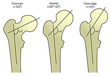
Coxa vara is a deformity of the hip, whereby the angle between the head and the shaft of the femur is reduced to less than 120 degrees. This results in the leg being shortened and the development of a limp. It may be congenital and is commonly caused by injury, such as a fracture. It can also occur when the bone tissue in the neck of the femur is softer than normal, causing it to bend under the weight of the body. This may either be congenital or the result of a bone disorder. The most common cause of coxa vara is either congenital or developmental. Other common causes include metabolic bone diseases, post-Perthes deformity, osteomyelitis, and post traumatic. Shepherd's Crook deformity is a severe form of coxa vara where the proximal femur is severely deformed with a reduction in the neck shaft angle beyond 90 degrees. It is most commonly a sequela of osteogenesis imperfecta, Paget's disease, osteomyelitis, tumour and tumour-like conditions.

Genu valgum, commonly called "knock-knee", is a condition in which the knees angle in and touch each other when the legs are straightened. Individuals with severe valgus deformities are typically unable to touch their feet together while simultaneously straightening the legs. The term originates from Latin genu 'knee' and valgus 'bent outwards', but is also used to describe the distal portion of the knee joint which bends outwards and thus the proximal portion seems to be bent inwards.

Genu varum is a varus deformity marked by (outward) bowing at the knee, which means that the lower leg is angled inward (medially) in relation to the thigh's axis, giving the limb overall the appearance of an archer's bow. Usually medial angulation of both lower limb bones is involved.

A varus deformity is an excessive inward angulation of the distal segment of a bone or joint. The opposite of varus is called valgus.
A valgus deformity is a condition in which the bone segment distal to a joint is angled outward, that is, angled laterally, away from the body's midline. The opposite deformation, where the twist or angulation is directed medially, toward the center of the body, is called varus.

In vertebrate anatomy, the hip, or coxa in medical terminology, refers to either an anatomical region or a joint on the outer (lateral) side of the pelvis.

The asymmetrical tonic neck reflex (ATNR) is a primitive reflex found in newborn humans that normally vanishes around 6 months of age. It is also known as the bow and arrow or "fencing reflex" because of the characteristic position of the infant's arms and head, which resembles that of a fencer. When the face is turned to one side, the arm and leg on that side extend, and the arm and leg on the opposite side flex. It is more likely to be seen in premature infants than full-term babies. It is rare in newborns but can be stimulated from infants to up to 3 months old. It is believed to help develop hand-eye coordination and help with awareness of both sides of the body.
A selective dorsal rhizotomy (SDR), also known as a rhizotomy, dorsal rhizotomy, or a selective posterior rhizotomy, is a neurosurgical procedure that selectively cuts problematic nerve roots in the spinal cord. This procedure has been well-established in the literature as a surgical intervention and is used to relieve negative symptoms of neuromuscular conditions such as spastic diplegia and other forms of spastic cerebral palsy. The specific sensory nerves inducing spasticity are identified using electromyographic (EMG) stimulation and graded on a scale of 1 (mild) to 4. Abnormal nerve responses are isolated and cut, thereby reducing symptoms of spasticity.
Epiphysiodesis is a pediatric orthopedic surgery procedure that aims at altering or stopping the bone growth naturally occurring through the growth plate also known as the physeal plate. There are two types of epiphysiodesis: temporary hemiepiphysiodesis and permanent epiphysiodesis. Temporary hemiepiphysiodesis is also known as guided growth surgery or growth modulation surgery. Temporary hemiepiphysiodesis is reversible i.e. the metal implants used to achieve epiphysiodesis can be removed after the desired correction is achieved and the growth plate can thus resume its normal growth and function. In contrast, permanent epiphysiodesis is irreversible and the growth plate function cannot be restored after surgery. Both temporary hemiepiphysiodesis and permanent epiphysiodesis are used to treat a diverse array of pediatric orthopedic disorders but the exact indications for each procedure are different.

Singleton Merten syndrome is an autosomal dominant genetic disorder with variable expression with an onset of symptoms during childhood.

Over time, the approach to cerebral palsy management has shifted away from narrow attempts to fix individual physical problems – such as spasticity in a particular limb – to making such treatments part of a larger goal of maximizing the person's independence and community engagement. Much of childhood therapy is aimed at improving gait and walking. Approximately 60% of people with CP are able to walk independently or with aids at adulthood. However, the evidence base for the effectiveness of intervention programs reflecting the philosophy of independence has not yet caught up: effective interventions for body structures and functions have a strong evidence base, but evidence is lacking for effective interventions targeted toward participation, environment, or personal factors. There is also no good evidence to show that an intervention that is effective at the body-specific level will result in an improvement at the activity level, or vice versa. Although such cross-over benefit might happen, not enough high-quality studies have been done to demonstrate it.

Genu recurvatum is a deformity in the knee joint, so that the knee bends backwards. In this deformity, excessive extension occurs in the tibiofemoral joint. Genu recurvatum is also called knee hyperextension and back knee. This deformity is more common in women and people with familial ligamentous laxity. Hyperextension of the knee may be mild, moderate or severe.
Dyskinetic cerebral palsy (DCP) is a subtype of cerebral palsy (CP) and is characterized by impaired muscle tone regulation, coordination and movement control. Dystonia and choreoathetosis are the two most dominant movement disorders in patients with DCP.
The Gross Motor Function Classification System or GMFCS is a 5 level clinical classification system that describes the gross motor function of people with cerebral palsy on the basis of self-initiated movement abilities. Particular emphasis in creating and maintaining the GMFCS scale rests on evaluating sitting, walking, and wheeled mobility. Distinctions between levels are based on functional abilities; the need for walkers, crutches, wheelchairs, or canes / walking sticks; and to a much lesser extent, the actual quality of movement.

The ADELI Suit is derived from a suit originally designed for the Soviet space program in the late 1960s that was first tested in 1971. The purpose then was to give the cosmonauts in space a way to counter the effects of long-term weightlessness on the body. The ADELI Suit is currently used to treat children with physical disabilities resulting from cerebral palsy, other neurological conditions originating from brain damage or spinal cord injury.

Orthopedic surgery is the branch of surgery concerned with conditions involving the musculoskeletal system. Orthopedic surgeons use both surgical and nonsurgical means to treat musculoskeletal injuries, sports injuries, degenerative diseases, infections, bone tumours, and congenital limb deformities. Trauma surgery and traumatology is a sub-specialty dealing with the operative management of fractures, major trauma and the multiply-injured patient.
X-rays of hip dysplasia are one of the two main methods of medical imaging to diagnose hip dysplasia, the other one being medical ultrasonography. Ultrasound imaging yields better results defining the anatomy until the cartilage is ossified. When the infant is around 3 months old a clear roentgenographic image can be achieved. Unfortunately the time the joint gives a good x-ray image is also the point at which nonsurgical treatment methods cease to give good results.