Trigonocephaly

Last updated
Trigonocephaly
Trigonocephaly (2).png
Trigonocephaly in a boy with 1q22–1q23.1 duplication
Specialty Medical genetics

Trigonocephaly is a congenital condition due to premature fusion of the metopic suture (from the Greek metopon, "forehead"), leading to a triangular forehead. The premature merging of the two frontal bones leads to transverse growth restriction and parallel growth expansion. It may occur as one component of a syndrome together with other abnormalities, or in isolated form. The term is from the Greek trigonon, "triangle", and kephale, "head".

Contents

Cause

Fused suture in trigonocephaly Trigonocephaly.png
Fused suture in trigonocephaly
Trigonocephaly as a kind of craniosynostosis Single suture synostosis.png
Trigonocephaly as a kind of craniosynostosis

Trigonocephaly can either occur in a syndrome or isolated, all by itself. Trigonocephaly is associated with the following syndromes: Bohring-Opitz syndrome, Muenke syndrome, Jacobsen syndrome, Baller–Gerold syndrome and Say–Meyer syndrome. The etiology of trigonocephaly is mostly unknown although there are three main theories. [1] Trigonocephaly is probably a multifactorial congenital condition, but due to limited proof of these theories this cannot safely be concluded. [2]

Intrinsic bone malformation

The first theory assumes that the origin of pathological synostosis lies within disturbed bone formation early on in the pregnancy. Causes can either be genetic [3] [4] [5] [6] (9p22–24, 11q23, 22q11, FGFR1 mutation), [7] metabolic [8] (TSH suppletion in hypothyroidism) [9] [10] or pharmaceutical [11] (valproate in epilepsy). [12] [13]

Fetal-head constraint

The second theory says that synostosis begins when the fetal head gets hindered in the pelvic outlet during birth. [14] [15]

Intrinsic brain malformation

The third theory predominates disturbed brain formation of the two frontal lobes as the main issue behind synostosis. [16] [17] [18] Limited growth of the frontal lobes leads to an absence of stimuli for cranial growth, therefore causing premature fusion of the metopic suture.[ citation needed ]

Other conditions and syndromes

Microcephalics (inset, D).jpg
Microcephalic idiot with local myxedema.jpg
Trigonocephaly in two cases of microcephaly

Other conditions and syndromes with trigonocephaly include:

Diagnosis

Diagnosis can be characterized by typical facial and cranial deformities. [2] [19]

Observatory signs of trigonocephaly are:

Imaging techniques (3D-CT, Röntgenography, MRI) show:

The neuropsychological development is not always affected. These effects are only visible in a small percentage of children with trigonocephaly or other suture synostoses.

Neuropsychological signs are:

Treatment

Treatment is surgical with attention to form and volume. Surgery usually takes place before the age of one since it has been reported that the intellectual outcome is better. [27] [28] [20] [21] [22] [29] [30] [31] [32] [ excessive citations ]

Wiki trigonocephaly1.jpg
A. Fronto-supraorbital advancement and remodelling – before remodelling
Wiki trigonocephaly2.jpg
B. Fronto-supraorbital advancement and remodelling – after remodelling

Fronto-supraorbital advancement and remodelling

A form of surgery is the so-called fronto-supraorbital advancement and remodelling. [33] Firstly, the supraorbital bar is remodelled by a wired greenstick fracture to straighten it. Secondly, the supraorbital bar is moved 2 cm. forward and fixed only to the frontal process of the zygoma without fixation to the cranium. Lastly, the frontal bone is divided into two, rotated and attached to the supraorbital bar causing a nude area (craniectomy) between the parietal bone and frontal bone. Bone will eventually regenerate since the dura mater lies underneath (the dura mater has osteogenic capabilities). This results in an advancement and straightening of the forehead.

'Floating forehead technique'

The so-called 'floating forehead technique' [2] combined with the remodelling of the supraorbital bar is derived from the fronto-supraorbital advancement and remodelling. The supraorbital bar is remodelled as described above. [34] [35] The frontal bone is split in two pieces. Instead of using both pieces as in fronto-supraorbital advancement and remodelling, only one piece is rotated and attached to the supraorbital bar. This technique also leaves a craniectomy behind. [36]

Other

The simplest form of surgery for trigonocephaly was suturectomy. [37] [21] [34] [38] [39] However, as this technique was insufficient to correct the deformities, it is not used anymore.

Distraction osteogenesis is based on creating more cranial space for the brain by gradually moving the bones apart. This can be achieved by using springs. [40]

These approaches are 2D solutions for a 3D problem, therefore the results are not optimal. Distraction osteogenesis and minimal invasive endoscopic surgery are yet in experimental phase.

Outcomes

Surgical

Trigonocephaly seems to be the most compliant form of craniosynostosis for surgery. [22] Because of standardization of current surgical approaches there is no surgical mortality and complications are few to none. [2] [41] [42] The simple suturectomy is presently insufficient to adjust the complicated growth restrictions caused by metopic synostosis. [2] On the other hand, the fronto-supraorbital advancement and remodelling and the 'floating forehead technique' create sufficient space for brain growth and result in a normal horizontal axis of the orbits and supraorbital bar. The fronto-supraorbital advancement and remodelling is the most used method nowadays. [41] Over the past few years[ when? ] distraction osteogenesis has been gradually acknowledged since it has a positive effect on hypotelorism.[ citation needed ] Expanding the distance between the orbits using springs seems to be successful. [2] [43] [44] [45] However, there are discussions whether hypotelorism really needs to be corrected. [46] The minimal invasive endoscopic surgery has been gaining attention since the early '90s, however, it has technical limitations (only strip craniectomy is possible). [2] Attempts have been made to reach beyond these limits. [47] [48] [49] [50] [51]

Aesthetic

Aesthetic outcome of metopic synostosis surgery is persistently good with reoperation hazards below 20%. [52] [53] In 1981 Anderson advised that craniofacial operations for synostosis should be as extensive as necessary after a study of 107 cases of metopic and coronal synostosis. [21] Surgery does not provide a 100% natural outcome, mostly there will be minor irregularities. Reoperations are usually performed on more severe cases (including syndromic metopic synostosis). The hypotelorism and temporal hollowing are the most difficult to correct: the hypotelorism usually remains under corrected and a second operation is often needed for correction of temporal hollowing. [52] [54]

Neurological

The highest rate of neurological problems of single suture synostosis are seen in patients with trigonocephaly. [2] Surgery is performed generally before the age of one because of claims of better intellectual outcome. [27] [28] [20] [21] [22] [29] [30] [31] [32] Seemingly surgery does not influence the high incidence of neurodevelopment problems in patients with metopic synostosis. Neurological disorders such as ADHD, ASD, ODD and CD are seen in patients with trigonocephaly. These disorders are usually also associated with decreased IQ. The presence of ADHD, ASD and ODD is higher in cases with an IQ below 85. This is not the case with CD which showed an insignificant increase at an IQ below 85. [2]

Epidemiology

The incidence of metopic synostosis is roughly between 1:700 and 1:15,000 newborns globally (differs per country). [11] [55] Trigonocephaly is seen more in males than females ranging from 2:1 to 6.5:1. [37] [56] [27] [28] Hereditary relations in metopic synostosis have been found of which 5.5% were well defined syndromic. [11] Maternal age and a birth weight of less than 2500g may also play a role in trigonocephaly. [57] These data are based on estimations and do not give factual information.

Only one article gives valuable and reliable information regarding the incidence of metopic synostosis in the Netherlands. The incidence in the Netherlands showed an increase from 0.6 (1997) to 1.9 (2007) for every 10,000 live births. [58]

History

In former times people born with malformed skulls were rejected based upon their appearance. [1] [59] This still persists today in various parts of the world even though the intellectual development is often normal. [2] The Austrian physician Franz Joseph Gall presented the science of phrenology in the early 19th century through his work The Anatomy and Physiology of the Nervous System in General, and of the Brain in Particular. [60]

Hippocrates described trigonocephaly as follows: Men's heads are by no means all like to one another, nor are the sutures of the head of all men constructed in the same form. Thus, whoever has a prominence in the anterior part of the head (by prominence is meant the round protuberant part of the bone which projects beyond the rest of it), in him the sutures of the head take the form of the Greek letter 'tau', τ . [61] [62]

Hermann Welcker coined the term trigonocephaly in 1862. He described a child with a V-shaped skull and a cleft lip. [63]

Via a photo shown on a Facebook page, the mother of a child previously diagnosed with this condition recognised the symptoms and reported them to the family involved, resulting in an immediate diagnosis that medical professionals had overlooked in all earlier consultations. [64]

Related Research Articles

<span class="mw-page-title-main">Say–Meyer syndrome</span> X-linked recessive disorder characterised by developmental delay

Say–Meyer syndrome is a rare X-linked genetic disorder that is mostly characterized as developmental delay. It is one of the rare causes of short stature. It is closely related with trigonocephaly. People with Say–Meyer syndrome have impaired growth, deficits in motor skills development and mental state.

<span class="mw-page-title-main">Scaphocephaly</span> Cephalic disorder involving premature fusion of the sagittal suture

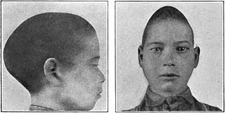
Scaphocephaly, or sagittal craniosynostosis, is a type of cephalic disorder which occurs when there is a premature fusion of the sagittal suture. Premature closure results in limited lateral expansion of the skull resulting in a characteristic long, narrow head. The skull base is typically spared.

<span class="mw-page-title-main">Rhinoplasty</span> Surgical procedure to enhance or reconstruct a human nose

Rhinoplasty, commonly called nose job, medically called nasal reconstruction is a plastic surgery procedure for altering and reconstructing the nose. There are two types of plastic surgery used – reconstructive surgery that restores the form and functions of the nose and cosmetic surgery that changes the appearance of the nose. Reconstructive surgery seeks to resolve nasal injuries caused by various traumas including blunt, and penetrating trauma and trauma caused by blast injury. Reconstructive surgery can also treat birth defects, breathing problems, and failed primary rhinoplasties. Rhinoplasty may remove a bump, narrow nostril width, change the angle between the nose and the mouth, or address injuries, birth defects, or other problems that affect breathing, such as a deviated nasal septum or a sinus condition. Surgery only on the septum is called a septoplasty.

Oral and maxillofacial surgery is a surgical specialty focusing on reconstructive surgery of the face, facial trauma surgery, the oral cavity, head and neck, mouth, and jaws, as well as facial cosmetic surgery/facial plastic surgery including cleft lip and cleft palate surgery.

Crouzon syndrome is an autosomal dominant genetic disorder known as a branchial arch syndrome. Specifically, this syndrome affects the first branchial arch, which is the precursor of the maxilla and mandible. Since the branchial arches are important developmental features in a growing embryo, disturbances in their development create lasting and widespread effects.

<span class="mw-page-title-main">Apert syndrome</span> Congenital disorder of the skull and digits

Apert syndrome is a form of acrocephalosyndactyly, a congenital disorder characterized by malformations of the skull, face, hands and feet. It is classified as a branchial arch syndrome, affecting the first branchial arch, the precursor of the maxilla and mandible. Disturbances in the development of the branchial arches in fetal development create lasting and widespread effects.

<span class="mw-page-title-main">Craniosynostosis</span> Premature fusion of bones in the skull

Craniosynostosis is a condition in which one or more of the fibrous sutures in a young infant's skull prematurely fuses by turning into bone (ossification), thereby changing the growth pattern of the skull. Because the skull cannot expand perpendicular to the fused suture, it compensates by growing more in the direction parallel to the closed sutures. Sometimes the resulting growth pattern provides the necessary space for the growing brain, but results in an abnormal head shape and abnormal facial features. In cases in which the compensation does not effectively provide enough space for the growing brain, craniosynostosis results in increased intracranial pressure leading possibly to visual impairment, sleeping impairment, eating difficulties, or an impairment of mental development combined with a significant reduction in IQ.

<span class="mw-page-title-main">Microtia</span> Medical condition

Microtia is a congenital deformity where the auricle is underdeveloped. A completely undeveloped pinna is referred to as anotia. Because microtia and anotia have the same origin, it can be referred to as microtia-anotia. Microtia can be unilateral or bilateral. Microtia occurs in 1 out of about 8,000–10,000 births. In unilateral microtia, the right ear is most commonly affected. It may occur as a complication of taking Accutane (isotretinoin) during pregnancy.

Craniofacial surgery is a surgical subspecialty that deals with congenital and acquired deformities of the head, skull, face, neck, jaws and associated structures. Although craniofacial treatment often involves manipulation of bone, craniofacial surgery is not tissue-specific; craniofacial surgeons deal with bone, skin, nerve, muscle, teeth, and other related anatomy.

<span class="mw-page-title-main">Frontal suture</span> Midline joint in the frontal bone of the forehead

The frontal suture is a fibrous joint that divides the two halves of the frontal bone of the skull in infants and children. Typically, it completely fuses between three and nine months of age, with the two halves of the frontal bone being fused together. It is also called the metopic suture, although this term may also refer specifically to a persistent frontal suture.

<span class="mw-page-title-main">Acrocephalosyndactyly</span> Group of diseases

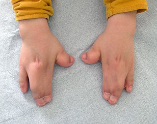
Acrocephalosyndactyly is a group of congenital conditions characterized by irregular features of the face and skull (craniosynostosis) and hands and feet (syndactyly). Craniosynostosis occurs when the cranial sutures, the fibrous tissue connecting the skull bones, fuse the cranial bones early in development. Cranial sutures allow the skull bones to continue growing until they fuse at age 24. Premature fusing of the cranial sutures can result in alterations to the skull shape and interfere with brain growth. Syndactyly occurs when digits of the hands or feet are fused together. When polydactyly is also present, the classification is acrocephalopolysyndactyly. Polydactyly occurs when the hands or feet possess additional digits. Acrocephalosyndactyly is usually diagnosed after birth, although prenatal diagnosis is sometimes possible if the genetic variation is present in family members, as the conditions are typically inherited in an autosomal dominant pattern Treatment often involves surgery in early childhood to correct for craniosynostosis and syndactyly.

Migraine surgery is a surgical operation undertaken with the goal of reducing or preventing migraines. Migraine surgery most often refers to surgical nerve decompression of one or several nerves in the head and neck which have been shown to trigger migraine symptoms in many migraine sufferers. Following the development of nerve decompression techniques for the relief of migraine pain in the year 2000, these procedures have been extensively studied and shown to be effective in appropriate candidates. The nerves that are most often addressed in migraine surgery are found outside of the skull, in the face and neck, and include the supra-orbital and supra-trochlear nerves in the forehead, the zygomaticotemporal nerve and auriculotemporal nerves in the temple region, and the greater occipital, lesser occipital, and third occipital nerves in the back of the neck. Nerve impingement in the nasal cavity has additionally been shown to be a trigger of migraine symptoms.

<span class="mw-page-title-main">Frontonasal dysplasia</span> Medical condition

Frontonasal dysplasia (FND) is a congenital malformation of the midface. For the diagnosis of FND, a patient should present at least two of the following characteristics: hypertelorism, a wide nasal root, vertical midline cleft of the nose and/or upper lip, cleft of the wings of the nose, malformed nasal tip, encephalocele or V-shaped hair pattern on the forehead. The cause of FND remains unknown. FND seems to be sporadic (random) and multiple environmental factors are suggested as possible causes for the syndrome. However, in some families multiple cases of FND were reported, which suggests a genetic cause of FND.

Karin Marie Muraszko is an American pediatric neurosurgeon.

A facial cleft is an opening or gap in the face, or a malformation of a part of the face. Facial clefts is a collective term for all sorts of clefts. All structures like bone, soft tissue, skin etc. can be affected. Facial clefts are extremely rare congenital anomalies. There are many variations of a type of clefting and classifications are needed to describe and classify all types of clefting. Facial clefts hardly ever occur isolated; most of the time there is an overlap of adjacent facial clefts.

<span class="mw-page-title-main">McGillivray syndrome</span> Medical condition

McGillivray syndrome is a rare syndrome characterized mainly by heart defects, skull and facial abnormalities and ambiguous genitalia. The symptoms of this syndrome are ventricular septal defect, patent ductus arteriosus, small jaw, undescended testes, and webbed fingers. Beside to these symptoms there are more symptoms which is related with bone structure and misshape.

<span class="mw-page-title-main">Carpal tunnel surgery</span>

Carpal tunnel surgery, also called carpal tunnel release (CTR) and carpal tunnel decompression surgery, is a nerve decompression in which the transverse carpal ligament is divided. It is a surgical treatment for carpal tunnel syndrome (CTS) and recommended when there is constant (not just intermittent) numbness, muscle weakness, or atrophy, and when night-splinting no longer controls intermittent symptoms of pain in the carpal tunnel. In general, milder cases can be controlled for months to years, but severe cases are unrelenting symptomatically and are likely to result in surgical treatment. Approximately 500,000 surgical procedures are performed each year, and the economic impact of this condition is estimated to exceed $2 billion annually.

<span class="mw-page-title-main">Metopism</span> Medical condition

Metopism is the condition of having a persistent metopic suture, or persistence of the frontal metopic suture in the adult human skull. Metopism is the opposite of craniosynostosis. The main factor of the metopic suture is to increase the volume of the anterior cranial fossa. The frontal bone includes the forehead, and the roofs of the orbits of the eyes. The frontal bone has vertical portion (squama) and horizontal portion. Some adults have a metopic or frontal suture in the vertical portion. In uterine period in right and left half of frontal region of the fetus there is a membrane tissue. On each half a primary ossification center appears about the end of the second month of the fetus. Primary ossification center extends to form the corresponding half of the vertical part (squama) and horizontal part of the frontal bone.

Neuroplastic or neuroplastic and reconstructive surgery is the surgical specialty involved in reconstruction or restoration of patients who undergo surgery of the central or peripheral nervous system. The field includes a wide variety of surgical procedures that seek to restore or replace a patient's skull, face, scalp, dura, the spine and/or its overlying tissues.

<span class="mw-page-title-main">Leptocephaly</span> Medical condition

Leptocephaly is a rare form of complex craniosynostosis in which the sagittal and metopic suture simultaneously close. Leptocephaly is characterized by equal narrowing of the head and a tall and narrow head shape. Leptocephaly is usually nonsyndromic, but has been seen in individual cases such as Neu-Laxova syndrome.

References

  1. 1 2 Strickler M, van der Meulen J, Rahael B, Mazolla R. Craniofacial malformations. Edinburgh, London, Melbourne, New York: Churchill Livingston, 1990.
  2. 1 2 3 4 5 6 7 8 9 10 11 12 13 Meulen, Jacobus Josephus Nicolaas Marie van der (2009). On trigonocephaly. Optima Grafische Communicatie. ISBN   978-90-8559-601-1.
  3. Wilkie, AO (1997). "Craniosynostosis: Genes and mechanisms". Human Molecular Genetics. 6 (10): 1647–56. doi: 10.1093/hmg/6.10.1647 . PMID   9300656.
  4. Wilkie, AO; Bochukova, EG; Hansen, RM; Taylor, IB; Rannan-Eliya, SV; Byren, JC; Wall, SA; Ramos, L; Venâncio, M; Hurst, Jane A.; O'Rourke, Anthony W.; Williams, Louise J.; Seller, Anneke; Lester, Tracy (2007). "Clinical dividends from the molecular genetic diagnosis of craniosynostosis". American Journal of Medical Genetics Part A. 143A (16): 1941–9. doi:10.1002/ajmg.a.31905. PMID   17621648. S2CID   35697095.
  5. Frydman M, Kauschansky A, Elian E. Trigonocephaly; a new familiail syndrome. Am J Med Genet 1984: 18: 55-9.Frydman, M; Kauschansky, A; Elian, E; Opitz, John M. (1984). "Trigonocephaly: A new familial syndrome". American Journal of Medical Genetics. 18 (1): 55–9. doi:10.1002/ajmg.1320180109. PMID   6741996.
  6. Hennekam, RC; Van Den Boogaard, MJ (1990). "Autosomal dominant craniosynostosis of the sutura metopica". Clinical Genetics. 38 (5): 374–7. doi:10.1111/j.1399-0004.1990.tb03598.x. PMID   2282717. S2CID   27026160.
  7. Kress, W; Petersen, B; Collmann, H; Grimm, T (2000). "An unusual FGFR1 mutation (fibroblast growth factor receptor 1 mutation) in a girl with non-syndromic trigonocephaly". Cytogenetics and Cell Genetics. 91 (1–4): 138–40. doi:10.1159/000056834. PMID   11173846. S2CID   37239155.
  8. Penfold, JL; Simpson, DA (1975). "Premature craniosynostosis-a complication of thyroid replacement therapy". The Journal of Pediatrics. 86 (3): 360–3. doi:10.1016/S0022-3476(75)80963-2. PMID   1113223.
  9. Johnsonbaugh, RE; Bryan, RN; Hierlwimmer, R; Georges, LP (1978). "Premature craniosynostosis: A common complication of juvenile thyrotoxicosis". The Journal of Pediatrics. 93 (2): 188–91. doi:10.1016/S0022-3476(78)80493-4. PMID   209162.
  10. Rasmussen, SA; Yazdy, MM; Carmichael, SL; Jamieson, DJ; Canfield, MA; Honein, MA (2007). "Maternal thyroid disease as a risk factor for craniosynostosis". Obstetrics and Gynecology. 110 (2 Pt 1): 369–77. doi:10.1097/01.AOG.0000270157.88896.76. PMID   17666613. S2CID   28956479.
  11. 1 2 3 Lajeunie, E; Le Merrer, M; Marchac, D; Renier, D (1998). "Syndromal and nonsyndromal primary trigonocephaly: Analysis of a series of 237 patients". American Journal of Medical Genetics. 75 (2): 211–5. doi:10.1002/(SICI)1096-8628(19980113)75:2<211::AID-AJMG19>3.0.CO;2-S. PMID   9450889.
  12. Valentin, M; Ducarme, G; Yver, C; Vuillard, E; Belarbi, N; Renier, D; Luton, D (2008). "Trigonocephaly and valproate: A case report and review of literature". Prenatal Diagnosis. 28 (3): 259–61. doi:10.1002/pd.1948. PMID   18264949. S2CID   206345527.
  13. Ardinger, HH; Atkin, JF; Blackston, RD; Elsas, LJ; Clarren, SK; Livingstone, S; Flannery, DB; Pellock, JM; Harrod, MJ; Lammer, Edward J.; Majewski, Frank; Schinzel, Albert; Toriello, Helga V.; Hanson, James W.; Optiz, John M.; Reynolds, James F. (1988). "Verification of the fetal valproate syndrome phenotype". American Journal of Medical Genetics. 29 (1): 171–85. doi:10.1002/ajmg.1320290123. PMID   3125743.
  14. Graham Jr, JM; Smith, DW (1980). "Metopic craniostenosis as a consequence of fetal head constraint: Two interesting experiments of nature". Pediatrics. 65 (5): 1000–2. doi:10.1542/peds.65.5.1000. PMID   7367110. S2CID   27668507.
  15. Smartt Jr, JM; Karmacharya, J; Gannon, FH; Teixeira, C; Mansfield, K; Hunenko, O; Shapiro, IM; Kirschner, RE (2005). "Intrauterine fetal constraint induces chondrocyte apoptosis and premature ossification of the cranial base". Plastic and Reconstructive Surgery. 116 (5): 1363–9. doi:10.1097/01.prs.0000182224.98761.cf. PMID   16217480. S2CID   23430674.
  16. Riemenschneider, PA (1957). "Trigonocephaly". Radiology. 68 (6): 863–5. doi:10.1148/68.6.863. PMID   13441914.
  17. Moss, ML (1959). "The pathogenesis of premature cranial synostosis in man". Acta Anatomica. 37 (4): 351–70. doi:10.1159/000141479. PMID   14424622.
  18. Kapp-Simon, KA; Speltz, ML; Cunningham, ML; Patel, PK; Tomita, T (2007). "Neurodevelopment of children with single suture craniosynostosis: A review". Child's Nervous System. 23 (3): 269–81. doi:10.1007/s00381-006-0251-z. PMID   17186250. S2CID   29722887.
  19. Anderson, Frank M; Gwinn, John L; Todt, John C (September 1962). "Trigonocephaly. Identity and surgical treatment". Journal of Neurosurgery. 19 (9): 723–30. doi: 10.3171/jns.1962.19.9.0723 . PMID   13861226.
  20. 1 2 3 Shillito Jr, J; Matson, DD (1968). "Craniosynostosis: A review of 519 surgical patients". Pediatrics. 41 (4): 829–53. doi:10.1542/peds.41.4.829. PMID   5643989. S2CID   2187741.
  21. 1 2 3 4 5 Anderson, FM (1981). "Treatment of coronal and metopic synostosis: 107 cases". Neurosurgery. 8 (2): 143–9. doi:10.1227/00006123-198102000-00001. PMID   7207779.
  22. 1 2 3 4 Collmann, H; Sörensen, N; Krauss, J (1996). "Consensus: Trigonocephaly". Child's Nervous System. 12 (11): 664–8. doi:10.1007/BF00366148. PMID   9118128. S2CID   32346174.
  23. Aryan, HE; Jandial, R; Ozgur, BM; Hughes, SA; Meltzer, HS; Park, MS; Levy, ML (2005). "Surgical correction of metopic synostosis". Child's Nervous System. 21 (5): 392–8. doi:10.1007/s00381-004-1108-y. PMID   15714353. S2CID   22517024.
  24. Oi, S; Matsumoto, S (1987). "Trigonocephaly (metopic synostosis). Clinical, surgical and anatomical concepts". Child's Nervous System. 3 (5): 259–65. doi:10.1007/BF00271819. PMID   3427566. S2CID   11548878.
  25. Sidoti Jr, EJ; Marsh, JL; Marty-Grames, L; Noetzel, MJ (1996). "Long-term studies of metopic synostosis: Frequency of cognitive impairment and behavioral disturbances". Plastic and Reconstructive Surgery. 97 (2): 276–81. doi:10.1097/00006534-199602000-00002. PMID   8559809. S2CID   24545701.
  26. Kapp-Simon, KA (1998). "Mental development and learning disorders in children with single suture craniosynostosis". The Cleft Palate-Craniofacial Journal. 35 (3): 197–203. doi:10.1597/1545-1569(1998)035<0197:MDALDI>2.3.CO;2. PMID   9603552.
  27. 1 2 3 Dirocco, C; Caldarelli, M; Ceddia, A; Iannelli, A; Velardi, F (1989). "Craniostenosis. Analysis of 161 cases surgically treated during the first year of life". Minerva Pediatrica. 41 (8): 393–404. PMID   2601658.
  28. 1 2 3 Dhellemmes, P; Pellerin, P; Lejeune, JP; Lepoutre, F (1986). "Surgical treatment of trigonocephaly. Experience with 30 cases". Child's Nervous System. 2 (5): 228–32. doi:10.1007/BF00272491. PMID   3791279. S2CID   44728949.
  29. 1 2 Marchac, D; Renier, D; Broumand, S (1994). "Timing of treatment for craniosynostosis and facio-craniosynostosis: A 20-year experience". British Journal of Plastic Surgery. 47 (4): 211–22. doi: 10.1016/0007-1226(94)90001-9 . PMID   8081607.
  30. 1 2 McCarthy, JG; Epstein, F; Sadove, M; Grayson, B; Zide, B; McCarthy, Joseph G. (1984). "Early surgery for craniofacial synostosis: An 8-year experience". Plastic and Reconstructive Surgery. 73 (4): 521–33. doi:10.1097/00006534-198404000-00001. PMID   6709733. S2CID   19795497.
  31. 1 2 Renier, D; Lajeunie, E; Arnaud, E; Marchac, D (2000). "Management of craniosynostoses". Child's Nervous System. 16 (10–11): 645–58. doi:10.1007/s003810000320. PMID   11151714. S2CID   22876385.
  32. 1 2 Whitaker, LA; Bartlett, SP; Schut, L; Bruce, D (1987). "Craniosynostosis: An analysis of the timing, treatment, and complications in 164 consecutive patients". Plastic and Reconstructive Surgery. 80 (2): 195–212. doi:10.1097/00006534-198708000-00006. PMID   3602170.
  33. Van Der Meulen, JJ; Nazir, PR; Mathijssen, IM; Van Adrichem, LN; Ongkosuwito, E; Stolk-Liefferink, SA; Vaandrager, MJ (2008). "Bitemporal depressions after cranioplasty for trigonocephaly: A long-term evaluation of (supra) orbital growth in 92 patients". The Journal of Craniofacial Surgery. 19 (1): 72–9. doi:10.1097/scs.0b013e31815c8a68. PMID   18216668. S2CID   41247691.
  34. 1 2 Marchac, D (June 1978). "Radical forehead remodeling for craniostenosis". Plastic and Reconstructive Surgery. 61 (6): 823–35. doi:10.1097/00006534-197861060-00001. PMID   662945.
  35. Meling, TR; Due-Tønnessen, BJ; Helseth, E (2000). "Metotopic craniosynostoses". Tidsskrift for den Norske Laegeforening. 120 (26): 3147–50. PMID   11109361.
  36. Marchac, D; Renier, D; Jones, BM (1988). "Experience with the "floating forehead"". British Journal of Plastic Surgery. 41 (1): 1–15. doi: 10.1016/0007-1226(88)90137-3 . PMID   3345401.
  37. 1 2 Friede, H; Alberius, P; Lilja, J; Lauritzen, C (1990). "Trigonocephaly: Clinical and cephalometric assessment of craniofacial morphology in operated and nontreated patients". The Cleft Palate-Craniofacial Journal. 27 (4): 362–7, discussion 368. doi:10.1597/1545-1569(1990)027<0362:TCACAO>2.3.CO;2. PMID   2253382. S2CID   39803020.
  38. Hoffman, HJ; Mohr, G (1976). "Lateral canthal advancement of the supraorbital margin. A new corrective technique in the treatment of coronal synostosis". Journal of Neurosurgery. 45 (4): 376–81. doi:10.3171/jns.1976.45.4.0376. PMID   956873.
  39. Delashaw, JB; Persing, JA; Park, TS; Jane, JA (1986). "Surgical approaches for the correction of metopic synostosis". Neurosurgery. 19 (2): 228–34. doi:10.1227/00006123-198608000-00008. PMID   3748350.
  40. Akai, Takuya; Iizuka, Hideaki; Kawakami, Shigehiko (2006). "Treatment of craniosynostosis by distraction osteogenesis". Pediatric Neurosurgery. 42 (5): 288–92. doi:10.1159/000094064. PMID   16902340. S2CID   31884062.
  41. 1 2 Di Rocco, C; Velardi, F; Ferrario, A; Marchese, E (1996). "Metopic synostosis: In favour of a "simplified" surgical treatment". Child's Nervous System. 12 (11): 654–63. doi:10.1007/BF00366147. PMID   9118127. S2CID   12026358.
  42. Galassi E, Giulioni M, Acciarri N, Cavina C, Pistorale T. Marchac procedure in the early treatment of metopic and coronal synostoses. Presented at the Consensus Conference on Craniosynostoses, Rome 1995.
  43. Lauritzen, CG; Davis, C; Ivarsson, A; Sanger, C; Hewitt, TD (2008). "The evolving role of springs in craniofacial surgery: The first 100 clinical cases". Plastic and Reconstructive Surgery. 121 (2): 545–54. doi:10.1097/01.prs.0000297638.76602.de. PMID   18300975. S2CID   24667119.
  44. Maltese, G; Tarnow, P; Lauritzen, CG (2007). "Spring-assisted correction of hypotelorism in metopic synostosis". Plastic and Reconstructive Surgery. 119 (3): 977–84. doi:10.1097/01.prs.0000252276.46113.ee. PMID   17312504. S2CID   42309644.
  45. Davis, C; Lauritzen, CG (2009). "Frontobasal suture distraction corrects hypotelorism in metopic synostosis". The Journal of Craniofacial Surgery. 20 (1): 121–4. doi:10.1097/SCS.0b013e318190e25d. PMID   19165007. S2CID   111447.
  46. Fearon, JA; Kolar, JC; Munro, IR (1996). "Trigonocephaly-associated hypotelorism: Is treatment necessary?". Plastic and Reconstructive Surgery. 97 (3): 503–9, discussion 510–11. doi:10.1097/00006534-199603000-00001. PMID   8596780. S2CID   19640552.
  47. Barone, CM; Jimenez, DF (1999). "Endoscopic craniectomy for early correction of craniosynostosis". Plastic and Reconstructive Surgery. 104 (7): 1965–73, discussion 1974–5. doi:10.1097/00006534-199912000-00003. PMID   11149758.
  48. Hinojosa, J; Esparza, J; García-Recuero, I; Romance, A (2007). "Endoscopically assisted fronto-orbitary correction in trigonocephaly". Cirugia Pediatrica. 20 (1): 33–8. PMID   17489491.
  49. Murad, GJ; Clayman, M; Seagle, MB; White, S; Perkins, LA; Pincus, DW (2005). "Endoscopic-assisted repair of craniosynostosis". Neurosurgical Focus. 19 (6): E6. doi: 10.3171/foc.2005.19.6.7 . PMID   16398483. S2CID   18876090.
  50. Jimenez, DF; Barone, CM (1998). "Endoscopic craniectomy for early surgical correction of sagittal craniosynostosis". Journal of Neurosurgery. 88 (1): 77–81. doi:10.3171/jns.1998.88.1.0077. PMID   9420076. S2CID   36281056.
  51. Hinojosa, J; Esparza, J; Muñoz, MJ (2007). "Endoscopic-assisted osteotomies for the treatment of craniosynostosis". Child's Nervous System. 23 (12): 1421–30. doi:10.1007/s00381-007-0473-8. PMID   17899127. S2CID   9318496.
  52. 1 2 Greenberg, BM; Schneider, SJ (2006). "Trigonocephaly: Surgical considerations and long term evaluation". The Journal of Craniofacial Surgery. 17 (3): 528–35. doi:10.1097/00001665-200605000-00024. PMID   16770193. S2CID   13150853.
  53. Cohen, SR; Maher, H; Wagner, JD; Dauser, RC; Newman, MH; Muraszko, KM (1994). "Metopic synostosis: Evaluation of aesthetic results". Plastic Surgery. 94 (6): 759–67. doi:10.1097/00006534-199411000-00002. PMID   7972420. S2CID   21873862.
  54. Posnick, JC; Lin, KY; Chen, P; Armstrong, D (1994). "Metopic synostosis: Quantitative assessment of presenting deformity and surgical results based on CT scans". Plastic and Reconstructive Surgery. 93 (1): 16–24. doi:10.1097/00006534-199401000-00003. PMID   8278471.
  55. Alderman, BW; Fernbach, SK; Greene, C; Mangione, EJ; Ferguson, SW (1997). "Diagnostic practice and the estimated prevalence of craniosynostosis in Colorado". Archives of Pediatrics & Adolescent Medicine. 151 (2): 159–64. doi:10.1001/archpedi.1997.02170390049009. PMID   9041871.
  56. Genitori, L; Cavalheiro, S; Lena, G; Dollo, C; Choux, M (1991). "Skull base in trigonocephaly". Pediatric Neurosurgery. 17 (4): 175–81. doi:10.1159/000120591. PMID   1822130.
  57. Boulet, SL; Rasmussen, SA; Honein, MA (2008). "A population-based study of craniosynostosis in metropolitan Atlanta, 1989-2003". American Journal of Medical Genetics Part A. 146A (8): 984–91. doi:10.1002/ajmg.a.32208. PMID   18344207. S2CID   33500062.
  58. Kweldam, CF; Van Der Vlugt, JJ; Van Der Meulen, JJ (2010). "The incidence of craniosynostosis in the Netherlands, 1997-2007". Journal of Plastic, Reconstructive & Aesthetic Surgery. 64 (5): 583–588. doi:10.1016/j.bjps.2010.08.026. PMID   20888312.
  59. Vermeij-Keers, C; Mazzola, RF; Van Der Meulen, JC; Strickler, M (1983). "Cerebro-craniofacial and craniofacial malformations: An embryological analysis". The Cleft Palate Journal. 20 (2): 128–45. PMID   6406099.
  60. Phrenology. http://www.phrenology.org/
  61. Hippocrates. On injuries of the head. Med classics 1938: 3: 145-60.
  62. Dimopoulos, VG; Machinis, TG; Fountas, KN; Robinson, JS (2005). "Head injury management algorithm as described in Hippocrates' "peri ton en cephali traumaton"". Neurosurgery. 57 (6): 1303–5, discussion 1303–5. doi:10.1227/01.NEU.0000187321.13149.B9. PMID   16331180. S2CID   26216660.
  63. Welcker H. Untersugungen uber wachtsum und bau des menschlischen Schädels. Leipzig: Engelmann, 1862.
  64. "British mum learns baby son has rare condition thanks to stranger on Facebook". News.com.au. 9 May 2012. Archived from the original on 11 June 2012. Retrieved 31 May 2012.