
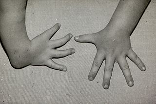
Cenani–Lenz syndactylism, also known as Cenani–Lenz syndrome or Cenani–syndactylism, is an autosomal recessive congenital malformation syndrome involving both upper and lower extremities.

Brachycephaly is the shape of a skull shorter than average in its species. It is perceived as a cosmetically desirable trait in some domesticated dog and cat breeds, notably the pug and Persian, and can be normal or abnormal in other animal species.

In human anatomy, the wrist is variously defined as (1) the carpus or carpal bones, the complex of eight bones forming the proximal skeletal segment of the hand; (2) the wrist joint or radiocarpal joint, the joint between the radius and the carpus and; (3) the anatomical region surrounding the carpus including the distal parts of the bones of the forearm and the proximal parts of the metacarpus or five metacarpal bones and the series of joints between these bones, thus referred to as wrist joints. This region also includes the carpal tunnel, the anatomical snuff box, bracelet lines, the flexor retinaculum, and the extensor retinaculum.
The forearm is the region of the upper limb between the elbow and the wrist. The term forearm is used in anatomy to distinguish it from the arm, a word which is used to describe the entire appendage of the upper limb, but which in anatomy, technically, means only the region of the upper arm, whereas the lower "arm" is called the forearm. It is homologous to the region of the leg that lies between the knee and the ankle joints, the crus.

Hereditary multiple osteochondromas (HMO), also known as hereditary multiple exostoses, is a disorder characterized by the development of multiple benign osteocartilaginous masses (exostoses) in relation to the ends of long bones of the lower limbs such as the femurs and tibias and of the upper limbs such as the humeri and forearm bones. They are also known as osteochondromas. Additional sites of occurrence include on flat bones such as the pelvic bone and scapula. The distribution and number of these exostoses show a wide diversity among affected individuals. Exostoses usually present during childhood. The vast majority of affected individuals become clinically manifest by the time they reach adolescence. A small percentage of affected individuals are at risk for development of sarcomas as a result of malignant transformation. The incidence of hereditary multiple exostoses is around 1 in 50,000 individuals. Hereditary multiple osteochondromas is the preferred term used by the World Health Organization.
Crouzon syndrome is an autosomal dominant genetic disorder known as a branchial arch syndrome. Specifically, this syndrome affects the first branchial arch, which is the precursor of the maxilla and mandible. Since the branchial arches are important developmental features in a growing embryo, disturbances in their development create lasting and widespread effects.

Craniosynostosis is a condition in which one or more of the fibrous sutures in a young infant's skull prematurely fuses by turning into bone (ossification), thereby changing the growth pattern of the skull. Because the skull cannot expand perpendicular to the fused suture, it compensates by growing more in the direction parallel to the closed sutures. Sometimes the resulting growth pattern provides the necessary space for the growing brain, but results in an abnormal head shape and abnormal facial features. In cases in which the compensation does not effectively provide enough space for the growing brain, craniosynostosis results in increased intracranial pressure leading possibly to visual impairment, sleeping impairment, eating difficulties, or an impairment of mental development combined with a significant reduction in IQ.

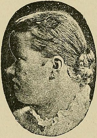
Saethre–Chotzen syndrome (SCS), also known as acrocephalosyndactyly type III, is a rare congenital disorder associated with craniosynostosis. This affects the shape of the head and face, resulting in a cone-shaped head and an asymmetrical face. Individuals with SCS also have droopy eyelids (ptosis), widely spaced eyes (hypertelorism), and minor abnormalities of the hands and feet (syndactyly). Individuals with more severe cases of SCS may have mild to moderate intellectual or learning disabilities. Depending on the level of severity, some individuals with SCS may require some form of medical or surgical intervention. Most individuals with SCS live fairly normal lives, regardless of whether medical treatment is needed or not.

Madelung's deformity is usually characterized by malformed wrists and wrist bones and is often associated with Léri-Weill dyschondrosteosis. It can be bilateral or just in the one wrist. It has only been recognized within the past hundred years. Named after Otto Wilhelm Madelung (1846–1926), a German surgeon, who described it in detail, it was noted by others. Guillaume Dupuytren mentioned it in 1834, Auguste Nélaton in 1847, and Joseph-François Malgaigne in 1855.

The distal radioulnar articulation is a synovial pivot joint between the two bones in the forearm; the radius and ulna. It is one of two joints between the radius and ulna, the other being the proximal radioulnar articulation. The joint features an articular disc, and is reinforced by the palmar and dorsal radioulnar ligaments.
Muenke syndrome, also known as FGFR3-related craniosynostosis, is a human specific condition characterized by the premature closure of certain bones of the skull during development, which affects the shape of the head and face. First described by Maximilian Muenke, the syndrome occurs in about 1 in 30,000 newborns. This condition accounts for an estimated 8 percent of all cases of craniosynostosis.

Dysmelia is a congenital disorder of a limb resulting from a disturbance in embryonic development.

The elbow is the region between the upper arm and the forearm that surrounds the elbow joint. The elbow includes prominent landmarks such as the olecranon, the cubital fossa, and the lateral and the medial epicondyles of the humerus. The elbow joint is a hinge joint between the arm and the forearm; more specifically between the humerus in the upper arm and the radius and ulna in the forearm which allows the forearm and hand to be moved towards and away from the body. The term elbow is specifically used for humans and other primates, and in other vertebrates forelimb plus joint is used.
Femur-fibula-ulna syndrome is a very rare syndrome characterized by abnormalities of the femur, fibula and the ulna. There have been suggestions that FFU complex may be the same as proximal femoral focal deficiency (PFFD) although authors are currently in disagreement over whether or not the disorders are in fact separate. The breadth of the abnormality and number of limbs involved is considered sporadic although upper limbs are more affected than lower limbs and right side malformation is more prevalent than the left. The condition was first noted by Lenz and Feldman in 1977.

McGillivray syndrome is a rare syndrome characterized mainly by heart defects, skull and facial abnormalities and ambiguous genitalia. The symptoms of this syndrome are ventricular septal defect, patent ductus arteriosus, small jaw, undescended testes, and webbed fingers. Beside to these symptoms there are more symptoms which is related with bone structure and misshape.
Radial dysplasia, also known as radial club hand or radial longitudinal deficiency, is a congenital difference occurring in a longitudinal direction resulting in radial deviation of the wrist and shortening of the forearm. It can occur in different ways, from a minor anomaly to complete absence of the radius, radial side of the carpal bones and thumb. Hypoplasia of the distal humerus may be present as well and can lead to stiffness of the elbow. Radial deviation of the wrist is caused by lack of support to the carpus, radial deviation may be reinforced if forearm muscles are functioning poorly or have abnormal insertions. Although radial longitudinal deficiency is often bilateral, the extent of involvement is most often asymmetric.
Liebenberg syndrome is a rare autosomal genetic disease that involves a deletion mutation upstream of the PITX1 gene, which is one that's responsible for the body's organization, specifically in forming lower limbs. In animal studies, when this deletion was introduced to developing birds, their wing buds were noted to take on limb-like structures.
Baller–Gerold syndrome (BGS) is a rare genetic syndrome that involves premature fusion of the skull bones and malformations of facial, forearm and hand bones. The symptoms of Baller–Gerold syndrome overlap with features of a few other genetics disorders: Rothmund–Thomson syndrome and RAPADILINO syndrome. The prevalence of BGS is unknown, as there have only been a few reported cases, but it is estimated to be less than 1 in a million. The name of the syndrome comes from the researchers Baller and Gerold who discovered the first three cases.

Radioulnar synostosis is a rare condition where there is an abnormal connection between the radius and ulna bones of the forearm. This can be present at birth (congenital), when it is a result of a failure of the bones to form separately, or following an injury (post-traumatic).
