Rib

Last updated
Rib
Image122.gif
Detail of a single human rib
Gray112.png
The human rib cage (Source: Gray's Anatomy of the Human Body , 20th ed. 1918)
Details
Identifiers
Latin costae
MeSH D012272
TA98 A02.3.01.001
A02.3.02.001
TA2 1105, 1118
FMA 7574
Anatomical terminology

In vertebrate anatomy, ribs (Latin : costae) are the long curved bones which form the rib cage, part of the axial skeleton. [1] In most tetrapods, ribs surround the chest, enabling the lungs to expand and thus facilitate breathing by expanding the chest cavity. They serve to protect the lungs, heart, and other internal organs of the thorax. In some animals, especially snakes, ribs may provide support and protection for the entire body.

Contents

Human anatomy

Rib details

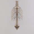
Human ribs are flat bones that form part of the rib cage to help protect internal organs. Humans usually have 24 ribs, in 12 pairs. [2] 1 in 500 people have an extra rib known as a cervical rib. People may have a cervical rib on the right, left or both sides. [3] All are attached at the back to the thoracic vertebrae and are numbered from 1 to 12 according to the vertebrae to which they attach. The first rib is attached to thoracic vertebra 1 (T1). At the front of the body, most of the ribs are joined by costal cartilage to the sternum. Ribs connect to vertebrae at the costovertebral joints. [4]

The parts of a rib includes the head, neck, body (or shaft), tubercle, and angle.

The head of the rib lies next to a vertebra. The ribs connect to the vertebrae with two costovertebral joints, one on the head and one on the neck. The head of the rib has a superior and an inferior articulating region, separated by a crest. These articulate with the superior and inferior costal facets on the connecting vertebrae. [5] The crest gives attachment to the intra-articulate ligament that joins the rib to the vertebra of the same number, at the intervertebral disc. Another ligament, the radiate ligament joins the head of the rib to both the body of the upper vertebra and to the body of the lower vertebra. The smaller middle part of the ligament connects to the intervertebral disc. This plane joint is known as the articulation of the head of the rib.

The other costovertebral joint is that between the tubercle on the neck and the transverse process of the joining thoracic vertebra of the same rib number, and this is known as the costotransverse joint. The superior costotransverse ligament attaches from the non-articular facet of the tubercle to the transverse process of the vertebra.

The neck of the rib is a flattened part that extends laterally from the head. The neck is about 3 cm long. Its anterior surface is flat and smooth, whilst its posterior is perforated by numerous foramina and its surface rough, to give attachment to the ligament of the neck. Its upper border presents a rough crest (crista colli costae) for the attachment of the anterior costotransverse ligament; its lower border is rounded.

A tubercle of rib on the posterior surface of the neck of the rib, has two facets (surfaces) one articulating and one non-articulating. The articular facet, is small and oval and is the lower and more medial of the two, and connects to the transverse costal facet on the thoracic vertebra of the same rib number. [5] The transverse costal facet is on the end of the transverse process of the lower of the two vertebrae to which the head is connected. The non-articular portion is a rough elevation and affords attachment to the ligament of the tubercle. The tubercle is much more prominent in the upper ribs than in the lower ribs.


Rib cage

X-ray image of human chest, with ribs labelled Ribs labeled.png
X-ray image of human chest, with ribs labelled

The first seven sets of ribs, known as "true ribs", are attached to the sternum by the costal cartilages. The first rib is unique and easier to distinguish than other ribs. It is a short, flat, C-shaped bone, and attaches to the manubrium. [6] The vertebral attachment can be found just below the neck at the first thoracic vertebra, and the majority of this bone can be found above the level of the clavicle. Ribs 2 through 7 then become longer and less curved as they progress downwards. [7] The following five sets are known as "false ribs", three of these sharing a common cartilaginous connection to the sternum, while the last two (eleventh and twelfth ribs) are termed floating ribs. [2] They are attached to the vertebrae only, and not to the sternum or cartilage coming off of the sternum.

In general, human ribs increase in length from ribs 1 through 7 and decrease in length again through rib 12. Along with this change in size, the ribs become progressively oblique (slanted) from ribs 1 through 9, then less slanted through rib 12. [7]

The rib cage is separated from the lower abdomen by the thoracic diaphragm which controls breathing. When the diaphragm contracts, the thoracic cavity is expanded, reducing intra-thoracic pressure and drawing air into the lungs. This happens through one of two actions (or a mix of the two): when the lower ribs the diaphragm connects to are stabilized by muscles and the central tendon is mobile, when the muscle contracts the central tendon is drawn down, compressing the cavity underneath and expanding the thoracic cavity downward. When the central tendon is stabilized and the lower ribs are mobile, a contraction of the diaphragm elevates the ribs, which works in conjunction with other muscles to expand the thoracic indent upward.

Development

Early in the developing embryo, somites form and soon subdivide into three mesodermal components – the myotome, dermatome, and the sclerotome. The vertebrae and ribs develop from the sclerotomes. [8]

During the fourth week (fertilization age) costal processes have formed on the vertebral bodies. These processes are small, lateral protrusions of mesenchyme that develop in association with the vertebral arches. During the fifth week the costal processes on the thoracic vertebrae become longer to form the ribs. In the sixth week, the costovertebral joints begin to develop and separate the ribs from the vertebrae. The first seven pairs of ribs, the true ribs join at the front to the sternal bars. By the fetal stage the sternal bars have completely fused. [8]

The ribs begin as cartilage that later ossifies – a process called endochondral ossification. Primary ossification centers are located near the angle of each rib, and ossification continues in the direction away from the head and neck. During adolescence secondary ossification centers are formed in the tubercles and heads of the ribs. [8]

Other animals

Skeleton of a dog showing the location of the ribs Dog anatomy lateral skeleton view.jpg
Skeleton of a dog showing the location of the ribs
Rib cage of the big brown bat (Eptesicus fuscus) Eptesicus fuscus ribcage.jpg
Rib cage of the big brown bat (Eptesicus fuscus)

In jawed fish, there are often two sets of ribs attached to the vertebral column. One set, the dorsal ribs, are found in the dividing septum between the upper and lower parts of the main muscle segments, projecting roughly sideways from the vertebral column. The second set, the ventral ribs arise from the vertebral column just below the dorsal ribs, and enclose the lower body, often joining at the tips. Not all species possess both types of rib, with the dorsal ribs being most commonly absent. Sharks, for example, have no ventral ribs, and only very short dorsal ribs. In some teleosts, there may be additional rib-like bones within the muscle mass. [9]

Tetrapods, however, only ever have a single set of ribs which are probably homologous with the dorsal ribs of fishes. In the earlier choanates, every vertebra bore a pair of ribs, although those on the thoracic vertebrae are typically the longest. The sacral ribs were stout and short, since they formed part of the pelvis, connecting the backbone to the hip bones. [9]

In most true tetrapods, many of these early ribs have been lost, and in living amphibians and reptiles, there is great variation in rib structure and number. For example, turtles have only eight pairs of ribs, which are developed into a bony or cartilaginous carapace and plastron, while snakes have numerous ribs running along the full length of their trunk. Frogs typically have no ribs, aside from a sacral pair, which form part of the pelvis. [9]

In birds, ribs are present as distinct bones only on the thoracic region, although small fused ribs are present on the cervical vertebrae. The thoracic ribs of birds possess a wide projection to the rear; this uncinate process is an attachment for the shoulder muscles. [9] Usually dogs have 26 ribs. Mammals usually also only have distinct ribs on the thoracic vertebra, although fixed cervical ribs are also present in monotremes. In therian mammals, the cervical and lumbar ribs are found only as tiny remnants fused to the vertebrae, where they are referred to as transverse processes. In general, the structure and number of the true ribs in humans is similar to that in other mammals. Unlike reptiles, caudal ribs are never found in mammals. [9]

Ribs as food

Ribs as food are widely used from many animals, such as cows, pigs, hogs, sheep and pandas. The ribs are the less meaty part of the meat chop and they are often cooked as part of a slab; five or more is known as a rack, as in a rack of lamb. Short ribs are ribs of beef either served singly or several as a plate. A rib steak from beef is a popular choice used in many cuisines. Pork ribs, including spare ribs are popular in European and Asian cuisine.

Animated images

See also

Related Research Articles

<span class="mw-page-title-main">Atlas (anatomy)</span> First cervical vertebra of the spine which supports the skull

In anatomy, the atlas (C1) is the most superior (first) cervical vertebra of the spine and is located in the neck.

<span class="mw-page-title-main">Rib cage</span> Bone structure that protects the vital organs and major blood vessels

The rib cage is an endoskeletal enclosure in the thorax of most vertebrate animals that comprises the ribs, vertebral column and sternum, which protects vital organs such as the heart, lungs and great vessels. The circumferential enclosure formed by left and right rib cages, together known as the thoracic cage, is a semi-rigid bony and cartilaginous structure which surrounds the thoracic cavity and supports the shoulder girdles to form the core part of the axial skeleton.

<span class="mw-page-title-main">Clavicle</span> Long bone that serves as a strut between the scapula and the sternum

The clavicle, collarbone, or keybone is a slender, S-shaped long bone approximately 6 inches (15 cm) long that serves as a strut between the shoulder blade and the sternum (breastbone). There are two clavicles, one on the left and one on the right. The clavicle is the only long bone in the body that lies horizontally. Together with the shoulder blade, it makes up the shoulder girdle. It is a palpable bone and, in people who have less fat in this region, the location of the bone is clearly visible. It receives its name from Latin clavicula 'little key' because the bone rotates along its axis like a key when the shoulder is abducted. The clavicle is the most commonly fractured bone. It can easily be fractured by impacts to the shoulder from the force of falling on outstretched arms or by a direct hit.

<span class="mw-page-title-main">Scapula</span> Bone that connects the humerus (upper arm bone) with the clavicle (collar bone)

The scapula, also known as the shoulder blade, is the bone that connects the humerus with the clavicle. Like their connected bones, the scapulae are paired, with each scapula on either side of the body being roughly a mirror image of the other. The name derives from the Classical Latin word for trowel or small shovel, which it was thought to resemble.

Articles related to anatomy include:

<span class="mw-page-title-main">Sacrum</span> Triangular-shaped bone at the bottom of the spine

The sacrum, in human anatomy, is a large, triangular bone at the base of the spine that forms by the fusing of the sacral vertebrae (S1–S5) between ages 18 and 30.

<span class="mw-page-title-main">Lumbar vertebrae</span> Five vertebrae between the pelvis and the rib cage

The lumbar vertebrae are found between the lower ribs and pelvis. They form the lower part of the back in humans, and the tail end of the back in quadrupeds. The term is used to describe the anatomy of humans and quadrupeds, such as horses, pigs, or cattle. These bones are found in particular cuts of meat, including tenderloin or sirloin steak.

<span class="mw-page-title-main">Axis (anatomy)</span> Second cervical vertebra of the spine

In anatomy, the axis is the second cervical vertebra (C2) of the spine, immediately inferior to the atlas, upon which the head rests.

<span class="mw-page-title-main">Axial skeleton</span> The Part of the skeleton that consists of the bones of the head and trunk of a vertebrate

The axial skeleton is the part of the skeleton that consists of the bones of the head and trunk of a vertebrate. In the human skeleton, it consists of 80 bones and is composed of six parts; the skull, also the ossicles of the middle ear, the hyoid bone, the rib cage, sternum and the vertebral column. The axial skeleton together with the appendicular skeleton form the complete skeleton. Another definition of axial skeleton is the bones including the vertebrae, sacrum, coccyx, skull, ribs, and sternum.

<span class="mw-page-title-main">Cervical vertebrae</span> Vertebrae of the neck

In tetrapods, cervical vertebrae are the vertebrae of the neck, immediately below the skull. Truncal vertebrae lie caudal of cervical vertebrae. In sauropsid species, the cervical vertebrae bear cervical ribs. In lizards and saurischian dinosaurs, the cervical ribs are large; in birds, they are small and completely fused to the vertebrae. The vertebral transverse processes of mammals are homologous to the cervical ribs of other amniotes. Most mammals have seven cervical vertebrae, with the only three known exceptions being the manatee with six, the two-toed sloth with five or six, and the three-toed sloth with nine.

<span class="mw-page-title-main">Thoracic vertebrae</span> Vertebrae between the cervical vertebrae and the lumbar vertebrae

In vertebrates, thoracic vertebrae compose the middle segment of the vertebral column, between the cervical vertebrae and the lumbar vertebrae. In humans, there are twelve thoracic vertebrae and they are intermediate in size between the cervical and lumbar vertebrae; they increase in size going towards the lumbar vertebrae, with the lower ones being much larger than the upper. They are distinguished by the presence of facets on the sides of the bodies for articulation with the heads of the ribs, as well as facets on the transverse processes of all, except the eleventh and twelfth, for articulation with the tubercles of the ribs. By convention, the human thoracic vertebrae are numbered T1–T12, with the first one (T1) located closest to the skull and the others going down the spine toward the lumbar region.

<span class="mw-page-title-main">Scalene muscles</span> Muscles on the sides of the neck

The scalene muscles are a group of three muscles on each side of the neck, identified as the anterior, the middle, and the posterior. They are innervated by the third to the eighth cervical spinal nerves (C3-C8).

<span class="mw-page-title-main">Deep cervical fascia</span>

The deep cervical fascia lies under cover of the platysma, and invests the muscles of the neck; it also forms sheaths for the carotid vessels, and for the structures situated in front of the vertebral column. Its attachment to the hyoid bone prevents the formation of a dewlap.

<span class="mw-page-title-main">Costotransverse joint</span>

The costotransverse joint is the joint formed between the facet of the tubercle of the rib and the adjacent transverse process of a thoracic vertebra. The costotransverse joint is a plane type of synovial joint which, under physiological conditions, allows only gliding movement.

<span class="mw-page-title-main">Skeletal system of the horse</span> The skeletal system is made of many interconnected tissues including bone, cartilage, and tendons

The skeletal system of the horse is a skeletal system of a horse that has three major functions in the body. It protects vital organs, provides framework, and supports soft parts of the body. Horses typically have 205 bones. The pelvic limb typically contains 19 bones, while the thoracic limb contains 20 bones.

<span class="mw-page-title-main">Outline of human anatomy</span> Overview of and topical guide to human anatomy

The following outline is provided as an overview of and topical guide to human anatomy:

<span class="mw-page-title-main">Vertebral column</span> Bony structure found in vertebrates

The vertebral column, also known as the backbone, spine, or spinal column, is the core part of the axial skeleton in vertebrate animals. The vertebral column is the defining characteristic of vertebrate endoskeleton in which the notochord found in all chordates has been replaced by a segmented series of mineralized irregular bones called vertebrae, separated by fibrocartilaginous intervertebral discs. The dorsal portion of the vertebral column houses the spinal canal, a cavity formed by alignment of the neural arches that encloses and protects the spinal cord.

This glossary explains technical terms commonly employed in the description of dinosaur body fossils. Besides dinosaur-specific terms, it covers terms with wider usage, when these are of central importance in the study of dinosaurs or when their discussion in the context of dinosaurs is beneficial. The glossary does not cover ichnological and bone histological terms, nor does it cover measurements.

<span class="mw-page-title-main">Vertebra</span> Bone in the vertebral column

Each vertebra is an irregular bone with a complex structure composed of bone and some hyaline cartilage, that make up the vertebral column or spine, of vertebrates. The proportions of the vertebrae differ according to their spinal segment and the particular species.

References

  1. Gillen, Glen (2016-01-01), Gillen, Glen (ed.), "Chapter 18 - Trunk Control: Supporting Functional Independence", Stroke Rehabilitation (Fourth Edition), Mosby, pp. 360–393, doi:10.1016/b978-0-323-17281-3.00018-6, ISBN   978-0-323-17281-3 , retrieved 2020-11-03
  2. 1 2 Sly, Peter D.; Collins, Rachel A. (2008-01-01), Taussig, Lynn M.; Landau, Louis I. (eds.), "Chapter 7 - Applied Clinical Respiratory Physiology", Pediatric Respiratory Medicine (Second Edition), Philadelphia: Mosby, pp. 73–88, doi:10.1016/b978-032304048-8.50011-6, ISBN   978-0-323-04048-8 , retrieved 2020-11-03
  3. Oner, Zulal; Oner, Serkan; Sahin, Necati Emre; Cay, Mahmut (26 January 2023). "Evaluation of congenital rib anomalies with multi-detector computed tomography in the Turkish population". Folia Morphologica. doi: 10.5603/FM.a2023.0006 . PMID   36794687. S2CID   256899032.
  4. Moore, Keith L.; Dalley, Arthur F.; Agur, Anne M. R. (2018). Clinically Oriented Anatomy (8th ed.). Philadelphia: Wolters Kluwer. pp. 293–297. ISBN   9781496347213.
  5. 1 2 Netter, Frank (2014). Atlas of human anatomy (Sixth ed.). Saunders. pp. 183–184. ISBN   9781455704187.
  6. Sly, Peter D.; Collins, Rachel A. (2008-01-01), Taussig, Lynn M.; Landau, Louis I. (eds.), "Chapter 7 - Applied Clinical Respiratory Physiology", Pediatric Respiratory Medicine (Second Edition), Philadelphia: Mosby, pp. 73–88, doi:10.1016/b978-032304048-8.50011-6, ISBN   978-0-323-04048-8 , retrieved 2020-11-03
  7. 1 2 Saladin, K. S. (2010). Anatomy and Physiology: The Unity of Form and Function (5th ed.). New York, NY: McGraw-Hill.
  8. 1 2 3 Larsen, William (2001). Human embryology (3rd ed.). Churchill Livingstone. pp. 80–85. ISBN   0443065837.
  9. 1 2 3 4 5 Romer, Alfred Sherwood; Parsons, Thomas S. (1977). The Vertebrate Body. Philadelphia, PA: Holt-Saunders International. pp. 170–173. ISBN   0-03-910284-X.