Holoprosencephaly

Last updated
Holoprosencephaly
EmbryonicBrain.svg
Diagram depicting the main subdivisions of the embryonic vertebrate brain.
Specialty Medical genetics

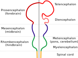
Holoprosencephaly (HPE) is a cephalic disorder in which the prosencephalon (the forebrain of the embryo) fails to develop into two hemispheres, typically occurring between the 18th and 28th day of gestation. [1] Normally, the forebrain is formed and the face begins to develop in the fifth and sixth weeks of human pregnancy. The condition also occurs in other species.

Contents

Holoprosencephaly is estimated to occur in approximately 1 in every 250 conceptions [1] and most cases are not compatible with life and result in fetal death in utero due to deformities to the skull and brain. [2] However, holoprosencephaly is still estimated to occur in approximately 1 in every 8,000 live births. [3]

When the embryo's forebrain does not divide to form bilateral cerebral hemispheres (the left and right halves of the brain), it causes defects in the development of the face and in brain structure and function.

The severity of holoprosencephaly is highly variable. In less severe cases, babies are born with normal or near-normal brain development and facial deformities that may affect the eyes, nose, and upper lip. [1]

Signs and symptoms

Symptoms of holoprosencephaly range from mild (no facial/organ defects, anosmia, or only a single central incisor) to moderate to severe (cyclopia). The symptoms are dependent upon the classification type. [3]

There are four classifications of holoprosencephaly as well as a mild "microform".

Gross pathology specimen from a case of alobar holoprosencephaly Alobar holoprosencephaly.jpg
Gross pathology specimen from a case of alobar holoprosencephaly

Diagnosis

Holoprosencephaly is typically diagnosed during fetal development when there are abnormalities found on fetal brain imaging, however it can also be diagnosed after birth. The protocol for diagnosis includes neuroimaging (Ultrasound or fetal MRI prior to birth or Ultrasound, MRI or CT post birth), syndrome evaluation, cytogenetics, molecular testing, and genetic counseling. [3]

There are four classifications of holoprosencephaly as well as a “microform". [3] These classifications can be distinguished by their anatomical differences. [1]

Causes

In holoprosencephaly, the neural tube fails to segment, resulting in incomplete separation of the prosencephalon at the fifth week of gestation. [5] According to one hypothesis, the holoprosencephalic brain is due to an incomplete axial twisting. [6] According to the axial twist theory, each side of the brain represents its opposite body side because the anterior part of the head, including the forebrain, is turned around by a twisting along the body axis during early development. [6] [7] Accordingly, holoprosencephaly is possiby an extreme form of Yakovlevian torque.

The exact cause(s) of HPE are yet to be determined. Mutations in the gene encoding the SHH protein, which is involved in the development of the central nervous system (CNS), can cause holoprosencephaly. [8] [9] [10] In other cases, it often seems that there is no specific cause at all. [11]

Ultrasound scan of a fetal head at 14 weeks of pregnancy with partial absence of the midline Holoprosencephaly fetus 14 weks US by Dr. W. Moroder.jpg
Ultrasound scan of a fetal head at 14 weeks of pregnancy with partial absence of the midline

Genetics

Armand Marie Leroi describes the cause of cyclopia as a genetic malfunctioning during the process by which the embryonic brain is divided into two. [12] Only later does the visual cortex take recognizable form, and at this point an individual with a single forebrain region will be likely to have a single, possibly rather large, eye (at such a time, individuals with separate cerebral hemispheres would form two eyes).

Increases in expression of such genes as Pax-2, as well as inhibition of Pax-6, from the notochord have been implicated in normal differentiation of cephalic midline structures. Inappropriate expression of any of these genes may result in mild to severe forms of holoprosencephaly.[ citation needed ] Other candidate genes have been located, including the SHH (holoprosencephaly type 3 a.k.a. HPE3), TGIF, ZIC2, SIX3 [13] and BOC genes. [14]

Although many children with holoprosencephaly have normal chromosomes, specific chromosomal abnormalities have been identified in some patients (trisomy of chromosome 13, also known as Patau syndrome). There is evidence that in some families, HPE is inherited (autosomal dominant as well as autosomal or X-linked recessive inheritance). [15] [16] [17] Features consistent with familial transmission of the disease (e.g., a single central maxillary incisor) should be carefully assessed in parents and family members. [18]

Non-genetic factors

Numerous possible risk factors have been identified, including gestational diabetes, transplacental infections (the "TORCH complex"), first trimester bleeding, and a history of miscarriage. [11] [19] As well, the disorder is found twice as often in female babies. [19] However, there appears to be no correlation between HPE and maternal age. [19]

There is evidence of a correlation between HPE and the use of various drugs classified as being potentially unsafe for pregnant and lactating mothers. These include insulin, birth control pills, aspirin, lithium, thorazine, retinoic acid, and anticonvulsants. [19] There is also a correlation between alcohol consumption and HPE, along with nicotine, the toxins in cigarettes and toxins in cigarette smoke when used during pregnancy. [19]

Prognosis

HPE is not a condition in which the brain deteriorates over time. Although serious seizure disorders, autonomic dysfunction, complicated endocrine disorders and other life-threatening conditions may sometimes be associated with HPE, the mere presence of HPE does not mean that these serious problems will occur or develop over time without any previous indication or warning. These abnormalities are usually recognized shortly after birth or early in life and only occur if areas of the brain controlling those functions are fused, malformed or absent. [1]

Prognosis is dependent upon the degree of fusion and malformation of the brain, as well as other health complications that may be present. [1]

The more severe forms of encephalopathy are usually fatal. This disorder consists of a spectrum of defects, malformations and associated abnormalities. Disability is based upon the degree in which the brain is affected. Moderate to severe defects may cause intellectual disability, spastic quadriparesis, athetoid movements, endocrine disorders, epilepsy and other serious conditions; mild brain defects may only cause learning or behavior problems with few motor impairments. [1]

Seizures may develop over time with the highest risk before 2 years of age and the onset of puberty. Most are managed with one medication or a combination of medications. Typically, seizures that are difficult to control appear soon after birth, requiring more aggressive medication combinations/doses.

Most children with HPE are at risk of having elevated blood sodium levels during moderate-severe illnesses, that alter fluid intake/output, even if they have no previous diagnosis of diabetes insipidus or hypernatremia. [1]

See also

Related Research Articles

Ethmocephaly is a type of cephalic disorder caused by holoprosencephaly. Ethmocephaly is the rarest phenotypic variant of a group of defects called the holoprosencephaly (HPE) malformation sequence; other variants include cebocephaly, cyclopia, and median cleft palate. It consists of a proboscis separating narrow-set eyes with an absent nose and microphthalmia. Cebocephaly, another facial anomaly, is characterized by a small, flattened nose with a single nostril situated below incomplete or underdeveloped closely set eyes.

<span class="mw-page-title-main">Lissencephaly</span> Medical condition

Lissencephaly is a set of rare brain disorders whereby the whole or parts of the surface of the brain appear smooth. It is caused by defective neuronal migration during the 12th to 24th weeks of gestation resulting in a lack of development of brain folds (gyri) and grooves (sulci). It is a form of cephalic disorder. Terms such as agyria and pachygyria are used to describe the appearance of the surface of the brain.

<span class="mw-page-title-main">Sonic hedgehog protein</span> Signaling molecule in animals

Sonic hedgehog protein (SHH) is encoded for by the SHH gene. The protein is named after the character Sonic the Hedgehog.

<span class="mw-page-title-main">Cyclopia</span> Congenital disorder

Cyclopia, also known as alobar holoprosencephaly, is the most extreme form of holoprosencephaly and is a congenital disorder characterized by the failure of the embryonic prosencephalon to properly divide the orbits of the eye into two cavities. Its incidence is 1 in 16,000 in born animals and 1 in 200 in miscarried fetuses.

Agenesis of the corpus callosum (ACC) is a rare birth defect in which there is a complete or partial absence of the corpus callosum. It occurs when the development of the corpus callosum, the band of white matter connecting the two hemispheres in the brain, in the embryo is disrupted. The result of this is that the fibers that would otherwise form the corpus callosum are instead longitudinally oriented along the ipsilateral ventricular wall and form structures called Probst bundles.

Pachygyria is a congenital malformation of the cerebral hemisphere. It results in unusually thick convolutions of the cerebral cortex. Typically, children have developmental delay and seizures, the onset and severity depending on the severity of the cortical malformation. Infantile spasms are common in affected children, as is intractable epilepsy.

<span class="mw-page-title-main">Otocephaly</span> Congenital first branchial arch defect

Otocephaly, also known as agnathia–otocephaly complex, is a very rare and lethal cephalic disorder characterized by the absence of the mandible (agnathia), with the ears fused together just below the chin (synotia). It is caused by a disruption to the development of the first branchial arch. It occurs in every 1 in 70,000 embryos.

<span class="mw-page-title-main">Cyclopamine</span> Chemical compound

Cyclopamine (11-deoxojervine) is a naturally occurring steroidal alkaloid. It is a teratogenic component of corn lily, which when consumed during gestation has been demonstrated to induce birth defects, including the development of a single eye (cyclopia) in offspring. The molecule was named after this effect, which was originally observed by Idaho lamb farmers in 1957 after their herds gave birth to cycloptic lambs. It then took more than a decade to identify corn lily as the culprit. Later work suggested that differing rain patterns had changed grazing behaviours, which led to a greater quantity of corn lily to be ingested by pregnant sheep. Cyclopamine interrupts the sonic hedgehog signalling pathway, instrumental in early development, ultimately causing birth defects.

<span class="mw-page-title-main">Simpson–Golabi–Behmel syndrome</span> Congenital disorder

Simpson–Golabi–Behmel syndrome (SGBS), is a rare inherited congenital disorder that can cause craniofacial, skeletal, vascular, cardiac, and renal abnormalities. There is a high prevalence of cancer associated in those with sgbs which includes wilms tumors, neuroblastoma, tumors of the adrenal gland, liver, lungs and abdominal organs. The syndrome is inherited in an X-linked recessive manner. Females that possess one copy of the mutation are considered to be carriers of the syndrome but may still express varying degrees of the phenotype, suffering mild to severe malady. Males experience a higher likelihood of fetal death.

<span class="mw-page-title-main">SIX3</span> Protein-coding gene in the species Homo sapiens

Homeobox protein SIX3 is a protein that in humans is encoded by the SIX3 gene.

<span class="mw-page-title-main">ZIC2</span> Protein-coding gene in the species Homo sapiens

Zinc finger protein ZIC2 is a protein that in humans is encoded by the ZIC2 gene. ZIC2 is a member of the Zinc finger of the cerebellum (ZIC) protein family.

<span class="mw-page-title-main">CDON</span> Protein-coding gene in the species Homo sapiens

Cell adhesion molecule-related/down-regulated by oncogenes is a protein that in humans is encoded by the CDON gene.

<span class="mw-page-title-main">Acrocallosal syndrome</span> Medical condition

Acrocallosal syndrome is an extremely rare autosomal recessive syndrome characterized by corpus callosum agenesis, polydactyly, multiple dysmorphic features, motor and intellectual disabilities, and other symptoms. The syndrome was first described by Albert Schinzel in 1979. Mutations in KIF7 are causative for ACLS, and mutations in GLI3 are associated with a similar syndrome.

<span class="mw-page-title-main">Frontonasal dysplasia</span> Medical condition

Frontonasal dysplasia (FND) is a congenital malformation of the midface. For the diagnosis of FND, a patient should present at least two of the following characteristics: hypertelorism, a wide nasal root, vertical midline cleft of the nose and/or upper lip, cleft of the wings of the nose, malformed nasal tip, encephalocele or V-shaped hair pattern on the forehead. The cause of FND remains unknown. FND seems to be sporadic (random) and multiple environmental factors are suggested as possible causes for the syndrome. However, in some families multiple cases of FND were reported, which suggests a genetic cause of FND.

<span class="mw-page-title-main">Congenital mirror movement disorder</span> Medical condition

Congenital mirror movement disorder(CMM disorder) is a rare genetic neurological disorder which is characterized by mirrored movement, sometimes referred to as associated or synkinetic movement, most often in the upper extremity of the body. These movements are voluntary intentional movements on one, ipsilateral, side of the body that are mirrored simultaneously by involuntary movements on the contralateral side.

<span class="mw-page-title-main">Young–Madders syndrome</span> Genetic disorder

Young–Madders syndrome, alternatively known as Pseudotrisomy 13 syndrome or holoprosencephaly–polydactyly syndrome, is a genetic disorder resulting from defective and duplicated chromosomes which result in holoprosencephaly, polydactyly, facial malformations and intellectual disability, with a significant variance in the severity of symptoms being seen across known cases. Many cases often suffer with several other genetic disorders, and some have presented with hypoplasia, cleft lip, cardiac lesions and other heart defects. In one case in 1991 and another in 2000 the condition was found in siblings who were the product of incest. Many cases are diagnosed prenatally and often in siblings. Cases are almost fatal in the prenatal stage with babies being stillborn.

Neu–Laxova syndrome is a rare autosomal recessive disorder characterized by severe intrauterine growth restriction and multiple congenital malformations. Neu–Laxova syndrome is a very severe disorder, leading to stillbirth or death shortly after birth. It was first described by Dr. Richard Neu in 1971 and Dr. Renata Laxova in 1972 as a lethal disorder in siblings with multiple malformations. Neu–Laxova syndrome is an extremely rare disorder with less than 100 cases reported in medical literature.

ZTTK syndrome is a rare multisystem disease caused in humans by a genetic mutation of the SON gene. Common symptoms include developmental delay and often mild to severe intellectual disability.

<span class="mw-page-title-main">Microlissencephaly</span> Microcephaly combined with lissencephaly

Microlissencephaly (MLIS) is a rare congenital brain disorder that combines severe microcephaly with lissencephaly. Microlissencephaly is a heterogeneous disorder, i.e. it has many different causes and a variable clinical course. Microlissencephaly is a malformation of cortical development (MCD) that occurs due to failure of neuronal migration between the third and fifth month of gestation as well as stem cell population abnormalities. Numerous genes have been found to be associated with microlissencephaly, however, the pathophysiology is still not completely understood.

XK aprosencephaly is an extremely rare congenital disorder characterized by the absence of the embryonic forebrain. Because the prosencephalon gives way to the cerebral cortex, survival with aprosencephaly is not possible outside utero. The external symptoms are similar to holoprosencephaly, a related disorder, including a smaller than normal head (microcephaly), small eyeballs (microphthalmia), a small mouth (microstomia), anal atresia, and abnormalities of the external genitalia, radius, nostrils, and pharynx (throat).

References

  1. 1 2 3 4 5 6 7 8 9 10 11 12 Dubourg, Christèle; Bendavid, Claude; Pasquier, Laurent; Henry, Catherine; Odent, Sylvie; David, Véronique (2007). "Holoprosencephaly". Orphanet Journal of Rare Diseases. 2 (1): 8. doi: 10.1186/1750-1172-2-8 . ISSN   1750-1172. PMC   1802747 . PMID   17274816.
  2. "Holoprosencephaly Information Page". National Institute of Neurological Disorders and Stroke. National Institutes of Health, U.S. Department of Health & Human Services.
  3. 1 2 3 4 5 6 7 8 9 Raam, Manu S; Solomon, Benjamin D; Muenke, Maximilian (June 2011). "Holoprosencephaly: A Guide to Diagnosis and Clinical Management". Indian Pediatrics. 48 (6): 457–466. doi:10.1007/s13312-011-0078-x. ISSN   0019-6061. PMC   4131946 . PMID   21743112.
  4. Rajalakshmi, P. Prathiba; Gadodia, Ankur; Priyatharshini, P. (2015). "Middle interhemispheric variant of holoprosencephaly: A rare midline malformation". Journal of Pediatric Neurosciences. 10 (3): 244–246. doi: 10.4103/1817-1745.165678 . ISSN   1817-1745. PMC   4611894 . PMID   26557166.
  5. Winter, Thomas C.; Kennedy, Anne M.; Woodward, Paula J. (2015-01-01). "Holoprosencephaly: A Survey of the Entity, with Embryology and Fetal Imaging". RadioGraphics. 35 (1): 275–290. doi:10.1148/rg.351140040. ISSN   0271-5333. PMID   25590404.
  6. 1 2 de Lussanet, M.H.E.; Osse, J.W.M. (2012). "An ancestral axial twist explains the contralateral forebain and the optic chiasm in vertebrates". Animal Biology. 62 (2): 193–216. arXiv: 1003.1872 . doi:10.1163/157075611X617102. S2CID   7399128.
  7. de Lussanet, M.H.E. (2019). "Opposite asymmetries of face and trunk and of kissing and hugging, as predicted by the axial twist hypothesis". PeerJ. 7: e7096. doi: 10.7717/peerj.7096 . PMC   6557252 . PMID   31211022.
  8. Chiang C, Litingtung Y, Lee E, Young KE, Corden JL, Westphal H, Beachy PA (October 1996). "Cyclopia and defective axial patterning in mice lacking Sonic hedgehog gene function". Nature. 383 (6599): 407–413. Bibcode:1996Natur.383..407C. doi:10.1038/383407a0. PMID   8837770. S2CID   4339131.
  9. Muenke M, Beachy PA (June 2000). "Genetics of ventral forebrain development and holoprosencephaly". Current Opinion in Genetics & Development. 10 (3): 262–269. doi:10.1016/s0959-437x(00)00084-8. PMID   10826992.
  10. Rash BG, Grove EA (October 2007). "Patterning the dorsal telencephalon: a role for sonic hedgehog?". The Journal of Neuroscience. 27 (43): 11595–11603. doi: 10.1523/jneurosci.3204-07.2007 . PMC   6673221 . PMID   17959802.
  11. 1 2 The Carter Centers for Brain Research in Holoprosencephaly and Related Malformations. "About Holoprosencephaly". Archived from the original on 2009-05-14.
  12. Leroi AM (2003). Mutants : on the form, varieties and errors of the human body. London: HarperCollins. ISBN   978-0-00-653164-7.
  13. "The Carter Center for Research in holoprosencephaly". Archived from the original on 2008-11-21.
  14. Hong M, Srivastava K, Kim S, Allen BL, Leahy DJ, Hu P, et al. (November 2017). "BOC is a modifier gene in holoprosencephaly". Human Mutation. 38 (11): 1464–1470. doi:10.1002/humu.23286. PMC   5673120 . PMID   28677295.
  15. Singh S, Tokhunts R, Baubet V, Goetz JA, Huang ZJ, Schilling NS, et al. (February 2009). "Sonic hedgehog mutations identified in holoprosencephaly patients can act in a dominant negative manner". Human Genetics. 125 (1): 95–103. doi:10.1007/s00439-008-0599-0. PMC   2692056 . PMID   19057928.
  16. Tekendo-Ngongang C, Muenke M, Kruszka P (1993). "Holoprosencephaly Overview". In Adam MP, Ardinger HH, Pagon RA, Wallace SE (eds.). GeneReviews®. Seattle (WA): University of Washington, Seattle. PMID   20301702 . Retrieved 2020-09-01.
  17. Nanni L, Ming JE, Bocian M, Steinhaus K, Bianchi DW, Die-Smulders C, et al. (December 1999). "The mutational spectrum of the sonic hedgehog gene in holoprosencephaly: SHH mutations cause a significant proportion of autosomal dominant holoprosencephaly". Human Molecular Genetics. 8 (13): 2479–2488. doi: 10.1093/hmg/8.13.2479 . PMID   10556296.
  18. Nanni L, Ming JE, Du Y, Hall RK, Aldred M, Bankier A, Muenke M (July 2001). "SHH mutation is associated with solitary median maxillary central incisor: a study of 13 patients and review of the literature". American Journal of Medical Genetics. 102 (1): 1–10. doi:10.1002/1096-8628(20010722)102:1<1::aid-ajmg1336>3.0.co;2-u. PMID   11471164.
  19. 1 2 3 4 5 Croen LA, Shaw GM, Lammer EJ (February 2000). "Risk factors for cytogenetically normal holoprosencephaly in California: a population-based case-control study". American Journal of Medical Genetics. 90 (4): 320–325. doi: 10.1002/(SICI)1096-8628(20000214)90:4<320::AID-AJMG11>3.0.CO;2-8 . PMID   10710231.