The coccyx, commonly referred to as the tailbone, is the final segment of the vertebral column in all apes, and analogous structures in certain other mammals such as horses. In tailless primates since Nacholapithecus, the coccyx is the remnant of a vestigial tail. In animals with bony tails, it is known as tailhead or dock, in bird anatomy as tailfan. It comprises three to five separate or fused coccygeal vertebrae below the sacrum, attached to the sacrum by a fibrocartilaginous joint, the sacrococcygeal symphysis, which permits limited movement between the sacrum and the coccyx.

A teratoma is a tumor made up of several types of tissue, such as hair, muscle, teeth, or bone. Teratomata typically form in the tailbone, ovary, or testicle.

Hirschsprung's disease is a birth defect in which nerves are missing from parts of the intestine. The most prominent symptom is constipation. Other symptoms may include vomiting, abdominal pain, diarrhea and slow growth. Most children develop signs and symptoms shortly after birth. However, others may be diagnosed later in infancy or early childhood. About half of all children with Hirschsprung's disease are diagnosed in the first year of life. Complications may include enterocolitis, megacolon, bowel obstruction and intestinal perforation.
In humans and other mammals, the caudal cell mass is the aggregate of undifferentiated cells at the caudal end on the spine. The caudal end of the spinal cord first begins to form after primary neurulation has taken place, indicating that it develops after the cranial portion of the spinal cord has developed. Following neurulation, the caudal tail begins to form a neurocoele as it develops a hollow core. After this, secondary neurulation occurs in which the medullary cord begins to form and is filled with many cavities that ultimately form the lumen. The cavities formed from the initial and secondary neurulation combine to form one uninterrupted cavity. There is still speculation on the formation of the caudal cell mass in humans with arguments being made for it arising from many cavities or the continuing growth of the neurocoele from the initial neurulation. The caudal cell mass will ultimately differentiate and form into many sacral structures such various nerve endings and the conus medullaris.

An ependymoma is a tumor that arises from the ependyma, a tissue of the central nervous system. Usually, in pediatric cases the location is intracranial, while in adults it is spinal. The common location of intracranial ependymomas is the floor of the fourth ventricle. Rarely, ependymomas can occur in the pelvic cavity.
Rapunzel syndrome is an extremely rare intestinal condition in humans resulting from ingesting hair (trichophagia). The syndrome is named after the long-haired girl Rapunzel in the fairy tale by the Brothers Grimm. Trichophagia is sometimes associated with the hair-pulling disorder trichotillomania. This syndrome is a rare and unusual form of trichobezoar. Since 1968, there have been fewer than 40 documented cases in the literature. This syndrome occurs when the trichobezoar (hairball) reaches past the small intestine, and sometimes even into the colon producing a long tail-like extension of hair.

Germ cell tumor (GCT) is a neoplasm derived from the primordial germ cells. Germ-cell tumors can be cancerous or benign. Germ cells normally occur inside the gonads. GCTs that originate outside the gonads may be birth defects resulting from errors during development of the embryo.

Congenital diaphragmatic hernia (CDH) is a birth defect of the diaphragm. The most common type of CDH is a Bochdalek hernia; other types include Morgagni hernia, diaphragm eventration and central tendon defects of the diaphragm. Malformation of the diaphragm allows the abdominal organs to push into the chest cavity, hindering proper lung formation.

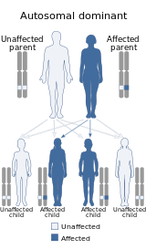
An imperforate anus or anorectal malformations (ARMs) are birth defects in which the rectum is malformed. ARMs are a spectrum of different congenital anomalies which vary from fairly minor lesions to complex anomalies. The cause of ARMs is unknown; the genetic basis of these anomalies is very complex because of their anatomical variability. In 8% of patients, genetic factors are clearly associated with ARMs. Anorectal malformation in Currarino syndrome represents the only association for which the gene HLXB9 has been identified.

The EXIT procedure, or ex utero intrapartum treatment procedure, is a specialized surgical delivery procedure used to deliver babies who have airway compression. Causes of airway compression in newborn babies result from a number of rare congenital disorders, including bronchopulmonary sequestration, congenital cystic adenomatoid malformation, mouth or neck tumor such as teratoma, and lung or pleural tumor such as pleuropulmonary blastoma. Airway compression discovered at birth is a medical emergency. In many cases, however, the airway compression is discovered during prenatal ultrasound exams, permitting time to plan a safe delivery using the EXIT procedure or other means.
Sacrococcygeal teratoma (SCT) is a type of tumor known as a teratoma that develops at the base of the coccyx (tailbone) and is thought to be primarily derived from remnants of the primitive streak. Sacrococcygeal teratomas are benign 75% of the time, malignant 12% of the time, and the remainder are considered "immature teratomas" that share benign and malignant features. Benign sacrococcygeal teratomas are more likely to develop in younger children who are less than 5 months old, and older children are more likely to develop malignant sacrococcygeal teratomas.
A neurectomy, or nerve resection is a neurosurgical procedure in which a peripheral nerve is cut or removed to alleviate neuropathic pain or permanently disable some function of a nerve. The nerve is not intended to grow back. For chronic pain it may be an alternative to a failed nerve decompression when the target nerve has no motor function and numbness is acceptable. Neurectomies have also been used to permanently block autonomic function, and special sensory function not related to pain.

Motor neuron and pancreas homeobox 1 (MNX1), also known as Homeobox HB9 (HLXB9), is a human protein encoded by the MNX1 gene.

Pulmonary hypoplasia is an incomplete development of the lungs, resulting in an abnormally low number or small size of bronchopulmonary segments or alveoli. A congenital malformation, most often occurs secondary to other fetal abnormalities that interfere with normal development of the lungs. Primary (idiopathic) pulmonary hypoplasia is rare and usually not associated with other maternal or fetal abnormalities.
Esophagogastric dissociation is a surgical procedure that is sometimes used to treat gastroesophageal reflux, mainly in neurologically impaired children. It has been suggested as an alternative to Nissen fundoplication for these cases. Preliminary studies have shown it may have a lower failure rate and a lower incidence of recurrent reflux.
The MOMS Trial was a clinical trial that studied treatment of a birth defect called myelomeningocele, which is the most severe form of spina bifida. The study looked at prenatal and postnatal surgery to repair this defect. The first major phase concluded that prenatal surgery had strong, long-term benefits and some risks.
Rowena Spencer was an American physician who specialized in pediatric surgery at a time when it was unusual for a female to become a surgeon. She was the first female surgical intern at the Johns Hopkins Hospital, the first female appointed to the full-time surgery staff at Louisiana State University, and the first female surgeon in Louisiana.

William Edwards Ladd was an American surgeon, and is commonly regarded as one of the founders of pediatric surgery.
Metanephric dysplastic hematoma of the sacral region (MDHSR) has been described by Cozzutto and Lazzaroni-Fossati in 1980, by Posalaki et al. in 1981 and by Cozzutto et al. in 1982. Three additional cases were seen by Finegold.
Denis Browne Gold Medal is a medal that was first struck in 1968, one year after the death of the paediatric surgeon Denis Browne and is awarded for outstanding contributions to paediatric surgery worldwide and is an honour bestowed by The British Association of Paediatric Surgeons.