Sacrococcygeal teratoma

Last updated
Sacrococcygeal teratoma
Specialty Oncology   OOjs UI icon edit-ltr-progressive.svg

Sacrococcygeal teratoma (SCT) is a type of tumor known as a teratoma that develops at the base of the coccyx (tailbone) and is thought to be primarily derived from remnants of the primitive streak. [1] Sacrococcygeal teratomas are benign 75% of the time, malignant 12% of the time, and the remainder are considered "immature teratomas" that share benign and malignant features. Benign sacrococcygeal teratomas are more likely to develop in younger children who are less than 5 months old, and older children are more likely to develop malignant sacrococcygeal teratomas.

Contents

The Currarino syndrome, due to an autosomal dominant mutation in the MNX1 gene, consists of a presacral mass (usually a mature teratoma or anterior meningocele), anorectal malformation and sacral dysgenesis.

Presentation

Complications

Maternal complications of pregnancy may include mirror syndrome. [2] Maternal complications of delivery may include a cesarean section or, alternatively, a vaginal delivery with mechanical dystocia. [3]

Complications of the mass effect of a teratoma in general are addressed on the teratoma page. Complications of the mass effect of a large SCT may include hip dysplasia, bowel obstruction, urinary obstruction, hydronephrosis and hydrops fetalis. Even a small SCT can produce complications of mass effect, if it is presacral (Altman Type IV). [4] In the fetus, severe hydronephrosis may contribute to inadequate lung development. Also in the fetus and newborn, the anus may be imperforate.[ citation needed ]

Later complications of the mass effect and/or surgery may include neurogenic bladder, other forms of urinary incontinence, fecal incontinence, and other chronic problems resulting from accidental damage to or sacrifice of nerves and muscles within the pelvis. [5] Removal of the coccyx may include additional complications. In one review of 25 patients, [6] however, the most frequent complication was an unsatisfactory appearance of the surgical scar.

Late effects

Late effects are of two kinds: consequences of the tumor itself, and consequences of surgery and other treatments for the tumor.[ citation needed ]

Complications of not removing the coccyx may include both recurrence of the teratoma [7] and metastatic cancer. [7] [8] Late malignancies usually involve incomplete excision of the coccyx and are adenocarcinoma.[ citation needed ]Although functional disability in survivors is common, [9] a small comparative study [10] found a nonsignificant difference between SCT survivors and a matched control group.

In rare cases, pelvic scarring may necessitate that a pregnant woman who is a SCT survivor deliver her baby by cesarean section. [11]

Cause

SCT is seen in 1 in every 35,000 live births, and is the most common tumor presenting in newborn humans. Most SCTs are found in babies and children, but SCTs have been reported in adults [12] and the increasingly routine use of prenatal ultrasound exams has dramatically increased the number of diagnosed SCTs presenting in fetuses. Like other teratomas, an SCT can grow very large. Unlike other teratomas, an SCT sometimes grows larger than the rest of the fetus.

Sacrococcygeal teratomas are the most common type of germ cell tumors (both benign and malignant) diagnosed in neonates, infants, and children younger than 4 years. [13] SCTs occur more often in girls than in boys; ratios of 3:1 to 4:1 have been reported. [14]

Historically, sacrococcygeal teratomas present in 2 clinical patterns related to the child's age, tumor location, and likelihood of tumor malignancy. With the advent of routine prenatal ultrasound examinations, a third clinical pattern is emerging.

Diagnosis

During prenatal ultrasound, an SCT having an external component may appear as a fluid-filled cyst or a solid mass sticking out from the fetus' body. Fetal SCTs that are entirely internal may be undetected if they are small; detection (or at least suspicion) is possible when the fetal bladder is seen in an abnormal position, due to the SCT pushing other organs out of place.

At birth, the usual presentation is a visible lump or mass under the skin at the top of the buttocks crease. If not visible, it can sometimes be felt; gently prodded, it feels somewhat like a hardboiled egg. A small SCT, if it is entirely inside the body, may not present for years, until it grows large enough to cause pain, constipation and other symptoms of a large mass inside the pelvis, or until it begins to extend out of the pelvis. Even a relatively large SCT may be missed, if it is internal, because the bony pelvis conceals and protects it. Mediastinal tumors, including teratomas, are similarly concealed and protected by the rib cage.

Some SCTs are discovered when a child begins to talk at about age 2 years and complains of their bottom hurting or feeling "poopy" when they ride in a car seat.

Other tumors can occur in the sacrococcygeal and/or presacral regions [16] and hence must be ruled out to obtain a differential diagnosis. These include extraspinal ependymoma, [17] ependymoblastoma, [18] neuroblastoma and rhabdomyosarcoma.

Smaller SCTs with an external component, seen in prenatal ultrasounds or at birth, often are mistaken for spina bifida.[ citation needed ] Cystic SCT and terminal myelocystocele are especially difficult to distinguish; for more accurate diagnosis, MRI has been recommended. [19]

Treatment

The preferred first treatment for SCT is complete surgical removal (i.e., complete resection). The preferred approach to a small SCT is through the perineum; a large SCT may require an additional approach through the abdomen. Resection should include the coccyx and may also include portions of the sacrum. The surgery should include reattachment of the small muscles and ligaments formerly attached to the coccyx, in effect reconstructing the posterior perineum. If not, there is an increased risk of perineal hernia later in life.

SCTs are classified morphologically according to their relative extent outside and inside the body:

The Altman type is significant in the contexts of management of labor and delivery, surgical approach, and complications of SCT. Serial ultrasound and MRI monitoring of SCTs in fetuses in utero has demonstrated that the Altman type can change over time. As the tumor grows, it can push between other organs and through the perineum to the body surface where the tumor appears as a bulge covered only by skin. Sometimes, the tumor bulge later slips back inside the perineum.

Like all teratomas, a sacrococcygeal teratoma has the potential to be malignant, and the standard of care requires long-term followup by an oncologist.

Management of fetal SCTs

Management of most fetal SCTs involves watchful waiting prior to any treatment. An often used decision tree is as follows:

Emergent problems include maternal mirror syndrome, polyhydramnios, and preterm labor. Poor management decisions, including interventions that are either premature or delayed, can have dire consequences. [20] [21] A very small retrospective study of 9 babies with SCTs greater than 10 cm diameter reported slightly higher survivorship in babies remaining in utero slightly longer. [22]

In many cases, a fetus with a small SCT (under 5 or 10 cm) may be delivered vaginally. [23] [24] [25] [26] Prior to the advent of prenatal detection and hence scheduled C-section, 90% of babies diagnosed with SCT were born full term. [27]

Management of adult SCTs

SCTs are very rare in adults, and as a rule these tumors are benign and have extremely low potential for malignancy. This estimation of potential is based on the idea that because the tumor existed for decades prior to diagnosis, without becoming malignant, it has little or no potential to ever become malignant. For this reason, and because coccygectomy in adults has greater risks than in babies, some surgeons prefer not to remove the coccyx of adult survivors of SCT. There are case reports of good outcomes. [28]

See also

Related Research Articles

<span class="mw-page-title-main">Amniocentesis</span> Sampling of amniotic fluid done mainly to detect fetal chromosomal abnormalities

Amniocentesis is a medical procedure used primarily in the prenatal diagnosis of genetic conditions. It has other uses such as in the assessment of infection and fetal lung maturity. Prenatal diagnostic testing, which includes amniocentesis, is necessary to conclusively diagnose the majority of genetic disorders, with amniocentesis being the gold-standard procedure after 15 weeks' gestation.

<span class="mw-page-title-main">Coccyx</span> Bone of the pelvis

The coccyx, commonly referred to as the tailbone, is the final segment of the vertebral column in all apes, and analogous structures in certain other mammals such as horses. In tailless primates since Nacholapithecus, the coccyx is the remnant of a vestigial tail. In animals with bony tails, it is known as tailhead or dock, in bird anatomy as tailfan. It comprises three to five separate or fused coccygeal vertebrae below the sacrum, attached to the sacrum by a fibrocartilaginous joint, the sacrococcygeal symphysis, which permits limited movement between the sacrum and the coccyx.

<span class="mw-page-title-main">Teratoma</span> Type of germ cell tumor

A teratoma is a tumor made up of several different types of tissue, such as hair, muscle, teeth, or bone. Teratomata typically form in the tailbone, ovary, or testicle.

<span class="mw-page-title-main">Rhabdomyoma</span> Medical condition

A rhabdomyoma is a benign tumor of striated muscle. Rhabdomyomas may be either cardiac or extracardiac. Extracardiac forms of rhabdomyoma are sub-classified into three distinct types: adult type, fetal type, and genital type.

A hysterotomy is an incision made in the uterus. This surgical incision is used in several medical procedures, including during termination of pregnancy in the second trimester and delivering the fetus during caesarean section. It is also used to gain access and perform surgery on a fetus during pregnancy to correct birth defects, and it is an option to achieve resuscitation if cardiac arrest occurs during pregnancy and it is necessary to remove the fetus from the uterus.

<span class="mw-page-title-main">Germ cell tumor</span> Medical condition

Germ cell tumor (GCT) is a neoplasm derived from the primordial germ cells. Germ-cell tumors can be cancerous or benign. Germ cells normally occur inside the gonads. GCTs that originate outside the gonads may be birth defects resulting from errors during development of the embryo.

<span class="mw-page-title-main">Hydrops fetalis</span> Human disease of fetuses

Hydrops fetalis or hydrops foetalis is a condition in the fetus characterized by an accumulation of fluid, or edema, in at least two fetal compartments. By comparison, hydrops allantois or hydrops amnion is an accumulation of excessive fluid in the allantoic or amniotic space, respectively.

<span class="mw-page-title-main">Hemangioendothelioma</span> Medical condition

Hemangioendotheliomas are a family of vascular neoplasms of intermediate malignancy.

<span class="mw-page-title-main">Fetal surgery</span> Growing branch of maternal-fetal medicine

Fetal surgery also known as antenatal surgery, prenatal surgery, is a growing branch of maternal-fetal medicine that covers any of a broad range of surgical techniques that are used to treat congenital abnormalities in fetuses who are still in the pregnant uterus. There are three main types: open fetal surgery, which involves completely opening the uterus to operate on the fetus; minimally invasive fetoscopic surgery, which uses small incisions and is guided by fetoscopy and sonography; and percutaneous fetal therapy, which involves placing a catheter under continuous ultrasound guidance.

<span class="mw-page-title-main">EXIT procedure</span>

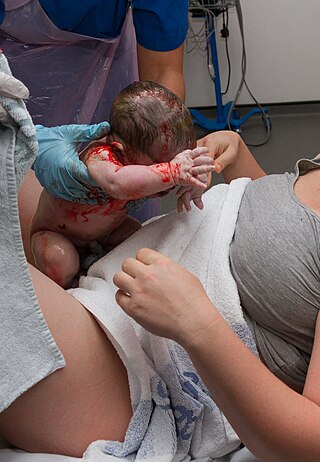
The EXIT procedure, or ex utero intrapartum treatment procedure, is a specialized surgical delivery procedure used to deliver babies who have airway compression. Causes of airway compression in newborn babies result from a number of rare congenital disorders, including bronchopulmonary sequestration, congenital cystic adenomatoid malformation, mouth or neck tumor such as teratoma, and lung or pleural tumor such as pleuropulmonary blastoma. Airway compression discovered at birth is a medical emergency. In many cases, however, the airway compression is discovered during prenatal ultrasound exams, permitting time to plan a safe delivery using the EXIT procedure or other means.

<span class="mw-page-title-main">Cystic hygroma</span> Human disease

A cystic hygroma is an abnormal growth that usually appears on a baby's neck or head. It consists of one or more cysts and tends to grow larger over time. The disorder usually develops while the fetus is still in the uterus, but can also appear after birth.

<span class="mw-page-title-main">Perineal hernia</span>

Perineal hernia is a hernia involving the perineum. The hernia may contain fluid, fat, any part of the intestine, the rectum, or the bladder. It is known to occur in humans, dogs, and other mammals, and often appears as a sudden swelling to one side of the anus.

<span class="mw-page-title-main">Primary tumors of the heart</span> Medical condition

Primary tumors of the heart are extremely rare tumors that arise from the normal tissues that make up the heart. The incidence of primary cardiac tumors has been found to be approximately 0.02%. This is in contrast to secondary tumors of the heart, which are typically either metastatic from another part of the body, or infiltrate the heart via direct extension from the surrounding tissues. Metastatic tumors to the heart are about 20 times more common than primary cardiac tumors.

<span class="mw-page-title-main">Currarino syndrome</span> Medical condition

Currarino syndrome is an inherited congenital disorder where either the sacrum is not formed properly, or there is a mass in the presacral space in front of the sacrum, and there are malformations of the anus or rectum. It occurs in approximately 1 in 100,000 people.

<span class="mw-page-title-main">Maternal–fetal medicine</span> Branch of medicine

Maternal–fetal medicine (MFM), also known as perinatology, is a branch of medicine that focuses on managing health concerns of the mother and fetus prior to, during, and shortly after pregnancy.

<span class="mw-page-title-main">Pulmonary hypoplasia</span> Congenital disorder of respiratory system

Pulmonary hypoplasia is incomplete development of the lungs, resulting in an abnormally low number or small size of bronchopulmonary segments or alveoli. A congenital malformation, it most often occurs secondary to other fetal abnormalities that interfere with normal development of the lungs. Primary (idiopathic) pulmonary hypoplasia is rare and usually not associated with other maternal or fetal abnormalities.

<span class="mw-page-title-main">Congenital pulmonary airway malformation</span> Medical condition

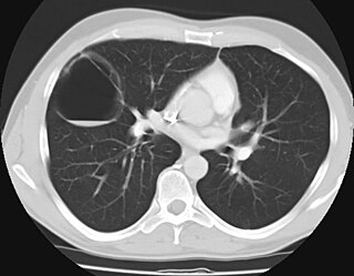
Congenital pulmonary airway malformation (CPAM), formerly known as congenital cystic adenomatoid malformation (CCAM), is a congenital disorder of the lung similar to bronchopulmonary sequestration. In CPAM, usually an entire lobe of lung is replaced by a non-working cystic piece of abnormal lung tissue. This abnormal tissue will never function as normal lung tissue. The underlying cause for CPAM is unknown. It occurs in approximately 1 in every 30,000 pregnancies.

Elevated alpha-fetoprotein refers to a state where alpha-fetoprotein levels are outside of the reference range.

<span class="mw-page-title-main">Scrotal ultrasound</span> Medical ultrasound examination of the scrotum.

Scrotalultrasound is a medical ultrasound examination of the scrotum. It is used in the evaluation of testicular pain, and can help identify solid masses.

Epignathus is a rare teratoma of the oropharynx. Epignathus is a form of oropharyngeal teratoma that arises from the palate and, in most cases, results in death. The pathology is thought to be due to unorganized and uncontrolled differentiation of somatic cells leading to formation of the teratoma; sometimes it is also referred to as "fetus-in-fetu", which is an extremely rare occurrence of an incomplete but parasitic fetus located in the body of its twin. This tumor is considered benign but life-threatening because of its atypical features and high risk of airway obstruction, which is the cause of death in 80-100% of the cases at the time of delivery.

References

  1. Sadler, T. W. (2010). Langman's medical embryology (11th ed.). Philadelphia: Lippincott Williams & Wilkins. p. 63. ISBN   9780781790697.
  2. Finamore PS, Kontopoulos E, Price M, Giannina G, Smulian JC (2007). "Mirror syndrome associated with sacrococcygeal teratoma: a case report". The Journal of Reproductive Medicine. 52 (3): 225–7. PMID   17465292.
  3. Nalbanski B, Markov D, Brankov O (2007). "Sacrococcygeal teratoma—a case report and literature review". Akusherstvo I Ginekologii͡a (in Bulgarian). 46 (2): 41–5. PMID   17469451.
  4. Galili O, Mogilner J (2005). "Type IV sacrococcygeal teratoma causing urinary retention: a rare presentation". J. Pediatr. Surg. 40 (2): E18–20. doi:10.1016/j.jpedsurg.2004.10.003. PMID   15750911.
  5. Engelskirchen R, Holschneider AM, Rhein R, Hecker WC, Höpner F (1987). "[Sacral teratomas in childhood. An analysis of long-term results in 87 children]". Zeitschrift für Kinderchirurgie (in German). 42 (6): 358–61. doi:10.1055/s-2008-1075622. PMID   3439358.
  6. Bittmann S, Bittmann V (2006). "Surgical experience and cosmetic outcomes in children with sacrococcygeal teratoma". Curr Surg. 63 (1): 51–4. doi:10.1016/j.cursur.2005.04.011. PMID   16373161.
  7. 1 2 Lahdenne P, Heikinheimo M, Nikkanen V, Klemi P, Siimes MA, Rapola J (1993). "Neonatal benign sacrococcygeal teratoma may recur in adulthood and give rise to malignancy". Cancer. 72 (12): 3727–31. doi: 10.1002/1097-0142(19931215)72:12<3727::AID-CNCR2820721227>3.0.CO;2-J . PMID   8252490. Synopsis: 45 survivors of infant SCT were followed up. Two reported recurrent benign teratoma and one reported metastatic adenocarcinoma originating from the residual coccyx. They were aged 21–43 at diagnosis.
  8. Lack EE, Glaun RS, Hefter LG, Seneca RP, Steigman C, Athari F (1993). "Late occurrence of malignancy following resection of a histologically mature sacrococcygeal teratoma. Report of a case and literature review". Arch. Pathol. Lab. Med. 117 (7): 724–8. PMID   8323438. Synopsis: A 40 year old man has widely metastatic adenocarcinoma arising from the residual coccyx remaining after surgical removal of an apparently benign SCT at age 2 months.
  9. Derikx JP, De Backer A, van de Schoot L, et al. (2007). "Long-term functional sequelae of sacrococcygeal teratoma: a national study in The Netherlands". J. Pediatr. Surg. 42 (6): 1122–6. doi:10.1016/j.jpedsurg.2007.01.050. PMID   17560233.
  10. Cozzi F, Schiavetti A, Zani A, Spagnol L, Totonelli G, Cozzi DA (2008). "The functional sequelae of sacrococcygeal teratoma: a longitudinal and cross-sectional follow-up study". J. Pediatr. Surg. 43 (4): 658–61. doi:10.1016/j.jpedsurg.2007.10.066. PMID   18405712.
  11. Kohlberger P, Helbich T, Schaller A (1997). "[Delivery following surgically treated sacrococcygeal teratoma in the mother]". Z Geburtshilfe Neonatol (in German). 201 (4): 148–51. PMID   9410520.
  12. Killen DA, Jackson LM (1964). "Sacrococcygeal teratoma in the adult". Archives of Surgery . 88 (3): 425–433. doi:10.1001/archsurg.1964.01310210099017. PMID   14088272.
  13. (PDQ) Sacrococcygeal Tumors in Children
  14. Rescorla FJ, Sawin RS, Coran AG, Dillon PW, Azizkhan RG (February 1998). "Long-term outcome for infants and children with sacrococcygeal teratoma: a report from the Childrens Cancer Group". J. Pediatr. Surg. 33 (2): 171–6. doi:10.1016/S0022-3468(98)90426-2. PMID   9498381.
  15. Rescorla FJ (March 1999). "Pediatric germ cell tumors". Semin Surg Oncol. 16 (2): 144–58. doi:10.1002/(SICI)1098-2388(199903)16:2<144::AID-SSU6>3.0.CO;2-M. PMID   9988869.
  16. Bale PM (1984). "Sacrococcygeal developmental abnormalities and tumors in children". Perspectives in Pediatric Pathology. 8 (1): 9–56. PMID   6366733.
  17. Aktuğ T, Hakgüder G, Sarioğlu S, Akgür FM, Olguner M, Pabuçcuoğlu U (2000). "Sacrococcygeal extraspinal ependymomas: the role of coccygectomy". J. Pediatr. Surg. 35 (3): 515–8. doi:10.1016/S0022-3468(00)90228-8. PMID   10726703.
  18. Santi M, Bulas D, Fasano R, et al. (2008). "Congenital ependymoblastoma arising in the sacrococcygeal soft tissue: a case study". Clin. Neuropathol. 27 (2): 78–82. doi:10.5414/npp27078. PMID   18402386.
  19. Yu JA, Sohaey R, Kennedy AM, Selden NR (2007). "Terminal myelocystocele and sacrococcygeal teratoma: a comparison of fetal ultrasound presentation and perinatal risk". AJNR Am J Neuroradiol. 28 (6): 1058–60. doi: 10.3174/ajnr.A0502 . PMC   8134136 . PMID   17569957.
  20. Mazneĭkova V, Dimitrova V (1999). "[Prenatal ultrasonographic diagnosis of four cases of sacrococcygeal teratoma]". Akusherstvo I Ginekologii͡a (in Bulgarian). 38 (1): 64–9. PMID   11965727.
  21. Sheil AT, Collins KA (2007). "Fatal birth trauma due to an undiagnosed abdominal teratoma: case report and review of the literature". The American Journal of Forensic Medicine and Pathology. 28 (2): 121–7. doi:10.1097/01.paf.0000257373.91126.0d. PMID   17525561. S2CID   26089472.
  22. Holcroft CJ, Blakemore KJ, Gurewitsch ED, Driggers RW, Northington FJ, Fischer AC (2008). "Large fetal sacrococcygeal teratomas: could early delivery improve outcome?". Fetal Diagn. Ther. 24 (1): 55–60. doi:10.1159/000132408. PMID   18504383. S2CID   35920064.
  23. Anteby EY, Yagel S (2003). "Route of delivery of fetuses with structural anomalies". Eur. J. Obstet. Gynecol. Reprod. Biol. 106 (1): 5–9. doi:10.1016/S0301-2115(02)00033-7. PMID   12475573.
  24. Ruangtrakool R, Nitipon A, Laohapensang M, et al. (2001). "Sacrococcygeal teratoma: 25 year experience". Journal of the Medical Association of Thailand = Chotmaihet Thangphaet. 84 (2): 265–73. PMID   11336088.
  25. McCurdy CM, Seeds JW (1993). "Route of delivery of infants with congenital anomalies". Clinics in Perinatology. 20 (1): 81–106. doi:10.1016/S0095-5108(18)30413-5. PMID   8458172.
  26. Kainer F, Winter R, Hofmann HM, Karpf EF (1990). "[Sacrococcygeal teratoma. Prenatal diagnosis and prognosis]". Zentralblatt für Gynäkologie (in German). 112 (10): 609–16. PMID   2205995.
  27. Gonzalez-Crussi F, Winkler RF, Mirkin DL (1978). "Sacrococcygeal teratomas in infants and children: relationship of histology and prognosis in 40 cases". Arch. Pathol. Lab. Med. 102 (8): 420–5. PMID   580884.
  28. Jucá M, de Oliveira FF, Gomes EG, Le Campion E (September 2006). "Sacrococcycygeal Teratoma in Adult: Report of a Case". Int J Gastrointest Cancer. 37 (2–3): 91–93. doi:10.1007/s12029-007-0004-6. PMID   17827528. S2CID   70971724.