Renal oncocytoma

Last updated
Renal oncocytoma
Renal oncocytoma3.jpg
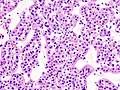
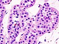
Micrograph of a renal oncocytoma.

A renal oncocytoma is a tumour of the kidney made up of oncocytes , epithelial cells with an excess amount of mitochondria. [1] [2]

Contents

Signs and symptoms

Renal oncocytoma of the right kidney at CT. Onkozytom der Niere.jpg
Renal oncocytoma of the right kidney at CT.

Renal oncocytomas are often asymptomatic and are frequently discovered by chance on a CT or ultrasound of the abdomen. Possible signs and symptoms of a renal oncocytoma include blood in the urine, flank pain, and an abdominal mass.

Pathophysiology

Renal oncocytoma is thought to arise from the intercalated cells of collecting ducts of the kidney. It represent 5% to 15% of surgically resected renal neoplasms. Ultrastructurally, the eosinophilic cells have numerous mitochondria.

Histologic appearance

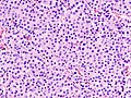
An oncocytoma is an epithelial tumor composed of oncocytes, large eosinophilic cells having small, round, benign-appearing nuclei with large nucleoli and excessive amounts of mitochondria.

Diagnosis

Gross appearance of a renal oncocytoma (left of image) and a slice of a normal kidney (right of image). Note the rounded contour, the mahogany colour and the central scar. Renal oncocytoma.jpg
Gross appearance of a renal oncocytoma (left of image) and a slice of a normal kidney (right of image). Note the rounded contour, the mahogany colour and the central scar.

In gross appearance, the tumors are tan or mahogany brown, well circumscribed and contain a central scar. They may achieve a large size (up to 12 cm in diameter).

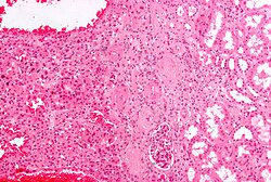
The main differential diagnosis of renal oncocytoma is chromophobe renal cell carcinoma oncocytic variant, which like the renal oncocytoma has eosinophilic cytoplasm, but has perinuclear clearing and, typically, some degree of nuclear atypia.

Immunohistochemical profile

[3]

Treatment

Renal oncocytoma is considered benign, cured by nephrectomy. There are some familial cases in which these tumors are multicentric rather than solitary. [4] However, they may be resected to exclude a malignant tumor, e.g. renal cell carcinoma.

Prognosis

Histopathologic types of kidney tumor, with relative incidences and prognoses. Oncocytoma is seen at right in the pie chart. Renal tumors by relative incidence and prognosis.png
Histopathologic types of kidney tumor, with relative incidences and prognoses. Oncocytoma is seen at right in the pie chart.

The overall five-year survival rate has been estimated to be 63%, with 100% disease-specific survival. [5]

Additional images

See also

Related Research Articles

<span class="mw-page-title-main">Thyroid neoplasm</span> Medical condition

Thyroid neoplasm is a neoplasm or tumor of the thyroid. It can be a benign tumor such as thyroid adenoma, or it can be a malignant neoplasm, such as papillary, follicular, medullary or anaplastic thyroid cancer. Most patients are 25 to 65 years of age when first diagnosed; women are more affected than men. The estimated number of new cases of thyroid cancer in the United States in 2010 is 44,670 compared to only 1,690 deaths. Of all thyroid nodules discovered, only about 5 percent are cancerous, and under 3 percent of those result in fatalities.

<span class="mw-page-title-main">Renal cell carcinoma</span> Medical condition

Renal cell carcinoma (RCC) is a kidney cancer that originates in the lining of the proximal convoluted tubule, a part of the very small tubes in the kidney that transport primary urine. RCC is the most common type of kidney cancer in adults, responsible for approximately 90–95% of cases. RCC occurrence shows a male predominance over women with a ratio of 1.5:1. RCC most commonly occurs between 6th and 7th decade of life.

<span class="mw-page-title-main">Kidney cancer</span> Medical condition

Kidney cancer, also known as renal cancer, is a group of cancers that starts in the kidney. Symptoms may include blood in the urine, lump in the abdomen, or back pain. Fever, weight loss, and tiredness may also occur. Complications can include spread to the lungs or brain.

<span class="mw-page-title-main">Surface epithelial-stromal tumor</span> Medical condition

Surface epithelial-stromal tumors are a class of ovarian neoplasms that may be benign or malignant. Neoplasms in this group are thought to be derived from the ovarian surface epithelium or from ectopic endometrial or Fallopian tube (tubal) tissue. Tumors of this type are also called ovarian adenocarcinoma. This group of tumors accounts for 90% to 95% of all cases of ovarian cancer; however is mainly only found in postmenopausal women with the exception of the United States where 7% of cases occur in women under the age of 40. Serum CA-125 is often elevated but is only 50% accurate so it is not a useful tumor marker to assess the progress of treatment. 75% of women with epithelial ovarian cancer are found within the advanced-stages; however younger patients are more likely to have better prognoses than older patients.

<span class="mw-page-title-main">Birt–Hogg–Dubé syndrome</span> Rare autosomal dominant cancer syndrome

Birt–Hogg–Dubé syndrome (BHD), also Hornstein–Birt–Hogg–Dubé syndrome, Hornstein–Knickenberg syndrome, and fibrofolliculomas with trichodiscomas and acrochordons is a human, adult onset, autosomal dominant genetic disorder caused by the FLCN gene. It can cause susceptibility to kidney cancer, renal and pulmonary cysts, and noncancerous tumors of the hair follicles, called fibrofolliculomas. The symptoms seen in each family are unique, and can include any combination of the three symptoms. Fibrofolliculomas are the most common manifestation, found on the face and upper trunk in over 80% of people with BHD over the age of 40. Pulmonary cysts are equally common (84%), but only 24% of people with BHD eventually experience a collapsed lung. Kidney tumors, both cancerous and benign, occur in 14–34% of people with BHD; the associated kidney cancers are often rare hybrid tumors.

An oncocytoma is a tumor made up of oncocytes, epithelial cells characterized by an excessive amount of mitochondria, resulting in an abundant acidophilic, granular cytoplasm. The cells and the tumor that they compose are often benign but sometimes may be premalignant or malignant.

<span class="mw-page-title-main">Hürthle cell</span> Medical condition

A Hürthle cell is a cell in the thyroid that is often associated with Hashimoto's thyroiditis as well as benign and malignant tumors. This version is a relatively rare form of differentiated thyroid cancer, accounting for only 3-10% of all differentiated thyroid cancers. Oncocytes in the thyroid are often called Hürthle cells. Although the terms oncocyte, oxyphilic cell, and Hürthle cell are used interchangeably, Hürthle cell is used only to indicate cells of thyroid follicular origin.

<span class="mw-page-title-main">Warthin's tumor</span> Medical condition

Warthin's tumor, also known as papillary cystadenoma lymphomatosum, is a benign cystic tumor of the salivary glands containing abundant lymphocytes and germinal centers. It is named for pathologist Aldred Scott Warthin, who described two cases in 1929.

<span class="mw-page-title-main">Granular cell tumor</span> Medical condition

Granular cell tumor is a tumor that can develop on any skin or mucosal surface, but occurs on the tongue 40% of the time.

<span class="mw-page-title-main">Chromophobe cell</span>

A chromophobe is a histological structure that does not stain readily, and thus appears relatively pale under the microscope.

<span class="mw-page-title-main">Transitional cell carcinoma</span> Medical condition

Transitional cell carcinoma, also called urothelial carcinoma, is a type of cancer that typically occurs in the urinary system. It is the most common type of bladder cancer and cancer of the ureter, urethra, and urachus. Symptoms of urothelial carcinoma in the bladder include hematuria. Diagnosis includes urine analysis and imaging of the urinary tract (cystoscopy). Transitional cell carcinomas arise from the transitional epithelium, a tissue lining the inner surface of these hollow organs. When the term "urothelial" is used, it specifically refers to a carcinoma of the urothelium, meaning a transitional cell carcinomas of the urinary system.

<span class="mw-page-title-main">Neprilysin</span> Mammalian protein found in Homo sapiens

Neprilysin, also known as membrane metallo-endopeptidase (MME), neutral endopeptidase (NEP), cluster of differentiation 10 (CD10), and common acute lymphoblastic leukemia antigen (CALLA) is an enzyme that in humans is encoded by the MME gene. Neprilysin is a zinc-dependent metalloprotease that cleaves peptides at the amino side of hydrophobic residues and inactivates several peptide hormones including glucagon, enkephalins, substance P, neurotensin, oxytocin, and bradykinin. It also degrades the amyloid beta peptide whose abnormal folding and aggregation in neural tissue has been implicated as a cause of Alzheimer's disease. Synthesized as a membrane-bound protein, the neprilysin ectodomain is released into the extracellular domain after it has been transported from the Golgi apparatus to the cell surface.

<span class="mw-page-title-main">Oncocyte</span> Epithelial cell

An oncocyte is an epithelial cell characterized by an excessive number of mitochondria, resulting in an abundant acidophilic, granular cytoplasm. Oncocytes can be benign or malignant.

Juxtaglomerular cell tumor is an extremely rare kidney tumour of the juxtaglomerular cells, with less than 100 cases reported in literature. This tumor typically secretes renin, hence the former name of reninoma. It often causes severe hypertension that is difficult to control, in adults and children, although among causes of secondary hypertension it is rare. It develops most commonly in young adults, but can be diagnosed much later in life. It is generally considered benign, but its malignant potential is uncertain.

<span class="mw-page-title-main">Cystic nephroma</span> Medical condition

A cystic nephroma, also known as multilocular cystic nephroma, mixed epithelial stromal tumour (MEST) and renal epithelial stromal tumour (REST), is a type of rare benign kidney tumour.

<span class="mw-page-title-main">Metanephric adenoma</span> Medical condition

Metanephric adenoma (MA) is a rare, benign tumour of the kidney, that can have a microscopic appearance similar to a nephroblastoma, or a papillary renal cell carcinoma.

Large cell lung carcinoma with rhabdoid phenotype (LCLC-RP) is a rare histological form of lung cancer, currently classified as a variant of large cell lung carcinoma (LCLC). In order for a LCLC to be subclassified as the rhabdoid phenotype variant, at least 10% of the malignant tumor cells must contain distinctive structures composed of tangled intermediate filaments that displace the cell nucleus outward toward the cell membrane. The whorled eosinophilic inclusions in LCLC-RP cells give it a microscopic resemblance to malignant cells found in rhabdomyosarcoma (RMS), a rare neoplasm arising from transformed skeletal muscle. Despite their microscopic similarities, LCLC-RP is not associated with rhabdomyosarcoma.

Hybrid oncocytoma/chromophobe renal cell carcinoma is rare subtype of renal cell carcinoma. It arises from intercalated cells of cortical collecting ducts of the kidney. It is associated with extreme hypodiploidy with multiple losses of entire chromosomes 1, 2, 6, 10, 13, 17, 21 and Y.

<span class="mw-page-title-main">Papillary renal cell carcinoma</span> Medical condition

Papillary renal cell carcinoma (PRCC) is a malignant, heterogeneous tumor originating from renal tubular epithelial cells of the kidney, which comprises approximately 10-15% of all kidney neoplasms. Based on its morphological features, PRCC can be classified into two main subtypes, which are type 1 (basophilic) and type 2 (eosinophilic).

References

  1. Coburn V, Radfar A, Snook D, Mahalingam M (2007). "Cutaneous oncocytoma - a report of three cases and review of the literature". J. Cutan. Pathol. 34 (4): 355–9. doi:10.1111/j.1600-0560.2006.00620.x. PMID   17381809. S2CID   19955625.
  2. "Atlas of Genetics and Cytogenetics in Oncology and Haematology - Thyroid:oncocytic tumors" . Retrieved 2009-02-01.
  3. [Renal Neoplasms: An Update on Immunohistochemical and Histochemical Features http://www.dako.com/08066_12may10_webchapter26.pdf]
  4. Robbins pathology, page 1015[ full citation needed ]
  5. Gudbjartsson, Tomas; Hardarson, Sverrir; Petursdottir, Vigdis; Thoroddsen, Asgeir; Magnusson, Jonas; Einarsson, Gudmundur V. (2005). "Renal oncocytoma: a clinicopathological analysis of 45 consecutive cases". BJU International. 96 (9): 1275–1279. doi: 10.1111/j.1464-410X.2005.05827.x . ISSN   1464-4096. PMID   16287444. S2CID   19390518.