Metanephric adenoma

Last updated
Metanephric adenoma
Metanephric adenoma - very high mag.jpg
Micrograph of a metanephric adenoma with structures reminiscent of those seen in papillary renal cell carcinoma. H&E stain.
Specialty Oncology   OOjs UI icon edit-ltr-progressive.svg

Metanephric adenoma (MA) is a rare, benign tumour of the kidney, that can have a microscopic appearance similar to a nephroblastoma (Wilms tumours), [1] or a papillary renal cell carcinoma.

Contents

It should not be confused with the pathologically unrelated, yet similar sounding, mesonephric adenoma .

Symptoms

The symptoms may be similar to those classically associated with renal cell carcinoma, and may include polycythemia, abdominal pain, hematuria and a palpable mass. Mean age at onset is around 40 years with a range of 5 to 83 years and the mean size of the tumour is 5.5 cm with a range 0.3 to 15 cm (1). Polycythemia is more frequent in MA than in any other type of renal tumour. Of further relevance is that this tumour is more commonly calcified than any other kidney neoplasm. [2] Surgery is curative and no other treatment is recommended. There is so far no evidence of metastases or local recurrence.

Pathology

Histopathology

Micrograph of a metanephric adenoma (right of image). Normal kidney is seen on the left. Kidney biopsy. PAS stain. Metanephric adenoma high mag.jpg
Micrograph of a metanephric adenoma (right of image). Normal kidney is seen on the left. Kidney biopsy. PAS stain.
Micrograph of a metanephric adenoma, right of image, showing the characteristic features (round nuclear membrane, no nucleoli, and fine chromatin). Normal kidney is seen on the left of the image. Kidney biopsy. PAS stain. Metanephric adenoma high mag cropped.jpg
Micrograph of a metanephric adenoma, right of image, showing the characteristic features (round nuclear membrane, no nucleoli, and fine chromatin). Normal kidney is seen on the left of the image. Kidney biopsy. PAS stain.

Metanephric adenoma is diagnosed histologically. The tumours can be located at upper pole, lower pole and mid-hilar region of the kidney; they are well circumscribed but unencapsulated, tan pink, with possible cystic and hemorrhagic foci. They show a uniform architecture of closely packed acinar or tubular structures of mature and bland appearance with scanty interposed stroma. [3] [4] [5] [6] [7] Cells are small with dark staining nuclei and inconspicuous nucleoli. Blastema is absent whereas calcospherites may be present. Glomeruloid figures are a striking finding, reminiscent of early fetal metenephric tissue. The lumen of the acini may contain otherwise epithelial infoldings or fibrillary material but it is quite often empty. Mitoses are conspicuously absent. [3] [4] [5] [6] [7] In the series reported by Jones et al. tumour cells were reactive for Leu7 in 3 cases of 5, to vimentine in 4 of 6, to cytocheratin in 2 of 6, to epithelial membrane antigen in 1 of 6 cases and muscle specific antigen in 1 of 6. [5] Olgac et al. found that intense and diffuse immunoreactivity for alpha-methylacyl-CoA racemase (AMACR) is useful in differentiating renal cell carcinoma from MA but a panel including AMACR, CK7 and CD57 is better in this differential diagnosis. [8] Differential diagnosis may be quite difficult indeed as exemplified by the three malignancies initially diagnosed as MA that later metastasized, in the report by Pins et al. [9]

Cytogenetic characteristics

Brunelli et al. stated that genetic analysis of chromosome 7, 17, and Y may facilitate discrimination of MA from papillary renal cell carcinoma in difficult cases. Their study showed that MA lacks the frequent gain of chromosomes 7 and 17 and losses of the Y chromosome that are typical of papillary renal cell neoplasms, suggesting that MA is not related to renal cell carcinoma and papillary adenoma. [10]

Treatment

As metanephric adenomas are considered benign, they can be left in place, i.e. no treatment is needed. [11]

History

MA has been described in the past under other names such as néphrome néphronogène, metanephroider Nierentumor and nephroblastomartiges Nierenadenom (5) but the term metanephric adenoma was suggested by Brisigotti, Cozzutto et al. in 1992 and then widely accepted. Prior to this report, Nagashima et al. in 1991 had not offered a nosological innovation for their two cases [12] whereas the denomination of néphrome néphronogène proposed by Pages and Granier in 1980 had gone largely undetected. [13]

Related Research Articles

<span class="mw-page-title-main">Thyroid neoplasm</span> Medical condition

Thyroid neoplasm is a neoplasm or tumor of the thyroid. It can be a benign tumor such as thyroid adenoma, or it can be a malignant neoplasm, such as papillary, follicular, medullary or anaplastic thyroid cancer. Most patients are 25 to 65 years of age when first diagnosed; women are more affected than men. The estimated number of new cases of thyroid cancer in the United States in 2010 is 44,670 compared to only 1,690 deaths. Of all thyroid nodules discovered, only about 5 percent are cancerous, and under 3 percent of those result in fatalities.

<span class="mw-page-title-main">Renal cell carcinoma</span> Medical condition

Renal cell carcinoma (RCC) is a kidney cancer that originates in the lining of the proximal convoluted tubule, a part of the very small tubes in the kidney that transport primary urine. RCC is the most common type of kidney cancer in adults, responsible for approximately 90–95% of cases. RCC occurrence shows a male predominance over women with a ratio of 1.5:1. RCC most commonly occurs between 6th and 7th decade of life.

<span class="mw-page-title-main">Collecting duct system</span> Kidney system

The collecting duct system of the kidney consists of a series of tubules and ducts that physically connect nephrons to a minor calyx or directly to the renal pelvis. The collecting duct system is the last part of nephron and participates in electrolyte and fluid balance through reabsorption and excretion, processes regulated by the hormones aldosterone and vasopressin.

<span class="mw-page-title-main">Wilms' tumor</span> Rare childhood cancer of the kidneys

Wilms' tumor or Wilms tumor, also known as nephroblastoma, is a cancer of the kidneys that typically occurs in children, and occurs most commonly as a renal tumor in child patients. It is named after Max Wilms, the German surgeon (1867–1918) who first described it.

<span class="mw-page-title-main">Adrenocortical carcinoma</span> Medical condition

Adrenocortical carcinoma (ACC) is an aggressive cancer originating in the cortex of the adrenal gland.

An oncocytoma is a tumor made up of oncocytes, epithelial cells characterized by an excessive amount of mitochondria, resulting in an abundant acidophilic, granular cytoplasm. The cells and the tumor that they compose are often benign but sometimes may be premalignant or malignant.

Malignant rhabdoid tumour (MRT) is a very aggressive form of tumour originally described as a variant of Wilms' tumour, which is primarily a kidney tumour that occurs mainly in children.

<span class="mw-page-title-main">Acinic cell carcinoma</span> Medical condition

Acinic cell carcinoma is a malignant tumor representing 2% of all salivary tumors. 90% of the time found in the parotid gland, 10% intraorally on buccal mucosa or palate. The disease presents as a slow growing mass, associated with pain or tenderness in 50% of the cases. Often appears pseudoencapsulated.

<span class="mw-page-title-main">Transitional cell carcinoma</span> Medical condition

Transitional cell carcinoma, also called urothelial carcinoma, is a type of cancer that typically occurs in the urinary system. It is the most common type of bladder cancer and cancer of the ureter, urethra, and urachus. It accounts for 95% of bladder cancer cases.

<span class="mw-page-title-main">Papillary hidradenoma</span> Medical condition

A papillary hidradenoma, also termed hidradenoma papilliferum or mammary-like gland adenoma of the vulva, is a rare, but nonetheless most common benign tumor that occurs in and between anal and genital regions of females. These hidradenomas are sharply circumscribed, nodular tumors that usually develop in women's anogenital area but uncommonly occur in other sites in women and men. Papillary hidradenomas that develop outside of the anogenital region are termed ecctopic papillary hidradenomas or ectopic hidradenoma papilliferums.

<span class="mw-page-title-main">PRCC (gene)</span> Protein-coding gene in the species Homo sapiens

Proline-rich protein PRCC is a protein that, in humans, is encoded by the PRCC gene.

<span class="mw-page-title-main">Renal oncocytoma</span> Medical condition

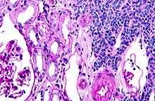
A renal oncocytoma is a tumour of the kidney made up of oncocytes, epithelial cells with an excess amount of mitochondria.

Juxtaglomerular cell tumor is an extremely rare kidney tumour of the juxtaglomerular cells, with less than 100 cases reported in literature. This tumor typically secretes renin, hence the former name of reninoma. It often causes severe hypertension that is difficult to control, in adults and children, although among causes of secondary hypertension it is rare. It develops most commonly in young adults, but can be diagnosed much later in life. It is generally considered benign, but its malignant potential is uncertain.

<span class="mw-page-title-main">Kidney tumour</span> Medical condition

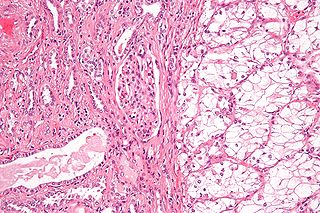
Kidney tumours are tumours, or growths, on or in the kidney. These growths can be benign or malignant.

<span class="mw-page-title-main">Mucinous tubular and spindle cell carcinoma</span> Medical condition

Mucinous tubular and spindle cell carcinoma (MTSCC) is a rare subtype of renal cell carcinoma (RCC), that is included in the 2004 WHO classification of RCC. MTSCC is a rare neoplasm and is considered as a low-grade entity. It may be a variant of papillary RCC. This tumor occurs throughout life and is more frequent in females.

<span class="mw-page-title-main">Epithelial-myoepithelial carcinoma</span> Medical condition

Epithelial-myoepithelial carcinoma (EMCa) is a rare malignant tumour that typically arises in a salivary gland and consists of both an epithelial and myoepithelial component. They are predominantly found in the parotid gland and represent approximately 1% of salivary gland tumours.

<span class="mw-page-title-main">Hyalinizing clear cell carcinoma</span> Medical condition

Hyalinizing clear cell carcinoma (HCCC) is a rare malignant salivary gland tumour, with a good prognosis, that is usually found on the tongue or palate.

A sialoblastoma is a low-grade salivary gland neoplasm that recapitulates primitive salivary gland anlage. It has previously been referred to as congenital basal cell adenoma, embryoma, or basaloid adenocarcinoma. It is an extremely rare tumor, with less than 100 cases reported worldwide.

<span class="mw-page-title-main">Endolymphatic sac tumor</span>

An endolymphatic sac tumor (ELST) is a very uncommon papillary epithelial neoplasm arising within the endolymphatic sac or endolymphatic duct. This tumor shows a very high association with Von Hippel–Lindau syndrome (VHL).

<span class="mw-page-title-main">Papillary renal cell carcinoma</span> Medical condition

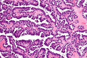
Papillary renal cell carcinoma (PRCC) is a malignant, heterogeneous tumor originating from renal tubular epithelial cells of the kidney, which comprises approximately 10-15% of all kidney neoplasms. Based on its morphological features, PRCC can be classified into two main subtypes, which are type 1 (basophilic) and type 2 (eosinophilic).

References

  1. Bastos Netto JM, Esteves TC, Mattos R, Tibiriçá SH, Costa SM, Vieira LJ (August 2007). "Metanephric adenoma: a rare differential diagnosis of renal tumor in children". Journal of Pediatric Urology. 3 (4): 340–341. doi:10.1016/j.jpurol.2006.10.003. PMID   18947770.
  2. Davis CJ, Barton JH, Sesterhenn IA, Mostofi FK (October 1995). "Metanephric adenoma. Clinicopathological study of fifty patients". The American Journal of Surgical Pathology. 19 (10): 1101–14. doi:10.1097/00000478-199510000-00001. PMID   7573669.
  3. 1 2 Kovacs G, Akhtar M, Beckwith BJ, Bugert P, Cooper CS, Delahunt B, et al. (October 1997). "The Heidelberg classification of renal cell tumours". The Journal of Pathology. 183 (2): 131–133. doi: 10.1002/(SICI)1096-9896(199710)183:2<131::AID-PATH931>3.0.CO;2-G . PMID   9390023. S2CID   34796951.
  4. 1 2 Brisigotti M, Cozzutto C, Fabbretti G, Sergi C, Callea F (October 1992). "Metanephric adenoma". Histology and Histopathology. 7 (4): 689–692. PMID   1333853.
  5. 1 2 3 Jones EC, Pins M, Dickersin GR, Young RH (June 1995). "Metanephric adenoma of the kidney. A clinicopathological, immunohistochemical, flow cytometric, cytogenetic, and electron microscopic study of seven cases". The American Journal of Surgical Pathology. 19 (6): 615–626. doi:10.1097/00000478-199506000-00001. PMID   7755148.
  6. 1 2 Bostwick DG, Eble JN (2008). Urologic Surgical Pathology. St Louis: Mosby. p. 118.
  7. 1 2 Grignon DJ, Eble JN (February 1998). "Papillary and metanephric adenomas of the kidney". Seminars in Diagnostic Pathology. 15 (1): 41–53. PMID   9503505.
  8. Olgac S, Hutchinson B, Tickoo SK, Reuter VE (February 2006). "Alpha-methylacyl-CoA racemase as a marker in the differential diagnosis of metanephric adenoma". Modern Pathology. 19 (2): 218–24. doi:10.1038/modpathol.3800520. PMID   16424894. S2CID   30923148.
  9. Pins MR, Jones EC, Martul EV, Kamat BR, Umlas J, Renshaw AA (May 1999). "Metanephric adenoma-like tumors of the kidney: report of 3 malignancies with emphasis on discriminating features". Archives of Pathology & Laboratory Medicine. 123 (5): 415–20. doi:10.5858/1999-123-0415-MALTOT. PMID   10235500.
  10. Brunelli M, Eble JN, Zhang S, Martignoni G, Cheng L (October 2003). "Metanephric adenoma lacks the gains of chromosomes 7 and 17 and loss of Y that are typical of papillary renal cell carcinoma and papillary adenoma". Modern Pathology. 16 (10): 1060–3. doi: 10.1097/01.MP.0000090923.50509.55 . PMID   14559991. S2CID   11701480.
  11. Galmiche L, Vasiliu V, Poirée S, Hélénon O, Casanova JM, Brousse N (October 2007). "[Diagnosis of renal metanephric adenoma: relevance of immunohistochemistry and biopsy]". Annales de Pathologie (in French). 27 (5): 365–368. doi:10.1016/s0242-6498(07)78275-5. PMID   18185471.
  12. Nagashima Y, Arai N, Tanaka Y, Yoshida S, Sumino K, Ohaki Y, Matsushita K, Morita T, Misugi K (1991). "Two cases of a renal epithelial tumour resembling immature nephron". Virchows Archiv A. 418 (1): 77–81. doi:10.1007/BF01600247. PMID   1989379. S2CID   28924982.
  13. Pages A, Granier M (1980). "Le néphrome néphronogène". Arch Anat Cytol Pathol. 28: 99–103.