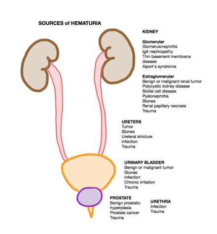
Hematuria

Last updated
Hematuria (differential diagnosis)
Other namesHaematuria, erythrocyturia, blood in the urine
HematuriaGross.jpg
Visible hematuria that is tea-colored
Specialty Nephrology, Urology
Symptoms Blood in the urine
Causes Urinary tract infection, kidney stone, bladder cancer, kidney cancer

Hematuria or haematuria is defined as the presence of blood or red blood cells in the urine. [1] "Gross hematuria" occurs when urine appears red, brown, or tea-colored due to the presence of blood. Hematuria may also be subtle and only detectable with a microscope or laboratory test. [2] Blood that enters and mixes with the urine can come from any location within the urinary system, including the kidney, ureter, urinary bladder, urethra, and in men, the prostate. [3] Common causes of hematuria include urinary tract infection (UTI), kidney stones, viral illness, trauma, bladder cancer, and exercise. [4] These causes are grouped into glomerular and non-glomerular causes, depending on the involvement of the glomerulus of the kidney. [1] But not all red urine is hematuria. [5] Other substances such as certain medications and foods (e.g. blackberries, beets, food dyes) can cause urine to appear red. [5] Menstruation in women may also cause the appearance of hematuria and may result in a positive urine dipstick test for hematuria. [6] A urine dipstick test may also give an incorrect positive result for hematuria if there are other substances in the urine such as myoglobin, a protein excreted into urine during rhabdomyolysis. A positive urine dipstick test should be confirmed with microscopy, where hematuria is defined by three or more red blood cells per high power field. [6] When hematuria is detected, a thorough history and physical examination with appropriate further evaluation (e.g. laboratory testing) can help determine the underlying cause. [1]

Contents

Differential diagnosis

Sources of Hematuria by Anatomic Location.png

Hematuria can be classified according to visibility, anatomical origin, and timing of blood during urination. [1] [6]

Many causes may present as either visible hematuria or microscopic hematuria, and so the differential diagnosis is frequently organized based on glomerular and non-glomerular causes. [4] [6]

Glomerular hematuria

Postrenal hematuria - the presence of blood in urine (because of damage to the urethra and prostate). The presence of blood in urine bag.jpg
Postrenal hematuria - the presence of blood in urine (because of damage to the urethra and prostate).

Glomerular causes include:

Hematuria due to a glomerular source commonly presents as dysmorphic red blood cells (misshapen red blood cells) or red cell casts (small tubular structures made up of red blood cell components) on urine microscopy. This occurs due to the red blood cells being deformed as they pass through the glomerular capillaries into the renal tubules and eventually into the urinary system. [9] Normally, red blood cells should never pass from the glomerular capillary into the renal tubule, and this is always a pathological process.

Non-glomerular hematuria

Visible blood clots in the urine indicate a non-glomerular cause. [6] Non-glomerular causes include:

Mimickers of hematuria

Pigmenturia

Not all red or brown urine is caused by hematuria. [3] Other substances such as certain medications and certain foods can cause urine to appear red. [3]

Medications that may cause urine to appear red include:

Foods that may cause urine to appear red include:

False positive urine dipstick

A urine dipstick may be falsely positive for hematuria due to other substances in the urine. [6] While the urine dipstick test is able to recognize heme in red blood cells, it also identifies free hemoglobin and myoglobin. [6] Free hemoglobin may be found in the urine resulting from hemolysis, and myoglobin may be found in the urine resulting from rhabdomyolysis (muscle breakdown). [6] [5] Thus, a positive dipstick test does not necessarily indicate hematuria; rather, microscopy of the urine showing three of more red blood cells per high power field confirms hematuria. [6] [3]

Menstruation

In women, menstruation may cause the appearance of hematuria and may result in a urine dipstick test positive for hematuria. [3] Menstruation can be ruled out as a cause of hematuria by inquiring about menstruation history and ensuring the urine specimen is collected without menstrual blood. [3]

In children

Common causes of hematuria in children [11] are: [12]

Evaluation

The evaluation of hematuria is dependent upon the visibility of the blood in the urine (i.e. visible/gross vs microscopic hematuria). [6] Visible hematuria must be investigated, as it may be due to a pathological cause. [1] [6] In those with visible hematuria, urological cancer (most frequently bladder or kidney cancer) is discovered in 20–25%. [3] Hematuria alone without accompanying symptoms should be raise suspicion of malignancy of the urinary tract until proven otherwise. [5] The initial evaluation of patients presenting with signs and symptoms that are consistent of hematuria include assessment of hemodynamic status, underlying cause of hematuria, and ensuring urinary drainage. These steps include assessment of the patient's heart rate, blood pressure, a physician exam taken by a healthcare professional, and blood work to ensure the patient's hemodynamic status is adequate. [13] It is important to obtain a detailed history from the patient (i.e. recreational, occupational, and medication exposures) as this information can be helpful in suggesting a cause of hematuria. [14] The physical exam can also be helpful in identifying a cause of the hematuria as certain signs found on the physical exam can suggest specific causes of the hematuria. [14] In the event the initial evaluation of hematuria does not reveal an underlying cause then evaluation by a physician who specializes in Urology may proceed. This medical evaluation may consist of, but is not limited too, a history and physical exam taken by healthcare personnel, laboratory studies (i.e. blood work), cystoscopy, and specialized imaging procedures (i.e. CT or MRI). [13]

Visible hematuria

The first step in evaluation of red or brown colored urine is to confirm true hematuria with urinalysis and urine microscopy, where hematuria is defined by three of more red blood cells per high power field. [3] Although a urine dipstick test may be used, it can give false positive or false negative results. [4] In gathering information, it is important to inquire about recent trauma, urologic procedures, menses, and culture-documented urinary tract infection. [3] If any of these are present, it is appropriate to repeat a urinalysis with urine microscopy in 1 to 2 weeks or after treatment of the infection. [6] [3] If the results of the urinalysis and urine microscopy reveal a glomerular origin of hematuria (indicated by proteinuria or red blood cell casts), consultation with a nephrologist should be made. [6] If the results of the urinalysis indicate a non-glomerular origin, a microbiological culture of the urine should be performed, if it has not been done already. [6] If the culture is positive (indicating a bladder infection), urinalysis and urine microscopy should be repeated following treatment to confirm resolution of the hematuria. [6] If the culture is negative or if hematuria persists after treatment, CT urogram or renal ultrasound and cystoscopy should be performed. [6] [9] Hemodynamic stability should be monitored and a complete blood count should be ordered to assess for anemia. [3]

Microscopic hematuria

Red blood cells seen on light microscopy on urinary cytology, next to benign urothelial cells (pap stain). Urine cytology with red blood cells.jpg
Red blood cells seen on light microscopy on urinary cytology, next to benign urothelial cells (pap stain).

After detecting and confirming hematuria with urinalysis and urine microscopy, the first step in evaluation of microhematuria is to rule out benign causes. [15] Benign causes include urinary tract infection, viral illness, kidney stone, recent intense exercise, menses, recent trauma, or recent urological procedure. [15] After benign causes have resolved or been treated, a repeat urinalysis and urine microscopy is warranted to ensure cessation of hematuria. [15] If hematuria persists (even if there is a suspected cause), the next step is to stratify the risk of the person for urothelial cancer into low, intermediate, or high risk to determine next steps. [16] To be in the low risk category, one must satisfy all of the following criteria: Has never smoked tobacco or smoked less than 10 pack-years; is a female less than 50 years old or a male less than 40 years old; has 3–10 red blood cells per high power field; has not had microscopic hematuria before; and has no other risk factors for urothelial cancer. [16] To be in the intermediate risk category, one must satisfy any of the following criteria: Has smoked 10–30 pack-years; is a female 50–59 years old or a male aged 40–59 years old; has 11–25 red blood cells per high power field; or was previously a low-risk patient with persistent microscopic hematuria and has 3–25 red blood cells per high power field. [16] To be in the high risk category, one must satisfy any of the following criteria: Has smoked more than 30 pack-years; is older than 60 years of age; or has above 25 red blood cells per high power field on any urinalysis. [16] For the low risk category, the next step is to either repeat a urinalysis with urine microscopy in 6 months or perform a cystoscopy and renal ultrasound. [16] For the intermediate risk category, the next step is to perform a cystoscopy and renal ultrasound. [16] For the high risk category, the next step is to perform a cystoscopy and CT urogram. [16] If an underlying cause for hematuria is discovered, it should be managed appropriately. [16] However, if no underlying cause is discovered, the hematuria should be re-evaluated with urinalysis and urine microscopy within 12 months. [16] Additionally, for all risk categories, if a nephrologic origin is suspected, consultation of a nephrologist should be made. [16]

Pathophysiology

The pathophysiology of hematuria can often be explained by damage to the structures of the urinary system, including the kidney, ureter, urinary bladder, and urethra, and in men, the prostate. [4] [1] Common mechanisms include structural disruption to the glomerular basement membrane and mechanical or chemical erosion of the mucosal surfaces of the genitourinary tract. [4]

Management

Medical emergency: acute clot retention

A 60cc/mL Toomey syringe.
Fill syringe with saline.
Connect syringe to a catheter port.
Instill 180cc of saline.
Draw back 180cc of bladder urine.
Dispose of medical waste.
Repeat until all clots are removed. Toomey syringe 60.jpg
A 60cc/mL Toomey syringe.
  1. Fill syringe with saline.
  2. Connect syringe to a catheter port.
  3. Instill 180cc of saline.
  4. Draw back 180cc of bladder urine.
  5. Dispose of medical waste.
  6. Repeat until all clots are removed.

Acute clot retention is one of three emergencies that can occur with hematuria. [17] The other two are anemia and shock. [17] Blood clots can prevent urine outflow through either ureter or the bladder. [17] This is known as acute urinary retention.

Blood clots that remain in the bladder are digested by urinary urokinase producing fibrin fragments. [17] These fibrin fragments are natural anticoagulants and promote ongoing bleeding from the urinary tract. [17] Removing all blood clots prevents the formation of this natural anticoagulant. [17] This in turns facilitates the cessation of bleeding from the urinary tract. [17]

The acute management of obstructing clots is the placement of a large (22–24 French) urethral Foley catheter. [17] Clots are evacuated with a Toomey syringe and saline irrigation. [17] If this does not control the bleeding, management should escalate to continuous bladder irrigation (CBI) via a three-port urethral catheter. [17] If both a large urethral Foley catheter and CBI fail, an urgent cystoscopy in the operating room will be necessary. [17] Lastly, a transfusion and/or a correction of a coexisting coagulopathy may be necessary. [17]

Medical emergency: urosepsis

Urosepsis is defined as sepsis caused by a urogenital tract infection and comprises about 25% of all sepsis cases. [18] Urosepsis is the result of a systemic inflammatory response to infection and can be identified by numerous signs and symptoms (e.g. fever, hypothermia, tachycardia, and leukocytosis). [18] Signs and symptoms that indicate a urogential tract infection is the source of the sepsis may include, but are not limited to, flank pain, costovertebral angle tenderness, pain with micturition, urinary retention, and scrotal pain. [18] In terms of the visibility, hematuria can be visible to the naked eye (termed "gross hematuria") and may appear red or brown (sometimes referred to as tea-colored), or it can be microscopic (i.e. not visible to the eye but detected of urosepsis. [18] In addition to imaging tests, patients may be treated with antibiotics to relieve the infection and intravenous fluids to maintain cardiovascular and renal perfusion. [18] Acute management of hemodynamic status, in the event intravenous fluids are unsuccessful, may include the use of vasopressor medications and the placement of a central venous line. [18]

Epidemiology

In the United States, microscopic hematuria has a prevalence of somewhere between 2% and 31%. [19] [9] Higher rates exist in individuals older than 60 years of age and those with a current or prior history of smoking. [19] Only a fraction of individuals with microhematuria are diagnosed with a urologic cancer. [19] When asymptomatic populations are screened with dipstick and/or microscopy medical testing about 2% to 3% of those with hematuria have a urologic malignancy. [19] Routine screening is not recommended. [19] [9] Individuals with risk factors who undergo repeated testing have higher rates of urologic malignancies. [19] These risks factors include age (> 40 years), male gender, previous or current smoking, chemical exposure (e.g., benzenes, hydrocarbons, aromatic amines), history of chemotherapy (alkylating agents, ifosfamide), prolonged foreign body in the bladder (such as a bladder catheter), prior pelvic radiation therapy, or greater than 25 red blood cells per high powered field on urine microscopy. [19] [9]

The prevalence of microscopic hematuria in North Africa is very high due to the high prevalence of the blood fluke schistosoma haematobium, which chronically infects the urinary tract. [9]

In pediatric populations, the prevalence is 0.5–2%. [20] Risks factor include older age and female gender. [21] About 5% of individuals with microscopic hematuria receive a cancer diagnosis. 40% of individuals with macroscopic hematuria (blood easily visible in the urine) receive a cancer diagnosis. [22]

Related Research Articles

<span class="mw-page-title-main">Urinary system</span> Human anatomical system consisting of the kidneys, ureters, urinary bladder, and the urethra

The urinary system, also known as the urinary tract or renal system, consists of the kidneys, ureters, bladder, and the urethra. The purpose of the urinary system is to eliminate waste from the body, regulate blood volume and blood pressure, control levels of electrolytes and metabolites, and regulate blood pH. The urinary tract is the body's drainage system for the eventual removal of urine. The kidneys have an extensive blood supply via the renal arteries which leave the kidneys via the renal vein. Each kidney consists of functional units called nephrons. Following filtration of blood and further processing, wastes exit the kidney via the ureters, tubes made of smooth muscle fibres that propel urine towards the urinary bladder, where it is stored and subsequently expelled from the body by urination. The female and male urinary system are very similar, differing only in the length of the urethra.

<span class="mw-page-title-main">Cystoscopy</span> Medical procedure; endoscopy of the urinary bladder via the urethra

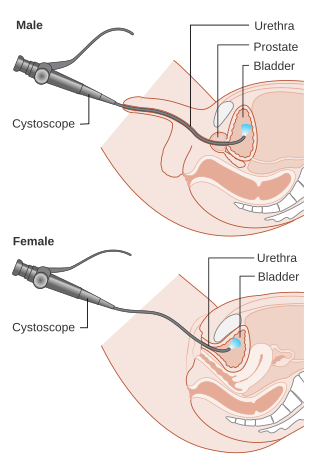
Cystoscopy is endoscopy of the urinary bladder via the urethra. It is carried out with a cystoscope.

<span class="mw-page-title-main">Urinary catheterization</span> Insertion of a catheter through the urethra to drain urine

In urinary catheterization, a latex, polyurethane, or silicone tube known as a urinary catheter is inserted into the bladder through the urethra to allow urine to drain from the bladder for collection. It may also be used to inject liquids used for treatment or diagnosis of bladder conditions. A clinician, often a nurse, usually performs the procedure, but self-catheterization is also possible. A catheter may be in place for long periods of time or removed after each use.

<span class="mw-page-title-main">Nephritis</span> Inflammation of the kidneys

Nephritis is inflammation of the kidneys and may involve the glomeruli, tubules, or interstitial tissue surrounding the glomeruli and tubules. It is one of several different types of nephropathy.

<span class="mw-page-title-main">Urinalysis</span> Array of tests performed on urine

Urinalysis, a portmanteau of the words urine and analysis, is a panel of medical tests that includes physical (macroscopic) examination of the urine, chemical evaluation using urine test strips, and microscopic examination. Macroscopic examination targets parameters such as color, clarity, odor, and specific gravity; urine test strips measure chemical properties such as pH, glucose concentration, and protein levels; and microscopy is performed to identify elements such as cells, urinary casts, crystals, and organisms.

<span class="mw-page-title-main">IgA nephropathy</span> Disease of the kidney

IgA nephropathy (IgAN), also known as Berger's disease, or synpharyngitic glomerulonephritis, is a disease of the kidney and the immune system; specifically it is a form of glomerulonephritis or an inflammation of the glomeruli of the kidney. Aggressive Berger's disease can attack other major organs, such as the liver, skin and heart.

Dysuria refers to painful or uncomfortable urination.

<span class="mw-page-title-main">Hydronephrosis</span> Medical condition

Hydronephrosis describes hydrostatic dilation of the renal pelvis and calyces as a result of obstruction to urine flow downstream. Alternatively, hydroureter describes the dilation of the ureter, and hydronephroureter describes the dilation of the entire upper urinary tract.

Microhematuria, also called microscopic hematuria, is a medical condition in which urine contains small amounts of blood; the blood quantity is too low to change the color of the urine. While not dangerous in itself, it may be a symptom of kidney disease, such as IgA nephropathy or Sickle cell trait, which should be monitored by a doctor.
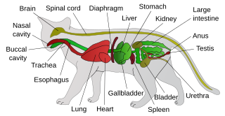
<span class="mw-page-title-main">Hemorrhagic cystitis</span> Medical condition

Hemorrhagic cystitis or haemorrhagic cystitis is an inflammation of the bladder defined by lower urinary tract symptoms that include dysuria, hematuria, and hemorrhage. The disease can occur as a complication of cyclophosphamide, ifosfamide and radiation therapy. In addition to hemorrhagic cystitis, temporary hematuria can also be seen in bladder infection or in children as a result of viral infection.

<span class="mw-page-title-main">Vesicoureteral reflux</span> Medical condition

Vesicoureteral reflux (VUR), also known as vesicoureteric reflux, is a condition in which urine flows retrograde, or backward, from the bladder into one or both ureters and then to the renal calyx or kidneys. Urine normally travels in one direction from the kidneys to the bladder via the ureters, with a one-way valve at the vesicoureteral (ureteral-bladder) junction preventing backflow. The valve is formed by oblique tunneling of the distal ureter through the wall of the bladder, creating a short length of ureter (1–2 cm) that can be compressed as the bladder fills. Reflux occurs if the ureter enters the bladder without sufficient tunneling, i.e., too "end-on".

<span class="mw-page-title-main">Ureteral stent</span>

A ureteral stent, or ureteric stent, is a thin tube inserted into the ureter to prevent or treat obstruction of the urine flow from the kidney. The length of the stents used in adult patients varies between 24 and 30 cm. Additionally, stents come in differing diameters or gauges, to fit different size ureters. The stent is usually inserted with the aid of a cystoscope. One or both ends of the stent may be coiled to prevent it from moving out of place; this is called a JJ stent, double J stent or pig-tail stent.

Pyelogram is a form of imaging of the renal pelvis and ureter.

<span class="mw-page-title-main">Loin pain hematuria syndrome</span> Medical condition

Loin pain hematuria syndrome (LPHS) is the combination of debilitating unilateral or bilateral flank pain and microscopic or macroscopic amounts of blood in the urine that is otherwise unexplained.

<span class="mw-page-title-main">Bacteriuria</span> Medical condition

Bacteriuria is the presence of bacteria in urine. Bacteriuria accompanied by symptoms is a urinary tract infection while that without is known as asymptomatic bacteriuria. Diagnosis is by urinalysis or urine culture. Escherichia coli is the most common bacterium found. People without symptoms should generally not be tested for the condition. Differential diagnosis include contamination.

Urologic diseases or conditions include urinary tract infections, kidney stones, bladder control problems, and prostate problems, among others. Some urologic conditions do not affect a person for that long and some are lifetime conditions. Kidney diseases are normally investigated and treated by nephrologists, while the specialty of urology deals with problems in the other organs. Gynecologists may deal with problems of incontinence in women.

<span class="mw-page-title-main">Urine test strip</span> Diagnostic tool used in urinalysis

A urine test strip or dipstick is a basic diagnostic tool used to determine pathological changes in a patient's urine in standard urinalysis.
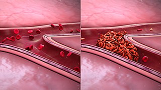
<span class="mw-page-title-main">Ureteral cancer</span> Medical condition

Ureteral cancer is cancer of the ureters, muscular tubes that propel urine from the kidneys to the urinary bladder. It is also known as ureter cancer, renal pelvic cancer, and rarely ureteric cancer or uretal cancer. Cancer in this location is rare. Ureteral cancer becomes more likely in older adults, usually ages 70–80, who have previously been diagnosed with bladder cancer.

<span class="mw-page-title-main">Urine cytology</span>

Urine cytology is a test that looks for abnormal cells in urine under a microscope. The test commonly checks for infection, inflammatory disease of the urinary tract, cancer, or precancerous conditions. It can be part of a broader urinalysis. If a cancerous condition is detected, other tests and procedures are usually recommended to diagnose cancers, including bladder cancer, ureteral cancer and cancer of the urethra. It is especially recommended when blood in the urine (hematuria) has been detected.

Normally, human urine color is straw-yellow. Urine color other than straw-yellow sometimes reflects an abnormality—an underlying pathological condition—in human beings.

References

  1. 1 2 3 4 5 6 7 8 9 Papadakis, Maxine A.; McPhee, Stephen J.; Rabow, Michael W. (14 September 2021). Current medical diagnosis & treatment 2022. 23-02: Hematuria. ISBN   978-1-264-26938-9. OCLC   1268130534.
  2. 1 2 Kirkpatrick, Wanda G. (1990), "Chapter 184 – Hematuria", in Walker, H. Kenneth; Hall, W. Dallas; Hurst, J. Willis (eds.), Clinical Methods: The History, Physical, and Laboratory Examinations (3rd ed.), Boston: Butterworths, ISBN   978-0-409-90077-4, PMID   21250137 , retrieved 2022-01-17
  3. 1 2 3 4 5 6 7 8 9 10 11 12 13 14 15 Partin, Alan W.; Dmochowski, Roger R.; Kavoussi, Louis R.; Peters, Craig, eds. (2021). "Evaluation and Management of Hematuria". Campbell-Walsh-Wein urology (Twelfth ed.). Philadelphia, Pennsylvania. ISBN   978-0-323-54642-3. OCLC   1130700336.{{cite book}}: CS1 maint: location missing publisher (link)
  4. 1 2 3 4 5 6 7 8 9 10 11 12 13 14 15 16 17 18 19 Saleem, Muhammad O.; Hamawy, Karim (2022), "Hematuria", StatPearls, Treasure Island, Florida: StatPearls Publishing, PMID   30480952 , retrieved 2022-01-17
  5. 1 2 3 4 5 6 7 8 McAninch, Jack W.; Lue, Tom (2013). "Chapter 3: Symptoms of Disorders of the Genitourinary Tract". Smith & Tanagho's General Urology. McGraw-Hill Education.
  6. 1 2 3 4 5 6 7 8 9 10 11 12 13 14 15 16 17 18 19 20 21 22 23 24 25 26 27 28 29 30 31 Stern, Scott D. C. Symptom to diagnosis: an evidence-based guide. Chapter 21-1: Approach to the Patient with Hematuria – Case 1. OCLC   1121597721.
  7. Hashmi, Mydah S.; Pandey, Jyotsna (2022), "Nephritic Syndrome", StatPearls, Treasure Island, Florida: StatPearls Publishing, PMID   32965911 , retrieved 2022-01-19
  8. Izzo, Joseph L.; Sica, Domenic A.; Black, Henry Richard (2008). Hypertension Primer. Lippincott Williams & Wilkins. p. 382. ISBN   978-0-7817-8205-0.
  9. 1 2 3 4 5 6 Ingelfinger, Julie R. (8 July 2021). "Hematuria in Adults". New England Journal of Medicine. 385 (2): 153–163. doi:10.1056/NEJMra1604481.
  10. "Changes in Urine; Symptoms, Causes & Treatment". Cleveland Clinic. Retrieved 2022-09-12.
  11. "Hematuria in Children". National Kidney Foundation. 2015-12-24. Retrieved 2023-03-11.
  12. Pade, Kathryn H.; Liu, Deborah R. (September 2014). "An evidence-based approach to the management of hematuria in children in the emergency department". Pediatric Emergency Medicine Practice. 11 (9): 1–13, quiz 14. ISSN   1549-9650. PMID   25296518.
  13. 1 2 Avellino, Gabriella J.; Bose, Sanchita; Wang, David S. (June 2016). "Diagnosis and Management of Hematuria". Surgical Clinics of North America. 96 (3): 503–515. doi:10.1016/j.suc.2016.02.007. PMID   27261791.
  14. 1 2 Yun, Edward J.; Meng, Maxwell V.; Carroll, Peter R. (March 2004). "Evaluation of the patient with hematuria". Medical Clinics of North America. 88 (2): 329–343. doi:10.1016/S0025-7125(03)00172-X. PMID   15049581.
  15. 1 2 3 Davis, Rodney; Jones, J. Stephen; Barocas, Daniel A.; Castle, Erik P.; Lang, Erich K.; Leveillee, Raymond J.; Messing, Edward M.; Miller, Scott D.; Peterson, Andrew C.; Turk, Thomas M. T.; Weitzel, William (2012-12-01). "Diagnosis, Evaluation and Follow-Up of Asymptomatic Microhematuria (AMH) in Adults: AUA Guideline". Journal of Urology (published 2012). 188 (6S): 2473–2481. doi:10.1016/j.juro.2012.09.078. PMID   23098784.
  16. 1 2 3 4 5 6 7 8 9 10 Barocas, Daniel A.; Boorjian, Stephen A.; Alvarez, Ronald D.; Downs, Tracy M.; Gross, Cary P.; Hamilton, Blake D.; Kobashi, Kathleen C.; Lipman, Robert R.; Lotan, Yair; Ng, Casey K.; Nielsen, Matthew E. (2020-10-01). "Microhematuria: AUA/SUFU Guideline". Journal of Urology. 204 (4): 778–786. doi: 10.1097/JU.0000000000001297 . PMID   32698717. S2CID   220717643.
  17. 1 2 3 4 5 6 7 8 9 10 11 12 Kaplan, Damara; Kohn, Taylor. "Urologic Emergencies: Gross Hematuria with Clot Retention". American Urological Association. Archived from the original on 2019-11-28. Retrieved 2019-12-11.
  18. 1 2 3 4 5 6 Wagenlehner, Florian M. E.; Lichtenstern, Christoph; Rolfes, Caroline; Mayer, Konstantin; Uhle, Florian; Weidner, Wolfgang; Weigand, Markus A. (June 2013). "Diagnosis and management for urosepsis: Items in urosepsis". International Journal of Urology. 20 (10): 963–970. doi: 10.1111/iju.12200 . PMID   23714209.
  19. 1 2 3 4 5 6 7 Coplen, D. E. (January 2013). "Diagnosis, Evaluation and Follow-Up of Asymptomatic Microhematuria (AMH) in Adults: AUA Guideline". Yearbook of Urology. 2013: 1–2. doi:10.1016/j.yuro.2013.07.019. ISSN   0084-4071.
  20. Shah, Samir; Ronan, Jeanine C.; Alverson, Brian (2014). Step-up to pediatrics (first ed.). Philadelphia: Wolters Kluwer/Lippincott Williams & Wilkins. pp. 175–176. ISBN   978-1451145809. OCLC   855779297.
  21. Cohen, Robert A.; Brown, Robert S. (2003-06-05). "Clinical practice. Microscopic hematuria". The New England Journal of Medicine. 348 (23): 2330–2338. doi:10.1056/NEJMcp012694. ISSN   1533-4406. PMID   12788998.
  22. Sharp, Victoria; Barnes, Kerri D.; Erickson, Bradley D. (December 1, 2013). "Assessment of Asymptomatic Microscopic Hematuria in Adults". American Family Physician. 88 (11): 747–754. PMID   24364522.

Commons-logo.svg Media related to Hematuria at Wikimedia Commons