Myoglobin (symbol Mb or MB) is an iron- and oxygen-binding protein found in the cardiac and skeletalmuscle tissue of vertebrates in general and in almost all mammals.[5][6][7][8][9] Myoglobin is distantly related to hemoglobin. Compared to hemoglobin, myoglobin has a higher affinity for oxygen and does not have cooperative binding with oxygen like hemoglobin does.[8][10] Myoglobin consists of non-polar amino acids at the core of the globulin, where the heme group is non-covalently bounded with the surrounding polypeptide of myoglobin. In humans, myoglobin is found in the bloodstream only after muscle injury.[11][12][13]
High concentrations of myoglobin in muscle cells allow organisms to hold their breath for a longer period of time. Diving mammals such as whales and seals have muscles with particularly high abundance of myoglobin.[13] Myoglobin is found in Type I muscle, Type II A, and Type II B; although many older texts describe myoglobin as not found in smooth muscle, this has proved erroneous: there is also myoglobin in smooth muscle cells.[14]
Myoglobin was the first protein to have its three-dimensional structure revealed by X-ray crystallography.[15] This achievement was reported in 1958 by John Kendrew and associates.[16] For this discovery, Kendrew shared the 1962 Nobel Prize in Chemistry with Max Perutz.[17][18] Despite being one of the most studied proteins in biology, its physiological function is not yet conclusively established: mice genetically engineered to lack myoglobin can be viable and fertile, but show many cellular and physiological adaptations to overcome the loss. Through observing these changes in myoglobin-depleted mice, it is hypothesised that myoglobin function relates to increased oxygen transport to muscle, and to oxygen storage; as well, it serves as a scavenger of reactive oxygen species.[19]
In humans, myoglobin is encoded by the MBgene.[20]
Myoglobin can take the forms oxymyoglobin (MbO2), carboxymyoglobin (MbCO), and metmyoglobin (met-Mb), analogously to hemoglobin taking the forms oxyhemoglobin (HbO2), carboxyhemoglobin (HbCO), and methemoglobin (met-Hb).[21]
Differences from hemoglobin
Like hemoglobin, myoglobin is a cytoplasmic protein that binds oxygen on a heme group. It harbors only one globulin group, whereas hemoglobin has four. Although its heme group is identical to those in Hb, Mb has a higher affinity for oxygen than does hemoglobin but fewer total oxygen-storage capacities. Research suggests that myoglobin facilitates oxygen diffusion down a gradient, enhancing oxygen transport in mitochondria.[22]
Role in cuisine
Myoglobin contains hemes, pigments responsible for the color of red meat. The color that meat takes is partly determined by the degree of oxidation of the myoglobin. In fresh meat the iron atom is in the ferrous (+2) oxidation state bound to an oxygen molecule (O2). Meat cooked well done is brown because the iron atom is now in the ferric (+3) oxidation state, having lost an electron. If meat has been exposed to nitrites, it will remain pink, because the iron atom is bound to NO, nitric oxide (true of, e.g., corned beef or cured hams). Grilled meats can also take on a reddish pink "smoke ring" that comes from the heme center binding to carbon monoxide.[23] Raw meat packed in a carbon monoxide atmosphere also shows this same pink "smoke ring" due to the same principles. Notably, the surface of this raw meat also displays the pink color, which is usually associated in consumers' minds with fresh meat. This artificially induced pink color can persist, reportedly up to one year.[24]Hormel and Cargill (meat processing companies in the US) are both reported to use this meat-packing process, and meat treated this way has been in the consumer market since 2003.[25] If fresh meat is left to an environment where the redox potential is high, oxymyoglobin will eventually turn into metmyoglobin (Fe+3) and the meat will turn into dark red naturally.[26]
Meat alternatives have used various ways to recreate the "meaty" taste associated with myoglobin. Impossible Foods uses leghemoglobin, a heme-containing globin from soy root nodule, produced as a recombinant protein in Komagataella ("Pichia pastoris") yeast.[27][28] Motif FoodWorks produces a recombinant bovine myoglobin using Komagataella yeast,[29] considered GRAS by the FDA.[30] Moolec Science has engineered a soybean that produces porcine myoglobin in its seeds called "Piggy Sooy"; it was approved by the USDA in April 2024.[31]
Role in disease
Myoglobin is released from damaged muscle tissue, which contain very high concentrations of myoglobin.[32] The released myoglobin enters the bloodstream, where high levels may indicate rhabdomyolysis. The myoglobin is filtered by the kidneys, but is toxic to the renal tubular epithelium and so may cause acute kidney injury.[33] It is not the myoglobin itself that is toxic (it is a protoxin), but the ferrihemate portion that is dissociated from myoglobin in acidic environments (e.g., acidic urine, lysosomes).[citation needed]
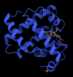
Myoglobin belongs to the globin superfamily of proteins, and as with other globins, consists of eight alpha helices connected by loops. Human myoglobin contains 154 amino acids.[36]
Myoglobin contains a porphyrin ring with an iron at its center. A proximalhistidine group (His-93) is attached directly to iron, and a distal histidine group (His-64) hovers near the opposite face.[36] The distal imidazole is not bonded to the iron, but is available to interact with the substrate O2. This interaction encourages the binding of O2, but not carbon monoxide (CO), which still binds about 240× more strongly than O2.[citation needed]
The binding of O2 causes substantial structural change at the Fe center, which shrinks in radius and moves into the center of N4 pocket. O2-binding induces "spin-pairing": the five-coordinate ferrous deoxy form is high spin and the six coordinate oxy form is low spin and diamagnetic.[citation needed]
Molecular orbital description of Fe-O2 interaction in myoglobin.[37]
This is an image of an oxygenated myoglobin molecule. The image shows the structural change when oxygen is bound to the iron atom of the heme prosthetic group. The oxygen atoms are colored in green, the iron atom is colored in red, and the heme group is colored in blue.
Myoglobin
Synthetic analogues
Many models of myoglobin have been synthesized as part of a broad interest in transition metal dioxygen complexes. A well known example is the picket fence porphyrin, which consists of a ferrous complex of a sterically bulky derivative of tetraphenylporphyrin.[38] In the presence of an imidazole ligand, this ferrous complex reversibly binds O2. The O2 substrate adopts a bent geometry, occupying the sixth position of the iron center. A key property of this model is the slow formation of the μ-oxo dimer, which is an inactive diferric state. In nature, such deactivation pathways are suppressed by protein matrix that prevents close approach of the Fe-porphyrin assemblies.[39]
A picket-fence porphyrin complex of Fe, with axial coordination sites occupied by methylimidazole (green) and dioxygen. The R groups flank the O2-binding site.
↑ Wick MR, Hornick JL (2011). "Immunohistology of Soft Tissue and Osseous Neoplasms". Diagnostic Immunohistochemistry. Elsevier. pp.83–136. doi:10.1016/b978-1-4160-5766-6.00008-x. ISBN978-1-4160-5766-6. Myoglobin is a 17.8-kD protein that is found in cardiac and skeletal muscle and that forms complexes with iron molecules.
↑ Feher J (2017). "Oxygen and Carbon Dioxide Transport". Quantitative Human Physiology. Elsevier. pp.656–664. doi:10.1016/b978-0-12-800883-6.00064-1. ISBN978-0-12-800883-6. Highly oxidative muscle fibers contain a lot of myoglobin. It has two functions in muscle: it stores oxygen for use during heavy exercise, and it enhances diffusion through the cytosol by carrying the oxygen. By binding O2, myoglobin (Mb) provides a second diffusive pathway for O2 through the cell cytosol.
1 2 Wilson MT, Reeder BJ (2006). "MYOGLOBIN". Encyclopedia of Respiratory Medicine. Elsevier. pp.73–76. doi:10.1016/b0-12-370879-6/00250-7. ISBN978-0-12-370879-3. Myoglobin (Mb) is a heme-containing globular protein that is found in abundance in myocyte cells of heart and skeletal muscle.
↑ Boncyk JC (2007). "Perioperative Hypoxia". Complications in Anesthesia. Elsevier. pp.193–199. doi:10.1016/b978-1-4160-2215-2.50052-1. ISBN978-1-4160-2215-2. Myoglobin serves both as an O2 buffer and to store O2 in muscle. All known vertebrate myoglobins and β-hemoglobin subunits are similar in structure, but myoglobin binds O2 more avidly at low Po2 (Fig. 47-5) because it is a monomer (i.e., it does not undergo a significant conformational change with oxygenation). Thus, myoglobin remains fully saturated at O2 tensions between 15 and 30 mm Hg and unloads its O2 to the muscle mitochondria only at very low O2 tensions.
↑ Sekhon N, Peacock WF (2019). "Biomarkers to Assist in the Evaluation of Chest Pain". Biomarkers in Cardiovascular Disease. Elsevier. pp.115–128. doi:10.1016/b978-0-323-54835-9.00011-9. ISBN978-0-323-54835-9. S2CID59548142. myoglobin is not specific for the death of cardiac myocytes, and levels can be elevated in renal disease as well as damage to skeletal muscle.
↑ Harvey JW (2008). "Iron Metabolism and Its Disorders". Clinical Biochemistry of Domestic Animals. Elsevier. pp.259–285. doi:10.1016/b978-0-12-370491-7.00009-x. ISBN978-0-12-370491-7. Myoglobin is an oxygen-binding protein located primarily in muscles. Myoglobin serves as a local oxygen reservoir that can temporarily provide oxygen when blood oxygen delivery is insufficient during periods of intense muscular activity. Iron within the heme group must be in the Fe+2 state to bind oxygen. If iron is oxidized to the Fe+3 state, metmyoglobin is formed.
↑ Berridge BR, Van Vleet JF, Herman E (2013). "Cardiac, Vascular, and Skeletal Muscle Systems". Haschek and Rousseaux's Handbook of Toxicologic Pathology. Elsevier. pp.1567–1665. doi:10.1016/b978-0-12-415759-0.00046-7. ISBN978-0-12-415759-0. Myoglobin is a low molecular weight oxygen binding heme protein that is found exclusively in heart and skeletal muscle cells. In blood, myoglobin is bound primarily to plasma globulins, a complex which is filtered by the kidneys. If the plasma concentration exceeds the plasma binding capacity (1.5 mg/dl in humans), myoglobin begins to appear in the urine. High concentrations of myoglobin can change the color of the urine to a dark red-brown color.
↑ Weber M, Rau M, Madlener K, Elsaesser A, Bankovic D, Mitrovic V, etal. (Nov 2005). "Diagnostic utility of new immunoassays for the cardiac markers cTnI, myoglobin and CK-MB mass". Clinical Biochemistry. 38 (11): 1027–30. doi:10.1016/j.clinbiochem.2005.07.011. PMID16125162.
↑ Dasgupta A, Wahed A (2014). "Cardiac Markers". Clinical Chemistry, Immunology and Laboratory Quality Control. Elsevier. pp.127–144. doi:10.1016/b978-0-12-407821-5.00008-5. ISBN978-0-12-407821-5. Myoglobin is a heme protein found in both skeletal and cardiac muscle. Myoglobin is typically released in the circulation as early as 1 h after myocardial infarction,... Myoglobin has poor clinical specificity due to the presence of large quantities of myoglobin in skeletal muscle. Some studies suggest adding the myoglobin test to the troponin I test in order to improve diagnostic value [4]. Myoglobin, being a small protein, is excreted in urine, and a high level of serum myoglobin is encountered in patients with acute renal failure (uremic syndrome). Acute renal failure is also a complication of rhabdomyolysis, ...
↑ Lippard SJ, Berg JM (1994). Principles of Bioinorganic Chemistry. Mill Valley, CA: University Science Books. ISBN0-935702-73-3.
Further reading
Collman JP, Boulatov R, Sunderland CJ, Fu L (Feb 2004). "Functional analogues of cytochrome c oxidase, myoglobin, and hemoglobin". Chemical Reviews. 104 (2): 561–88. doi:10.1021/cr0206059. PMID14871135.
Reeder BJ, Svistunenko DA, Cooper CE, Wilson MT (Dec 2004). "The radical and redox chemistry of myoglobin and hemoglobin: from in vitro studies to human pathology". Antioxidants & Redox Signaling. 6 (6): 954–66. doi:10.1089/ars.2004.6.954. PMID15548893.
Schlieper G, Kim JH, Molojavyi A, Jacoby C, Laussmann T, Flögel U, etal. (Apr 2004). "Adaptation of the myoglobin knockout mouse to hypoxic stress". American Journal of Physiology. Regulatory, Integrative and Comparative Physiology. 286 (4): R786-92. doi:10.1152/ajpregu.00043.2003. PMID14656764. S2CID24831969.
Takano T (Mar 1977). "Structure of myoglobin refined at 2-0 A resolution. II. Structure of deoxymyoglobin from sperm whale". Journal of Molecular Biology. 110 (3): 569–84. doi:10.1016/S0022-2836(77)80112-5. PMID845960.
Roy A, Sen S, Chakraborti AS (Feb 2004). "In vitro nonenzymatic glycation enhances the role of myoglobin as a source of oxidative stress". Free Radical Research. 38 (2): 139–46. doi:10.1080/10715160310001638038. PMID15104207. S2CID11631439.
Stewart JM, Blakely JA, Karpowicz PA, Kalanxhi E, Thatcher BJ, Martin BM (Mar 2004). "Unusually weak oxygen binding, physical properties, partial sequence, autoxidation rate and a potential phosphorylation site of beluga whale (Delphinapterus leucas) myoglobin". Comparative Biochemistry and Physiology B. 137 (3): 401–12. doi:10.1016/j.cbpc.2004.01.007. PMID15050527.
1mnh: INTERACTIONS AMONG RESIDUES CD3, E7, E10 AND E11 IN MYOGLOBINS: ATTEMPTS TO SIMULATE THE O2 AND CO BINDING PROPERTIES OF APLYSIA MYOGLOBIN
1mni: ALTERATION OF AXIAL COORDINATION BY PROTEIN ENGINEERING IN MYOGLOBIN. BIS-IMIDAZOLE LIGATION IN THE HIS64-->VAL(SLASH)VAL68-->HIS DOUBLE MUTANT
1mnj: INTERACTIONS AMONG RESIDUES CD3, E7, E10 AND E11 IN MYOGLOBINS: ATTEMPTS TO SIMULATE THE O2 AND CO BINDING PROPERTIES OF APLYSIA MYOGLOBIN
1mnk: INTERACTIONS AMONG RESIDUES CD3, E7, E10 AND E11 IN MYOGLOBINS: ATTEMPTS TO SIMULATE THE O2 AND CO BINDING PROPERTIES OF APLYSIA MYOGLOBIN
1mno: V68N MYOGLOBIN OXY FORM
1mwc: WILD TYPE MYOGLOBIN WITH CO
1mwd: WILD TYPE DEOXY MYOGLOBIN
1myg: HIGH RESOLUTION X-RAY STRUCTURES OF PIG METMYOGLOBIN AND TWO CD3 MUTANTS MB(LYS45-> ARG) AND MB(LYS45-> SER)
1myh: HIGH RESOLUTION X-RAY STRUCTURES OF PIG METMYOGLOBIN AND TWO CD3 MUTANTS MB(LYS45-> ARG) AND MB(LYS45-> SER)
1myi: HIGH RESOLUTION X-RAY STRUCTURES OF PIG METMYOGLOBIN AND TWO CD3 MUTANTS MB(LYS45-> ARG) AND MB(LYS45-> SER)
1myj: DISTAL POLARITY IN LIGAND BINDING TO MYOGLOBIN: STRUCTURAL AND FUNCTIONAL CHARACTERIZATION OF A THREONINE68(E11) MUTANT
1pmb: THE DETERMINATION OF THE CRYSTAL STRUCTURE OF RECOMBINANT PIG MYOGLOBIN BY MOLECULAR REPLACEMENT AND ITS REFINEMENT
1yca: DISTAL POCKET POLARITY IN LIGAND BINDING TO MYOGLOBIN: DEOXY AND CARBONMONOXY FORMS OF A THREONINE68 (E11) MUTANT INVESTIGATED BY X-RAY CRYSTALLOGRAPHY AND INFRARED SPECTROSCOPY
1ycb: DISTAL POCKET POLARITY IN LIGAND BINDING TO MYOGLOBIN: DEOXY AND CARBONMONOXY FORMS OF A THREONINE68 (E11) MUTANT INVESTIGATED BY X-RAY CRYSTALLOGRAPHY AND INFRARED SPECTROSCOPY
2mm1: X-RAY CRYSTAL STRUCTURE OF A RECOMBINANT HUMAN MYOGLOBIN MUTANT AT 2.8 ANGSTROMS RESOLUTION
This page is based on this Wikipedia article Text is available under the CC BY-SA 4.0 license; additional terms may apply. Images, videos and audio are available under their respective licenses.