Thyroid function tests | |
---|---|
MeSH | D013960 |
MedlinePlus | 003444 |
Thyroid function tests (TFTs) is a collective term for blood tests used to check the function of the thyroid. [1] TFTs may be requested if a patient is thought to suffer from hyperthyroidism (overactive thyroid) or hypothyroidism (underactive thyroid), or to monitor the effectiveness of either thyroid-suppression or hormone replacement therapy. It is also requested routinely in conditions linked to thyroid disease, such as atrial fibrillation and anxiety disorder.
A TFT panel typically includes thyroid hormones such as thyroid-stimulating hormone (TSH, thyrotropin) and thyroxine (T4), and triiodothyronine (T3) depending on local laboratory policy.
Thyroid-stimulating hormone (TSH, thyrotropin) is generally increased in hypothyroidism and decreased in hyperthyroidism, [2] making it the most important test for early detection of both of these conditions. [3] [4] The result of this assay is suggestive of the presence and cause of thyroid disease, since a measurement of elevated TSH generally indicates hypothyroidism, while a measurement of low TSH generally indicates hyperthyroidism. [2] However, when TSH is measured by itself, it can yield misleading results, so additional thyroid function tests must be compared with the result of this test for accurate diagnosis. [4] [5] [6]
TSH is produced in the pituitary gland. The production of TSH is controlled by thyrotropin-releasing hormone (TRH), which is produced in the hypothalamus. TSH levels may be suppressed by excess free T3 (fT3) or free T4 (fT4) in the blood.[ citation needed ]
First-generation TSH assays were done by radioimmunoassay and were introduced in 1965. [3] There were variations and improvements upon TSH radioimmunoassay, but their use declined as a new immunometric assay technique became available in the middle of the 1980s. [3] [4] The new techniques were more accurate, leading to the second, third, and even fourth generations of TSH assay, with each generation possessing ten times greater functional sensitivity than the last. [7] Third generation immunometric assay methods are typically automated. [3] Fourth generation TSH immunometric assay has been developed for use in research. [4]
Third generation TSH assay is the requirement for modern standards of care. TSH testing in the United States is typically carried out with automated platforms using advanced forms of immunometric assay. [3] Nonetheless, there is no international standard for measurement of thyroid-stimulating hormone. [4]
Accurate interpretation takes a variety of factors into account, such as the thyroid hormones i.e. thyroxine (T4) and triiodothyronine (T3), current medical status (such as pregnancy [3] ), [4] certain medications like propylthiouracil, [4] temporal effects including circadian rhythm [8] and hysteresis, [9] and other past medical history. [10]
Total thyroxine is rarely measured, having been largely superseded by free thyroxine tests. Total thyroxine (Total T4) is generally elevated in hyperthyroidism and decreased in hypothyroidism. [2] It is usually slightly elevated in pregnancy secondary to increased levels of thyroid binding globulin (TBG). [2]
Total T4 is measured to see the bound and unbound levels of T4. The total T4 is less useful in cases where there could be protein abnormalities. The total T4 is less accurate due to the large amount of T4 that is bound. The total T3 is measured in clinical practice since the T3 has decreased amount that is bound as compared to T4.[ citation needed ]
Reference ranges depend on the method of analysis. Results should always be interpreted using the range from the laboratory that performed the test. Example values are:
Lower limit | Upper limit | Unit |
4, [11] 5.5 [12] | 11, [11] 12.3 [12] | μg/dL |
60 [11] [13] | 140, [11] 160 [13] | nmol/L |
Free thyroxine (fT4 or free T4) is generally elevated in hyperthyroidism and decreased in hypothyroidism. [2]
Reference ranges depend on the method of analysis. Results should always be interpreted using the range from the laboratory that performed the test. Example values are:
Patient type | Lower limit | Upper limit | Unit |
Normal adult | 0.7, [14] 0.8 [12] | 1.4, [14] 1.5, [12] 1.8 [15] | ng/dL |
9, [16] [17] 10, [11] 12 [13] | 18, [16] [17] 23 [13] | pmol/L | |
Infant 0–3 d | 2.0 [14] | 5.0 [14] | ng/dL |
26 [17] | 65 [17] | pmol/L | |
Infant 3–30 d | 0.9 [14] | 2.2 [14] | ng/dL |
12 [17] | 30 [17] | pmol/L | |
Child/Adolescent 31 d – 18 y | 0.8 [14] | 2.0 [14] | ng/dL |
10 [17] | 26 [17] | pmol/L | |
Pregnant | 0.5 [14] | 1.0 [14] | ng/dL |
6.5 [17] | 13 [17] | pmol/L | |
Total triiodothyronine (Total T3) is rarely measured, having been largely superseded by free T3 tests. Total T3 is generally elevated in hyperthyroidism and decreased in hypothyroidism. [2]
Reference ranges depend on the method of analysis. Results should always be interpreted using the range from the laboratory that performed the test. Example values are:
Test | Lower limit | Upper limit | Unit |
Total triiodothyronine | 60, [12] 75 [11] | 175, [11] 181 [12] | ng/dL |
0.9, [16] 1.1 [11] | 2.5, [16] 2.7 [11] | nmol/L | |
Free triiodothyronine (fT3 or free T3) is generally elevated in hyperthyroidism and decreased in hypothyroidism. [2]
Reference ranges depend on the method of analysis. Results should always be interpreted using the range from the laboratory that performed the test. Example values are:
Patient type | Lower limit | Upper limit | Unit |
Normal adult | 3.0 [11] | 7.0 [11] | pg/mL |
3.1 [18] | 7.7 [18] | pmol/L | |
Children 2–16 y | 3.0 [19] | 7.0 [19] | pg/mL |
1.5 [18] | 15.2 [18] | pmol/L | |
An increased thyroxine-binding globulin results in an increased total thyroxine and total triiodothyronine without an actual increase in hormonal activity of thyroid hormones.
Reference ranges:
Lower limit | Upper limit | Unit |
12 [12] | 30 [12] | mg/L |
Reference ranges:
Lower limit | Upper limit | Unit |
1.5 [11] | 30 [11] | pmol/L |
1 [11] | 20 [11] | μg/L |
Thyroid hormone uptake (Tuptake or T3 uptake) is a measure of the unbound thyroxine binding globulins in the blood, that is, the TBG that is unsaturated with thyroid hormone. [2] Unsaturated TBG increases with decreased levels of thyroid hormones. It is not directly related to triiodothyronine, despite the name T3 uptake. [2]
Reference ranges:
Patient type | Lower limit | Upper limit | Unit |
Females | 25 [2] | 35 [2] | % |
In pregnancy | 15 [2] | 25 [2] | % |
Males | 25 [2] | 35 [2] | % |
The Free Thyroxine Index (FTI or T7) is obtained by multiplying the total T4 with T3 uptake. [2] FTI is considered to be a more reliable indicator of thyroid status in the presence of abnormalities in plasma protein binding. [2] This test is rarely used now that reliable free thyroxine and free triiodothyronine assays are routinely available.
FTI is elevated in hyperthyroidism and decreased in hypothyroidism. [2]
Patient type | Lower limit | Upper limit | Unit |
Females | 1.8 [2] | 5.0 [2] | |
Males | 1.3 [2] | 4.2 [2] | |
Derived structure parameters that describe constant properties of the overall feedback control system may add useful information for special purposes, e.g. in diagnosis of nonthyroidal illness syndrome or central hypothyroidism. [20] [21] [22] [23]
Thyroid's secretory capacity (GT, also referred to as SPINA-GT) is the maximum stimulated amount of thyroxine the thyroid can produce in one second. [24] GT is elevated in hyperthyroidism and reduced in hypothyroidism. [25]
GT is calculated with
or
: Dilution factor for T4 (reciprocal of apparent volume of distribution, 0.1 l−1)
: Clearance exponent for T4 (1.1e-6 sec−1)
K41: Dissociation constant T4-TBG (2e10 L/mol)
K42: Dissociation constant T4-TBPA (2e8 L/mol)
DT: EC50 for TSH (2.75 mU/L) [24]
Lower limit | Upper limit | Unit |
1.41 [24] | 8.67 [24] | pmol/s |
The sum activity of peripheral deiodinases (GD, also referred to as SPINA-GD) is reduced in nonthyroidal illness with hypodeiodination. [21] [22] [26]
GD is obtained with
or
: Dilution factor for T3 (reciprocal of apparent volume of distribution, 0.026 L−1)
: Clearance exponent for T3 (8e-6 sec−1)
KM1: Dissociation constant of type-1-deiodinase (5e-7 mol/L)
K30: Dissociation constant T3-TBG (2e9 L/mol) [24]
Lower limit | Upper limit | Unit |
20 [24] | 40 [24] | nmol/s |
Jostel's TSH index (JTI or TSHI) helps to determine thyrotropic function of anterior pituitary on a quantitative level. [27] It is reduced in thyrotropic insufficiency [27] and in certain cases of non-thyroidal illness syndrome. [26]
It is calculated with
.
Additionally, a standardized form of TSH index may be calculated with
. [27]
Parameter | Lower limit | Upper limit | Unit |
TSHI | 1.3 [27] | 4.1 [27] | |
sTSHI | -2 [27] | 2 [27] | |
The Thyrotroph Thyroid Hormone Sensitivity Index (TTSI, also referred to as Thyrotroph T4 Resistance Index or TT4RI) was developed to enable fast screening for resistance to thyroid hormone. [28] [29] Somewhat similar to the TSH Index it is calculated from equilibrium values for TSH and FT4, however with a different equation.
Lower limit | Upper limit | Unit |
100 | 150 | |
The Thyroid Feedback Quantile-based Index (TFQI) is another parameter for thyrotropic pituitary function. It was defined to be more robust to distorted data than JTI and TTSI. It is calculated with
from quantiles of FT4 and TSH concentration (as determined based on cumulative distribution functions). [30] Per definition the TFQI has a mean of 0 and a standard deviation of 0.37 in a reference population. [30] Higher values of TFQI are associated with obesity, metabolic syndrome, impaired renal function, diabetes, and diabetes-related mortality. [30] [31] [32] [33] [34] [35] [36] TFQI results are also elevated in takotsubo syndrome, [37] potentially reflecting type 2 allostatic load in the situation of psychosocial stress. Reductions have been observed in subjects with schizophrenia after initiation of therapy with oxcarbazepine, potentially reflecting declining allostatic load. [38]
Lower limit | Upper limit | Unit |
–0,74 | +0.74 | |
In healthy persons, the intra-individual variation of TSH and thyroid hormones is considerably smaller than the inter-individual variation. [39] [40] [41] This results from a personal set point of thyroid homeostasis. [42] In hypothyroidism, it is impossible to directly access the set point, [43] but it can be reconstructed with methods of systems theory. [44] [45] [46]
A computerised algorithm, called Thyroid-SPOT, which is based on this mathematical theory, has been implemented in software applications. [47] In patients undergoing thyroidectomy it could be demonstrated that this algorithm can be used to reconstruct the personal set point with sufficient precision. [48]
Drugs can profoundly affect thyroid function tests. Listed below is a selection of important effects.
Cause | Drug | Effect on hormone concentrations | Effect on structure parameters |
---|---|---|---|
Inhibited TSH secretion | Dopamine, L-DOPA, glucocorticoids, somatostatin | ↓T4; ↓T3; ↓TSH | ↔SPINA-GT; ↓JTI |
Inhibited synthesis or release of thyroid hormone | Iodine, lithium | ↓T4; ↓T3; ↑TSH | ↓SPINA-GT; ↔JTI |
Inhibited conversion of T4 to T3 (Step-up hypodeiodination) | Amiodarone, glucocorticoids, propranolol, propylthiouracil, radiographic contrast agents | ↓T3; ↑rT3; ↓, ↔, ↑T4 and fT4; ↔, ↑TSH | ↓SPINA-GD |
Inhibited binding of T4/T3 to serum proteins | Salicylates, phenytoin, carbamazepine, furosemide, nonsteroidal anti-inflammatory agents, heparin (in vitro effect) | ↓T4; ↓T3; ↓fT4E, ↔, ↑fT4; ↔TSH | ↓T4/fT4 ratio |
Stimulated metabolism of iodothyronines | Phenobarbital, phenytoin, carbamazepine, rifampicin | ↓T4; ↓fT4; ↔TSH | |
Inhibited absorption of ingested T4 | Aluminium hydroxide, ferrous sulfate, cholestyramine, colestipol, iron sucralfate, soybean preparations, kayexalate | ↓T4; ↓fT4; ↑TSH | |
Increase in concentration of T4-binding proteins | Estrogen, clofibrate, opiates (heroin, methadone), 5-fluorouracil, perphenazine | ↑T4; ↑T3; ↔fT4; ↔TSH | ↔SPINA-GT; ↔SPINA-GD; ↔JTI; ↑T4/fT4 ratio |
Decrease in concentration of T4-binding proteins | Androgens, glucocorticoids | ↓T4; ↓T3; ↔fT4; ↔TSH | ↔SPINA-GT; ↔SPINA-GD; ↔JTI; ↓T4/fT4 ratio |
↓: reduced serum concentration or structure parameter; ↑: increased serum concentration or structure parameter; ↔: no change; TSH: Thyroid-stimulating hormone; T3: Total triiodothyronine; T4: Total thyroxine; fT4: Free thyroxine; fT3: Free triiodothyronine; rT3: Reverse triiodothyronine
The thyroid, or thyroid gland, is an endocrine gland in vertebrates. In humans, it is in the neck and consists of two connected lobes. The lower two thirds of the lobes are connected by a thin band of tissue called the isthmus (pl.: isthmi). The thyroid gland is a butterfly-shaped gland located in the neck below the Adam's apple. Microscopically, the functional unit of the thyroid gland is the spherical thyroid follicle, lined with follicular cells (thyrocytes), and occasional parafollicular cells that surround a lumen containing colloid. The thyroid gland secretes three hormones: the two thyroid hormones – triiodothyronine (T3) and thyroxine (T4) – and a peptide hormone, calcitonin. The thyroid hormones influence the metabolic rate and protein synthesis and growth and development in children. Calcitonin plays a role in calcium homeostasis. Secretion of the two thyroid hormones is regulated by thyroid-stimulating hormone (TSH), which is secreted from the anterior pituitary gland. TSH is regulated by thyrotropin-releasing hormone (TRH), which is produced by the hypothalamus.
Hypothyroidism is a disorder of the endocrine system in which the thyroid gland does not produce enough thyroid hormones. It can cause a number of symptoms, such as poor ability to tolerate cold, extreme fatigue, muscle aches, constipation, slow heart rate, depression, and weight gain. Occasionally there may be swelling of the front part of the neck due to goitre. Untreated cases of hypothyroidism during pregnancy can lead to delays in growth and intellectual development in the baby or congenital iodine deficiency syndrome.
Iodothyronine deiodinases (EC 1.21.99.4 and EC 1.21.99.3) are a subfamily of deiodinase enzymes important in the activation and deactivation of thyroid hormones. Thyroxine (T4), the precursor of 3,5,3'-triiodothyronine (T3) is transformed into T3 by deiodinase activity. T3, through binding a nuclear thyroid hormone receptor, influences the expression of genes in practically every vertebrate cell. Iodothyronine deiodinases are unusual in that these enzymes contain selenium, in the form of an otherwise rare amino acid selenocysteine.
Thyroid-stimulating hormone (also known as thyrotropin, thyrotropic hormone, or abbreviated TSH) is a pituitary hormone that stimulates the thyroid gland to produce thyroxine (T4), and then triiodothyronine (T3) which stimulates the metabolism of almost every tissue in the body. It is a glycoprotein hormone produced by thyrotrope cells in the anterior pituitary gland, which regulates the endocrine function of the thyroid.
Thyroxine-binding globulin (TBG) is a globulin protein encoded by the SERPINA7 gene in humans. TBG binds thyroid hormones in circulation. It is one of three transport proteins (along with transthyretin and serum albumin) responsible for carrying the thyroid hormones thyroxine (T4) and triiodothyronine (T3) in the bloodstream. Of these three proteins, TBG has the highest affinity for T4 and T3 but is present in the lowest concentration relative to transthyretin and albumin, which also bind T3 and T4 in circulation. Despite its low concentration, TBG carries the majority of T4 in the blood plasma. Due to the very low concentration of T4 and T3 in the blood, TBG is rarely more than 25% saturated with its ligand. Unlike transthyretin and albumin, TBG has a single binding site for T4/T3. TBG is synthesized primarily in the liver as a 54-kDa protein. In terms of genomics, TBG is a serpin; however, it has no inhibitory function like many other members of this class of proteins.
Triiodothyronine, also known as T3, is a thyroid hormone. It affects almost every physiological process in the body, including growth and development, metabolism, body temperature, and heart rate.
Levothyroxine, also known as L-thyroxine, is a synthetic form of the thyroid hormone thyroxine (T4). It is used to treat thyroid hormone deficiency (hypothyroidism), including a severe form known as myxedema coma. It may also be used to treat and prevent certain types of thyroid tumors. It is not indicated for weight loss. Levothyroxine is taken orally (by mouth) or given by intravenous injection. Levothyroxine has a half-life of 7.5 days when taken daily, so about six weeks is required for it to reach a steady level in the blood.
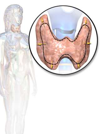
Thyroid disease is a medical condition that affects the function of the thyroid gland. The thyroid gland is located at the front of the neck and produces thyroid hormones that travel through the blood to help regulate many other organs, meaning that it is an endocrine organ. These hormones normally act in the body to regulate energy use, infant development, and childhood development.
Desiccated thyroid extract (DTE), is thyroid gland that has been dried and powdered for medical use. It is used to treat hypothyroidism., but less preferred than levothyroxine. It is taken by mouth. Maximal effects may take up to three weeks to occur.
The hypothalamic–pituitary–thyroid axis is part of the neuroendocrine system responsible for the regulation of metabolism and also responds to stress.
Reverse triiodothyronine, also known as rT3, is an isomer of triiodothyronine (T3).
Euthyroid sick syndrome (ESS) is a state of adaptation or dysregulation of thyrotropic feedback control wherein the levels of T3 and/or T4 are abnormal, but the thyroid gland does not appear to be dysfunctional. This condition may result from allostatic responses of hypothalamus-pituitary-thyroid feedback control, dyshomeostatic disorders, drug interferences, and impaired assay characteristics in critical illness.
Myxedema coma is an extreme or decompensated form of hypothyroidism and while uncommon, is potentially lethal. A person may have laboratory values identical to a "normal" hypothyroid state, but a stressful event precipitates the myxedema coma state, usually in the elderly. Primary symptoms of myxedema coma are altered mental status and low body temperature. Low blood sugar, low blood pressure, hyponatremia, hypercapnia, hypoxia, slowed heart rate, and hypoventilation may also occur. Myxedema, although included in the name, is not necessarily seen in myxedema coma. Coma is also not necessarily seen in myxedema coma, as patients may be obtunded without being comatose.
Thyroid hormones are any hormones produced and released by the thyroid gland, namely triiodothyronine (T3) and thyroxine (T4). They are tyrosine-based hormones that are primarily responsible for regulation of metabolism. T3 and T4 are partially composed of iodine, derived from food. A deficiency of iodine leads to decreased production of T3 and T4, enlarges the thyroid tissue and will cause the disease known as simple goitre.
Deiodinase (monodeiodinase) is a peroxidase enzyme that is involved in the activation or deactivation of thyroid hormones.
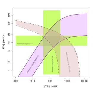
Thyroid's secretory capacity is the maximum stimulated amount of thyroxine that the thyroid can produce in a given time-unit.
The sum activity of peripheral deiodinases is the maximum amount of triiodothyronine produced per time-unit under conditions of substrate saturation. It is assumed to reflect the activity of deiodinases outside the central nervous system and other isolated compartments. GD is therefore expected to reflect predominantly the activity of type I deiodinase.
Jostel's TSH index, also referred to as Jostel's thyrotropin index or Thyroid Function index (TFI), is a method for estimating the thyrotropic function of the anterior pituitary lobe in a quantitative way. The equation has been derived from the logarithmic standard model of thyroid homeostasis. In a paper from 2014 further study was suggested to show if it is useful, but the 2018 guideline by the European Thyroid Association for the diagnosis of uncertain cases of central hypothyroidism regarded it as beneficial. It is also recommended for purposes of differential diagnosis in the sociomedical expert assessment.
SimThyr is a free continuous dynamic simulation program for the pituitary-thyroid feedback control system. The open-source program is based on a nonlinear model of thyroid homeostasis. In addition to simulations in the time domain the software supports various methods of sensitivity analysis. Its simulation engine is multi-threaded and supports multiple processor cores. SimThyr provides a GUI, which allows for visualising time series, modifying constant structure parameters of the feedback loop, storing parameter sets as XML files and exporting results of simulations in various formats that are suitable for statistical software. SimThyr is intended for both educational purposes and in-silico research.
The Thyrotroph Thyroid Hormone Sensitivity Index is a calculated structure parameter of thyroid homeostasis. It was originally developed to deliver a method for fast screening for resistance to thyroid hormone. Today it is also used to get an estimate for the set point of thyroid homeostasis, especially to assess dynamic thyrotropic adaptation of the anterior pituitary gland, including non-thyroidal illnesses.
The Centers for Disease Control and Prevention has published the following laboratory procedure manuals for measuring thyroid-stimulating hormone: