Luteinizing hormone

Last updated
Chorionic gonadotropin alpha
Protein CGA PDB 1dz7.png
Identifiers
Symbol CGA
Alt. symbolsHCG, GPHa, GPHA1
NCBI gene 1081
HGNC 1885
OMIM 118850
RefSeq NM_000735
UniProt P01215
Other data
Locus Chr. 6 q14-q21
Search for
Structures Swiss-model
Domains InterPro
Luteinizing hormone beta polypeptide
Identifiers
Symbol LHB
NCBI gene 3972
HGNC 6584
OMIM 152780
RefSeq NM_000894
UniProt P01229
Other data
Locus Chr. 19 q13.3
Search for
Structures Swiss-model
Domains InterPro

Luteinizing hormone (LH, also known as luteinising hormone, [1] lutropin and sometimes lutrophin [2] ) is a hormone produced by gonadotropic cells in the anterior pituitary gland. The production of LH is regulated by gonadotropin-releasing hormone (GnRH) from the hypothalamus. [3] In females, an acute rise of LH known as an LH surge, triggers ovulation [4] and development of the corpus luteum. In males, where LH had also been called interstitial cell–stimulating hormone (ICSH), [5] it stimulates Leydig cell production of testosterone. [4] It acts synergistically with follicle-stimulating hormone (FSH).

Contents

Etymology

The term luteinizing comes from the Latin "luteus," meaning "yellow." This is in reference to the corpus luteum, which is a mass of cells that forms in an ovary after an ovum (egg) has been discharged but remains unfertilized. The corpus luteum is so named because it often has a distinctive yellow color. The process of forming the corpus luteum is known as "luteinization," and thus the hormone that triggers this process is termed the "luteinizing" hormone.
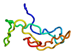
Structure

LH is a heterodimeric glycoprotein. Each monomeric unit is a glycoprotein molecule; one alpha and one beta subunit make the full, functional protein.

Its structure is similar to that of the other glycoprotein hormones, follicle-stimulating hormone (FSH), thyroid-stimulating hormone (TSH), and human chorionic gonadotropin (hCG). The protein dimer contains 2 glycopeptidic subunits (labeled alpha- and beta- subunits) that are non-covalently associated: [6]

The different composition of these oligosaccharides affects bioactivity and speed of degradation. The biologic half-life of LH is 20 minutes, shorter than that of FSH (3–4 hours) and hCG (24 hours).[ citation needed ] The biological half-life of LH is 23 hours subcutaneous [7] or terminal half life of 10-12 hours. [8]

Genes

The gene for the alpha subunit is located on chromosome 6q12.21.

The luteinizing hormone beta subunit gene is localized in the LHB/CGB gene cluster on chromosome 19q13.32. In contrast to the alpha gene activity, beta LH subunit gene activity is restricted to the pituitary gonadotropic cells. It is regulated by the gonadotropin-releasing hormone from the hypothalamus. Inhibin, activin, and sex hormones do not affect genetic activity for the beta subunit production of LH.

Function

Effects of LH on the body Figure 28 03 01.jpg
Effects of LH on the body

In both males and females, LH works upon endocrine cells in the gonads to produce androgens.

Effects in females

LH supports theca cells in the ovaries that provide androgens and hormonal precursors for estradiol production. At the time of menstruation, FSH initiates follicular growth, specifically affecting granulosa cells. [9] With the rise in estrogens, LH receptors are also expressed on the maturing follicle, which causes it to produce more estradiol. Eventually, when the follicle has fully matured, a spike in 17α-hydroxyprogesterone production by the follicle inhibits the production of estrogens. Previously, the preovulatory LH surge was attributed to a decrease in estrogen-mediated negative feedback of GnRH in the hypothalamus, subsequently stimulating the release of LH from the anterior pituitary. [10] More recent studies, however, attribute the LH surge to positive feedback from estradiol after production by the dominant follicle exceeds a certain threshold. Exceptionally high levels of estradiol induce hypothalamic production of progesterone, which stimulates elevated GnRH secretion, triggering a surge in LH. [11] The increase in LH production only lasts for 24 to 48 hours. This "LH surge" triggers ovulation, thereby not only releasing the egg from the follicle, but also initiating the conversion of the residual follicle into a corpus luteum that, in turn, produces progesterone to prepare the endometrium for a possible implantation. LH is necessary to maintain luteal function for the second two weeks of the menstrual cycle. If pregnancy occurs, LH levels will decrease, and luteal function will instead be maintained by the action of hCG (human chorionic gonadotropin), a hormone very similar to LH but secreted from the new placenta.

Gonadal steroids (estrogens and androgens) generally have negative feedback effects on GnRH-1 release at the level of the hypothalamus and at the gonadotropes, reducing their sensitivity to GnRH. Positive feedback by estrogens also occurs in the gonadal axis of female mammals and is responsible for the midcycle surge of LH that stimulates ovulation. Although estrogens inhibit kisspeptin (Kp) release from kiss1 neurons in the ARC, estrogens stimulate Kp release from the Kp neurons in the AVPV. As estrogens' levels gradually increase the positive effect predominates, leading to the LH surge. GABA-secreting neurons that innervate GnRH-1 neurons also can stimulate GnRH-1 release. These GABA neurons also possess ERs and may be responsible for the GnRH-1 surge. Part of the inhibitory action of endorphins on GnRH-1 release is through inhibition of these GABA neurons. Rupture of the ovarian follicle at ovulation causes a drastic reduction in estrogen synthesis and a marked increase in secretion of progesterone by the corpus luteum in the ovary, reinstating a predominantly negative feedback on hypothalamic secretion of GnRH-1. [12]

Effects in males

LH acts upon the Leydig cells of the testis and is regulated by gonadotropin-releasing hormone (GnRH). [13] The Leydig cells produce testosterone under the control of LH. LH binds to LH receptors on the membrane surface of Leydig cells. Binding to this receptor causes an increase in cyclic adenosine monophosphate (cAMP), a secondary messenger, which allows cholesterol to translocate into the mitochondria. Within the mitochondria, cholesterol is converted to pregnenolone by CYP11A1. [14] Pregnenolone is then converted to dehydroepiandrosterone (DHEA). [15] DHEA is then converted to androstenedione by 3β-hydroxysteroid dehydrogenase (3β-HSD) [16] and then finally converted to testosterone by 17β-hydroxysteroid dehydrogenase (HSD17B). The onset of puberty is controlled by two major hormones: FSH initiates spermatogenesis and LH signals the release of testosterone, [17] an androgen that exerts both endocrine activity and intratesticular activity on spermatogenesis.

LH is released from the pituitary gland, and is controlled by pulses of gonadotropin-releasing hormone. When bloodstream testosterone levels are low, the pituitary gland is stimulated to release LH. [13] As the levels of testosterone increase, it will act on the pituitary through a negative feedback loop and inhibit the release of GnRH and LH consequently. [18] Androgens (including testosterone and dihydrotestosterone) inhibit monoamine oxidase (MAO) in the pineal gland, leading to increased melatonin and reduced LH and FSH by melatonin-induced increase of Gonadotropin-Inhibitory Hormone (GnIH) [19] synthesis and secretion. Testosterone can also be aromatized into estradiol (E2) to inhibit LH. E2 decreases pulse amplitude and responsiveness to GnRH from the hypothalamus onto the pituitary. [20]

Changes in LH and testosterone blood levels and pulse secretions are induced by changes in sexual arousal in human males. [21]

Effects in the brain

Luteinizing hormone receptors are located in areas of the brain associated with cognitive function. [22] The role of LH role in the central nervous system (CNS) may be of relevance to understanding and treating post-menopausal cognitive decline. [23]

Recent research has observed an inverse relationship between circulating LH and CNS LH levels. [24] After ovariectomy (a procedure used to mimic menopause) in female mice, circulating LH levels surge while CNS levels of LH fall. [25] Treatments that lower circulating LH restore LH levels in the CNS. [25]

Normal levels

Reference ranges for the blood content of luteinizing hormone (LH) during the menstrual cycle.
The ranges denoted By biological stage may be used in closely monitored menstrual cycles in regard to other markers of its biological progression, with the time scale being compressed or stretched to how much faster or slower, respectively, the cycle progresses compared to an average cycle.
The ranges denoted Inter-cycle variability are more appropriate to use in non-monitored cycles with only the beginning of menstruation known, but where the woman accurately knows her average cycle lengths and time of ovulation, and that they are somewhat averagely regular, with the time scale being compressed or stretched to how much a woman's average cycle length is shorter or longer, respectively, than the average of the population.
The ranges denoted Inter-woman variability are more appropriate to use when the average cycle lengths and time of ovulation are unknown, but only the beginning of menstruation is given. Luteinizing hormone (LH) during menstrual cycle.png
Reference ranges for the blood content of luteinizing hormone (LH) during the menstrual cycle.
  • The ranges denoted By biological stage may be used in closely monitored menstrual cycles in regard to other markers of its biological progression, with the time scale being compressed or stretched to how much faster or slower, respectively, the cycle progresses compared to an average cycle.
  • The ranges denoted Inter-cycle variability are more appropriate to use in non-monitored cycles with only the beginning of menstruation known, but where the woman accurately knows her average cycle lengths and time of ovulation, and that they are somewhat averagely regular, with the time scale being compressed or stretched to how much a woman's average cycle length is shorter or longer, respectively, than the average of the population.
  • The ranges denoted Inter-woman variability are more appropriate to use when the average cycle lengths and time of ovulation are unknown, but only the beginning of menstruation is given.

LH levels are normally low during childhood and in women, high after menopause. Since LH is secreted as pulses, it is necessary to follow its concentration over a sufficient period of time to get proper information about its blood level.

During reproductive years, typical levels are between 1 and 20 IU/L. Physiologic high LH levels are seen during the LH surge (v.s.) and typically last 48 hours.

In males over 18 years of age, reference ranges have been estimated to be 1.8–8.6 IU/L. [27]

LH is measured in international units (IU). When quantifying the amount of LH in a sample in IUs, it is important to know which international standard your lot of LH was calibrated against since they can vary broadly from year to year. For human urinary LH, one IU is most recently defined as 1/189th of an ampule denoted 96/602 and distributed by the NIBSC, corresponding to approximately 0.04656 µg of LH protein for a single IU, but older standard versions are still widely in use. [28] [29]

Lateral flow test strip for urine LH used to predict ovulation. Ovulatietest.jpg
Lateral flow test strip for urine LH used to predict ovulation.

Predicting ovulation

Chance of fertilization by menstrual cycle day relative to ovulation Pregnancy chance by day near ovulation.jpg
Chance of fertilization by menstrual cycle day relative to ovulation

The detection of a surge in release of luteinizing hormone indicates impending ovulation. LH can be detected by urinary ovulation predictor kits (OPK, also LH-kit) that are performed, typically daily, around the time ovulation may be expected. [31] A conversion from a negative to a positive reading would suggest that ovulation is about to occur within 24–48 hours, giving women two days to engage in sexual intercourse or artificial insemination with the intention of conceiving. [32]

The recommended testing frequency differs between manufacturers. For example, the Clearblue test is taken daily, and an increased frequency does not decrease the risk of missing an LH surge. [33] On the other hand, the Chinese company Nantong Egens Biotechnology recommends using their test twice per day. [34] If testing once per day, no significant difference has been found between testing LH in the morning versus in the evening, in relation to conception rates, [35] and recommendations of what time in the day to take the test varies between manufacturers and healthcare workers. [36] Tests may be read manually using a color-change paper strip, or digitally with the assistance of reading electronics.

Tests for luteinizing hormone may be combined with testing for estradiol in tests such as the Clearblue fertility monitor.[ medical citation needed ]

The sensitivity of LH tests are measured in milli international unit, with tests commonly available in the range 10–40 m.i.u. (the lower the number, the higher the sensitivity).[ citation needed ]

As sperm can stay viable in the woman for several days, LH tests are not recommended for contraceptive practices, as the LH surge typically occurs after the beginning of the fertile window.[ citation needed ]

Disease states

Excess

In children with precocious puberty of pituitary or central origin, LH and FSH levels may be in the reproductive range instead of the low levels typical for their age.

During the reproductive years, relatively elevated LH is frequently seen in patients with polycystic ovary syndrome; however, it would be unusual for them to have LH levels outside of the normal reproductive range.

Persistently high LH levels are indicative of situations where the normal restricting feedback from the gonad is absent, leading to a pituitary production of both LH and FSH. While this is typical in menopause, it is abnormal in the reproductive years. There it may be a sign of:

  1. Premature menopause
  2. Gonadal dysgenesis, Turner syndrome, Klinefelter syndrome
  3. Castration
  4. Swyer syndrome
  5. Polycystic ovary syndrome
  6. Certain forms of congenital adrenal hyperplasia
  7. Testicular failure
  8. Pregnancy – BetaHCG can mimic LH so tests may show elevated LH

Note: A medical drug for inhibiting luteinizing hormone secretion is butinazocine. [37]

Deficiency

Diminished secretion of LH can result in failure of gonadal function (hypogonadism). This condition is typically manifest in males as failure in production of normal numbers of sperm. In females, amenorrhea is commonly observed. Conditions with very low LH secretions include:

  1. Pasqualini syndrome [38] [39]
  2. Kallmann syndrome
  3. Hypothalamic suppression
  4. Hypopituitarism
  5. Eating disorder
  6. Female athlete triad
  7. Hyperprolactinemia
  8. Hypogonadism
  9. Gonadal suppression therapy
    1. GnRH antagonist
    2. GnRH agonist (inducing an initial stimulation (flare up) followed by permanent blockage of the GnRH pituitary receptor)

As a medication

LH is available mixed with FSH in the form of menotropin, and other forms of urinary gonadotropins. More purified forms of urinary gonadotropins may reduce the LH portion in relation to FSH. Recombinant LH is available as lutropin alfa (Luveris). [40] All these medications have to be given parenterally. They are commonly used in infertility therapy to stimulate follicular development, the notable one being in IVF therapy.

Often, HCG medication is used as an LH substitute because it activates the same receptor. Medically used hCG is derived from urine of pregnant women, is less costly, and has a longer half-life than LH.

Role in phosphorylation

Phosphorylation is a biochemical process that involves the addition of phosphate to an organic compound. Steroidogenesis entails processes by which cholesterol is converted to biologically active steroid hormones. Recent study shows that LH via a PKA signaling pathway regulates the phosphorylation and localization of DRP1 within mitochondria of the steroidogenic cells of the ovary. [41]

Related Research Articles

<span class="mw-page-title-main">Menstrual cycle</span> Natural changes in the human female reproductive system

The menstrual cycle is a series of natural changes in hormone production and the structures of the uterus and ovaries of the female reproductive system that makes pregnancy possible. The ovarian cycle controls the production and release of eggs and the cyclic release of estrogen and progesterone. The uterine cycle governs the preparation and maintenance of the lining of the uterus (womb) to receive an embryo. These cycles are concurrent and coordinated, normally last between 21 and 35 days, with a median length of 28 days, and continue for about 30–45 years.

<span class="mw-page-title-main">Follicle-stimulating hormone</span> Gonadotropin that regulates the development of reproductive processes

Follicle-stimulating hormone (FSH) is a gonadotropin, a glycoprotein polypeptide hormone. FSH is synthesized and secreted by the gonadotropic cells of the anterior pituitary gland and regulates the development, growth, pubertal maturation, and reproductive processes of the body. FSH and luteinizing hormone (LH) work together in the reproductive system.

<span class="mw-page-title-main">Anterior pituitary</span> Anterior lobe of the pituitary gland

A major organ of the endocrine system, the anterior pituitary is the glandular, anterior lobe that together with the posterior lobe makes up the pituitary gland (hypophysis) which, in humans, is located at the base of the brain, protruding off the bottom of the hypothalamus.

<span class="mw-page-title-main">Gonadotropin-releasing hormone</span> Mammalian protein found in Homo sapiens

Gonadotropin-releasing hormone (GnRH) is a releasing hormone responsible for the release of follicle-stimulating hormone (FSH) and luteinizing hormone (LH) from the anterior pituitary. GnRH is a tropic peptide hormone synthesized and released from GnRH neurons within the hypothalamus. The peptide belongs to gonadotropin-releasing hormone family. It constitutes the initial step in the hypothalamic–pituitary–gonadal axis.

Anovulation is when the ovaries do not release an oocyte during a menstrual cycle. Therefore, ovulation does not take place. However, a woman who does not ovulate at each menstrual cycle is not necessarily going through menopause. Chronic anovulation is a common cause of infertility.

Gonadotropins are glycoprotein hormones secreted by gonadotropic cells of the anterior pituitary of vertebrates. This family includes the mammalian hormones follicle-stimulating hormone (FSH) and luteinizing hormone (LH), the placental/chorionic gonadotropins, human chorionic gonadotropin (hCG) and equine chorionic gonadotropin (eCG), as well as at least two forms of fish gonadotropins. These hormones are central to the complex endocrine system that regulates normal growth, sexual development, and reproductive function. LH and FSH are secreted by the anterior pituitary gland, while hCG and eCG are secreted by the placenta in pregnant humans and mares, respectively. The gonadotropins act on the gonads, controlling gamete and sex hormone production.

Gonadarche refers to the earliest gonadal changes of puberty. In response to pituitary gonadotropins, the ovaries in females and the testes in males begin to grow and increase the production of the sex steroids, especially estradiol and testosterone. The ovary and testis have receptors, follicle cells and leydig cells, respectively, where gonadotropins bind to stimulate the maturation of the gonads and secretion of estrogen and testosterone. Certain disorders can result in changes to timing or nature of these processes.

<span class="mw-page-title-main">Folliculogenesis</span> Process of maturation of primordial follicles

In biology, folliculogenesis is the maturation of the ovarian follicle, a densely packed shell of somatic cells that contains an immature oocyte. Folliculogenesis describes the progression of a number of small primordial follicles into large preovulatory follicles that occurs in part during the menstrual cycle.

<span class="mw-page-title-main">Hypothalamic–pituitary–gonadal axis</span> Concept of regarding the hypothalamus, pituitary gland and gonadal glands as a single entity

The hypothalamic–pituitary–gonadal axis refers to the hypothalamus, pituitary gland, and gonadal glands as if these individual endocrine glands were a single entity. Because these glands often act in concert, physiologists and endocrinologists find it convenient and descriptive to speak of them as a single system.

<span class="mw-page-title-main">Luteal phase</span> The latter part of the menstrual cycle associated with ovulation and an increase in progesterone

The menstrual cycle is on average 28 days in length. It begins with menses during the follicular phase, followed by ovulation and ending with the luteal phase. Unlike the follicular phase which can vary in length among individuals, the luteal phase is typically fixed at approximately 14 days and is characterized by changes to hormone levels, such as an increase in progesterone and estrogen levels, decrease in gonadotropins such as follicle-stimulating hormone (FSH) and luteinizing hormone (LH), changes to the endometrial lining to promote implantation of the fertilized egg, and development of the corpus luteum. In the absence of fertilization by sperm, the corpus luteum atrophies leading to a decrease in progesterone and estrogen, an increase in FSH and LH, and shedding of the endometrial lining (menses) to begin the menstrual cycle again.

<span class="mw-page-title-main">Follicular phase</span> Phase of the estrous or menstrual cycle

The follicular phase, also known as the preovulatory phase or proliferative phase, is the phase of the estrous cycle during which follicles in the ovary mature from primary follicle to a fully mature graafian follicle. It ends with ovulation. The main hormones controlling this stage are secretion of gonadotropin-releasing hormones, which are follicle-stimulating hormones and luteinising hormones. They are released by pulsatile secretion. The duration of the follicular phase can differ depending on the length of the menstrual cycle, while the luteal phase is usually stable, does not really change and lasts 14 days.

<span class="mw-page-title-main">Gonadotropin-releasing hormone agonist</span> Drug class affecting sex hormones

A gonadotropin-releasing hormone agonist is a type of medication which affects gonadotropins and sex hormones. They are used for a variety of indications including in fertility medicine and to lower sex hormone levels in the treatment of hormone-sensitive cancers such as prostate cancer and breast cancer, certain gynecological disorders like heavy periods and endometriosis, high testosterone levels in women, early puberty in children, as a part of transgender hormone therapy, and to delay puberty in transgender youth among other uses. It is also used in the suppression of spontaneous ovulation as part of controlled ovarian hyperstimulation, an essential component in IVF. GnRH agonists are given by injections into fat, as implants placed into fat, and as nasal sprays.

<span class="mw-page-title-main">Gonadotropin-releasing hormone antagonist</span> Class of medications

Gonadotropin-releasing hormone antagonists are a class of medications that antagonize the gonadotropin-releasing hormone receptor and thus the action of gonadotropin-releasing hormone (GnRH). They are used in the treatment of prostate cancer, endometriosis, uterine fibroids, female infertility in assisted reproduction, and for other indications.

The theca folliculi comprise a layer of the ovarian follicles. They appear as the follicles become secondary follicles.

Gonadotropin-releasing hormone (GnRH) insensitivity also known as Isolated gonadotropin-releasing hormone (GnRH)deficiency (IGD) is a rare autosomal recessive genetic and endocrine syndrome which is characterized by inactivating mutations of the gonadotropin-releasing hormone receptor (GnRHR) and thus an insensitivity of the receptor to gonadotropin-releasing hormone (GnRH), resulting in a partial or complete loss of the ability of the gonads to synthesize the sex hormones. The condition manifests itself as isolated hypogonadotropic hypogonadism (IHH), presenting with symptoms such as delayed, reduced, or absent puberty, low or complete lack of libido, and infertility, and is the predominant cause of IHH when it does not present alongside anosmia.

Hypogonadotropic hypogonadism (HH), is due to problems with either the hypothalamus or pituitary gland affecting the hypothalamic-pituitary-gonadal axis. Hypothalamic disorders result from a deficiency in the release of gonadotropic releasing hormone (GnRH), while pituitary gland disorders are due to a deficiency in the release of gonadotropins from the anterior pituitary. GnRH is the central regulator in reproductive function and sexual development via the HPG axis. GnRH is released by GnRH neurons, which are hypothalamic neuroendocrine cells, into the hypophyseal portal system acting on gonadotrophs in the anterior pituitary. The release of gonadotropins, LH and FSH, act on the gonads for the development and maintenance of proper adult reproductive physiology. LH acts on Leydig cells in the male testes and theca cells in the female. FSH acts on Sertoli cells in the male and follicular cells in the female. Combined this causes the secretion of gonadal sex steroids and the initiation of folliculogenesis and spermatogenesis. The production of sex steroids forms a negative feedback loop acting on both the anterior pituitary and hypothalamus causing a pulsatile secretion of GnRH. GnRH neurons lack sex steroid receptors and mediators such as kisspeptin stimulate GnRH neurons for pulsatile secretion of GnRH.

Pulsatile secretion is a biochemical phenomenon observed in a wide variety of cell and tissue types, in which chemical products are secreted in a regular temporal pattern. The most common cellular products observed to be released in this manner are intercellular signaling molecules such as hormones or neurotransmitters. Examples of hormones that are secreted pulsatilely include insulin, thyrotropin, TRH, gonadotropin-releasing hormone (GnRH) and growth hormone (GH). In the nervous system, pulsatility is observed in oscillatory activity from central pattern generators. In the heart, pacemakers are able to work and secrete in a pulsatile manner. A pulsatile secretion pattern is critical to the function of many hormones in order to maintain the delicate homeostatic balance necessary for essential life processes, such as development and reproduction. Variations of the concentration in a certain frequency can be critical to hormone function, as evidenced by the case of GnRH agonists, which cause functional inhibition of the receptor for GnRH due to profound downregulation in response to constant (tonic) stimulation. Pulsatility may function to sensitize target tissues to the hormone of interest and upregulate receptors, leading to improved responses. This heightened response may have served to improve the animal's fitness in its environment and promote its evolutionary retention.

<span class="mw-page-title-main">Premature thelarche</span> Medical condition

Premature thelarche (PT) is a medical condition, characterised by isolated breast development in female infants. It occurs in females younger than 8 years, with the highest occurrence before the age of 2. PT is rare, occurring in 2.2-4.7% of females aged 0 to 2 years old. The exact cause of the condition is still unknown, but it has been linked to a variety of genetic, dietary and physiological factors.

<span class="mw-page-title-main">Gonadotropin-releasing hormone modulator</span> Type of medication which modulates the GnRH receptor

A GnRH modulator, or GnRH receptor modulator, also known as an LHRH modulator or LHRH receptor modulator, is a type of medication which modulates the GnRH receptor, the biological target of the hypothalamic hormone gonadotropin-releasing hormone. They include GnRH agonists and GnRH antagonists. These medications may be GnRH analogues like leuprorelin and cetrorelix – peptides that are structurally related to GnRH – or small-molecules like elagolix and relugolix, which are structurally distinct from and unrelated to GnRH analogues.

Gonadotropin surge-attenuating factor (GnSAF) is a nonsteroidal ovarian hormone produced by the granulosa cells of small antral ovarian follicles in females. GnSAF is involved in regulating the secretion of luteinizing hormone (LH) from the anterior pituitary and the ovarian cycle. During the early to mid-follicular phase of the ovarian cycle, GnSAF acts on the anterior pituitary to attenuate LH release, limiting the secretion of LH to only basal levels. At the transition between follicular and luteal phase, GnSAF bioactivity declines sufficiently to permit LH secretion above basal levels, resulting in the mid-cycle LH surge that initiates ovulation. In normally ovulating women, the LH surge only occurs when the oocyte is mature and ready for extrusion. GnSAF bioactivity is responsible for the synchronised, biphasic nature of LH secretion.

References

  1. GCSE Science Revision Biology "The Menstrual Cycle" , retrieved 2022-03-23
  2. Ujihara M, Yamamoto K, Nomura K, Toyoshima S, Demura H, Nakamura Y, et al. (June 1992). "Subunit-specific sulphation of oligosaccharides relating to charge-heterogeneity in porcine lutrophin isoforms". Glycobiology. 2 (3): 225–231. doi:10.1093/glycob/2.3.225. PMID   1498420.
  3. Stamatiades GA, Kaiser UB (March 2018). "Gonadotropin regulation by pulsatile GnRH: Signaling and gene expression". Molecular and Cellular Endocrinology. Signaling Pathways Regulating Pituitary Functions. 463: 131–141. doi:10.1016/j.mce.2017.10.015. PMC   5812824 . PMID   29102564.
  4. 1 2 Nosek TM. "Section 5/5ch9/s5ch9_5". Essentials of Human Physiology. Archived from the original on 2016-03-24.
  5. Louvet JP, Harman SM, Ross GT (May 1975). "Effects of human chorionic gonadotropin, human interstitial cell stimulating hormone and human follicle-stimulating hormone on ovarian weights in estrogen-primed hypophysectomized immature female rats". Endocrinology. 96 (5): 1179–1186. doi:10.1210/endo-96-5-1179. PMID   1122882.
  6. Jiang X, Dias JA, He X (January 2014). "Structural biology of glycoprotein hormones and their receptors: insights to signaling". Molecular and Cellular Endocrinology. 382 (1): 424–451. doi: 10.1016/j.mce.2013.08.021 . PMID   24001578.
  7. Ezcurra D, Humaidan P (October 2014). "A review of luteinising hormone and human chorionic gonadotropin when used in assisted reproductive technology". Reproductive Biology and Endocrinology. 12 (1): 95. doi: 10.1186/1477-7827-12-95 . PMC   4287577 . PMID   25280580.
  8. le Cotonnec JY, Porchet HC, Beltrami V, Munafo A (February 1998). "Clinical pharmacology of recombinant human luteinizing hormone: Part I. Pharmacokinetics after intravenous administration to healthy female volunteers and comparison with urinary human luteinizing hormone". Fertility and Sterility. 69 (2): 189–194. doi:10.1016/S0015-0282(97)00501-3. PMID   9496327.
  9. Bowen R (13 May 2004). "Gonadotropins: Luteinizing and Follicle Stimulating Hormones". Colorado State University. Retrieved 12 March 2012.
  10. Mahesh VB (January 2012). "Hirsutism, virilism, polycystic ovarian disease, and the steroid-gonadotropin-feedback system: a career retrospective". American Journal of Physiology. Endocrinology and Metabolism. 302 (1): E4–E18. doi:10.1152/ajpendo.00488.2011. PMC   3328092 . PMID   22028409.
  11. Micevych P, Sinchak K (2 December 2011). "The Neurosteroid Progesterone Underlies Estrogen Positive Feedback of the LH Surge". Frontiers in Endocrinology. 2: 90. doi: 10.3389/fendo.2011.00090 . PMC   3356049 . PMID   22654832.
  12. Norris DO, Carr JA (2013). Vertebrate Endocrinology. Academic Press. p. 126. ISBN   978-0-12-396465-6.
  13. 1 2 Nedresky D, Singh G (September 2022). "Physiology, Luteinizing Hormone". StatPearls [Internet]. Treasure Island (FL): StatPearls Publishing. PMID   30969514.
  14. Zirkin BR, Papadopoulos V (July 2018). "Leydig cells: formation, function, and regulation". Biology of Reproduction. 99 (1): 101–111. doi:10.1093/biolre/ioy059. PMC   6044347 . PMID   29566165.
  15. Akhtar MK, Kelly SL, Kaderbhai MA (November 2005). "Cytochrome b(5) modulation of 17{alpha} hydroxylase and 17-20 lyase (CYP17) activities in steroidogenesis". The Journal of Endocrinology. 187 (2): 267–274. doi: 10.1677/joe.1.06375 . PMID   16293774.
  16. Liu L, Kang J, Ding X, Chen D, Zhou Y, Ma H (2015). "Dehydroepiandrosterone-Regulated Testosterone Biosynthesis via Activation of the ERK1/2 Signaling Pathway in Primary Rat Leydig Cells". Cellular Physiology and Biochemistry. 36 (5): 1778–1792. doi: 10.1159/000430150 . PMID   26184424. S2CID   13816368.
  17. Oduwole OO, Peltoketo H, Huhtaniemi IT (2018). "Role of Follicle-Stimulating Hormone in Spermatogenesis". Frontiers in Endocrinology. 9: 763. doi: 10.3389/fendo.2018.00763 . PMC   6302021 . PMID   30619093.
  18. Tilbrook, A.J.; Clarke, I.J. (2001-03-01). "Negative Feedback Regulation of the Secretion and Actions of Gonadotropin-Releasing Hormone in Males". Biology of Reproduction. 64 (3): 735–742. doi:10.1095/biolreprod64.3.735. ISSN   0006-3363.
  19. Ubuka T, Son YL, Tobari Y, Narihiro M, Bentley GE, Kriegsfeld LJ, Tsutsui K (2014). "Central and direct regulation of testicular activity by gonadotropin-inhibitory hormone and its receptor". Frontiers in Endocrinology. 5: 8. doi: 10.3389/fendo.2014.00008 . PMC   3902780 . PMID   24478760.
  20. Pitteloud N, Dwyer AA, DeCruz S, Lee H, Boepple PA, Crowley WF, Hayes FJ (March 2008). "Inhibition of luteinizing hormone secretion by testosterone in men requires aromatization for its pituitary but not its hypothalamic effects: evidence from the tandem study of normal and gonadotropin-releasing hormone-deficient men". The Journal of Clinical Endocrinology and Metabolism. 93 (3): 784–791. doi:10.1210/jc.2007-2156. PMC   2266963 . PMID   18073301.
  21. Stoléru SG, Ennaji A, Cournot A, Spira A (1993). "LH pulsatile secretion and testosterone blood levels are influenced by sexual arousal in human males". Psychoneuroendocrinology. 18 (3): 205–218. doi:10.1016/0306-4530(93)90005-6. PMID   8516424. S2CID   23595343.
  22. Blair JA, Bhatta S, McGee H, Casadesus G (November 2015). "Luteinizing hormone: Evidence for direct action in the CNS". Hormones and Behavior. 76: 57–62. doi:10.1016/j.yhbeh.2015.06.020. PMC   4741372 . PMID   26172857.
  23. Than S, Moran C, Beare R, Vincent A, Lane E, Collyer TA, et al. (2023). "Cognitive trajectories during the menopausal transition". Frontiers in Dementia. 2. doi: 10.3389/frdem.2023.1098693 . ISSN   2813-3919.
  24. Bhatta S, Blair JA, Casadesus G (2018-09-24). "Luteinizing Hormone Involvement in Aging Female Cognition: Not All Is Estrogen Loss". Frontiers in Endocrinology. 9: 544. doi: 10.3389/fendo.2018.00544 . PMC   6165885 . PMID   30319538.
  25. 1 2 Blair JA, Bhatta S, McGee H, Casadesus G (November 2015). "Luteinizing hormone: Evidence for direct action in the CNS". Hormones and Behavior. 76: 57–62. doi:10.1016/j.yhbeh.2015.06.020. PMC   4741372 . PMID   26172857.
  26. Häggström M (2014). "Reference ranges for estradiol, progesterone, luteinizing hormone and follicle-stimulating hormone during the menstrual cycle". WikiJournal of Medicine. 1 (1). doi: 10.15347/wjm/2014.001 . ISSN   2002-4436.
  27. "Test ID: LH, Luteinizing Hormone (LH), Serum". Mayo Medical Laboratories. Archived from the original on 2016-09-25. Retrieved 1 December 2012.
  28. WHO Expert Committee on Biological Standardization (2003). "Proposed International Standard for Luteinizing Hormone" (PDF). Geneva: World Health Organization.
  29. "WHO International Standard, Luteinizing Hormone, Human, Recombinant" (PDF). National Institute for Biological Standards and Control.
  30. Dunson DB, Baird DD, Wilcox AJ, Weinberg CR (July 1999). "Day-specific probabilities of clinical pregnancy based on two studies with imperfect measures of ovulation". Human Reproduction. 14 (7): 1835–1839. doi: 10.1093/humrep/14.7.1835 . PMID   10402400.
  31. Nielsen MS, Barton SD, Hatasaka HH, Stanford JB (August 2001). "Comparison of several one-step home urinary luteinizing hormone detection test kits to OvuQuick". Fertility and Sterility. 76 (2): 384–387. doi: 10.1016/S0015-0282(01)01881-7 . PMID   11476792.
  32. "Ovulation Predictor Kit Frequently Asked Questions". Fertility Plus. Archived from the original on March 12, 2012. Retrieved 12 March 2012.[ unreliable medical source? ]
  33. "Clear Blue Ovulation Test Instructions". Ovulation Guide. Retrieved 2018-01-19.
  34. "Advanced Ovulation Test" (PDF). Homehealth-UK. Retrieved 2018-01-19. Version 1.1 02/11/15
  35. Martinez AR, Bernardus RE, Vermeiden JP, Schoemaker J (March 1994). "Time schedules of intrauterine insemination after urinary luteinizing hormone surge detection and pregnancy results". Gynecological Endocrinology. 8 (1): 1–5. doi:10.3109/09513599409028450. PMID   8059611.
  36. Meniru GI (2001). Cambridge Guide to Infertility Management and Assisted Reproduction. Cambridge University Press. p.  67.
  37. US 4406904,Welle HB, Marko M,"Method of inhibiting luteinizing hormone secretion with 6,7-benzomorphan derivatives",issued 27 September 1983, assigned to ACF Chemiefarma NV.
  38. Weiss J, Axelrod L, Whitcomb RW, Harris PE, Crowley WF, Jameson JL (January 1992). "Hypogonadism caused by a single amino acid substitution in the beta subunit of luteinizing hormone". The New England Journal of Medicine. 326 (3): 179–183. doi: 10.1056/NEJM199201163260306 . PMID   1727547.
  39. Valdes-Socin H, Salvi R, Daly AF, Gaillard RC, Quatresooz P, Tebeu PM, et al. (December 2004). "Hypogonadism in a patient with a mutation in the luteinizing hormone beta-subunit gene". The New England Journal of Medicine. 351 (25): 2619–2625. doi:10.1056/NEJMoa040326. hdl: 2268/23485 . PMID   15602022.
  40. "Luveris information". Archived from the original on June 18, 2006.[ unreliable medical source? ]
  41. Plewes MR, Hou X, Talbott HA, Zhang P, Wood JR, Cupp AS, Davis JS (April 2020). "Luteinizing hormone regulates the phosphorylation and localization of the mitochondrial effector dynamin-related protein-1 (DRP1) and steroidogenesis in the bovine corpus luteum". FASEB Journal. Federation of American Societies for Experimental Biology. 34 (4): 5299–5316. doi: 10.1096/fj.201902958R . PMC   7136153 . PMID   32077149.