Granulosa cell

Last updated
Pig oocyte surrounded by granulosa cells. Fluorescence microscopy, colored with DAPI. Pig oocyte dapi 4.jpg
Pig oocyte surrounded by granulosa cells. Fluorescence microscopy, colored with DAPI.

A granulosa cell or follicular cell is a somatic cell of the sex cord that is closely associated with the developing female gamete (called an oocyte or egg) in the ovary of mammals.

Contents

Structure and function

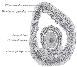
In the primordial ovarian follicle, and later in follicle development (folliculogenesis), granulosa cells advance to form a multilayered cumulus oophorus surrounding the oocyte in the preovulatory or antral (or Graafian) follicle.

The major functions of granulosa cells include the production of sex steroids, as well as myriad growth factors thought to interact with the oocyte during its development. The sex steroid production begins with follicle-stimulating hormone (FSH) from the anterior pituitary, stimulating granulosa cells to convert androgens (coming from the thecal cells) to estradiol by aromatase during the follicular phase of the menstrual cycle. [1] However, after ovulation the granulosa cells turn into granulosa lutein cells that produce progesterone. The progesterone may maintain a potential pregnancy and causes production of a thick cervical mucus that inhibits sperm entry into the uterus.

Embryology of ovarian granulosa cells

In the development of the urinary and reproductive organs, the oogonia become invaginated in the gonadal ridge.

The embryological origin of granulosa cells remains controversial. In the 1970s, evidence emerged that the first cells to make contact with the oogonia were of mesonephric origin. It was suggested that mesonephric cells already closely associated with the oogonia proliferated throughout development to form the granulosa cell layer. [2] [3] [4] Recently, this hypothesis has been challenged with some thorough histology. Sawyer et al. hypothesized that in sheep most of the granulosa cells develop from cells of the mesothelium (i.e., epithelial cells from the presumptive surface epithelium of the ovary). [5] In 2013, it was proposed that both granulosa cells and the ovarian surface epithelial cells are instead derived from a precursor cell called gonadal-ridge epithelial-like cell. [6]

Granulosa cell types

Cumulus cells (CC) vs mural granulosa cells (MGC)

Cumulus cells surround the oocyte. They provide nutrients to the oocyte and influence the development of the oocyte in a paracrine fashion. Mural granulosa cells line the follicular wall and surround the fluid-filled antrum. The oocyte secretes factors that determine the functional differences between CCs and MGGs. CCs primarily support growth and development of the oocyte whereas MGCs primarily serve an endocrine function and support the growth of the follicle. Cumulus cells aid in oocyte development and show higher expression of SLC38A3, a transporter for amino acids, and Aldoa, Eno1, Ldh1, Pfkp, Pkm2, and Tpi1, enzymes responsible for glycolysis. [7] MGCs are more steroidogenically active and have higher levels of mRNA expression of steroidogenic enzymes such as cytochrome P450. [8] MGCs produce an increasing amount of estrogen which leads to the LH surge. [9] Following the LH surge, cumulus cells undergo cumulus expansion, in which they proliferate at a ten-fold higher rate than MGCs in response to FSH. [10] During expansion CCs also produce a mucified matrix required for ovulation. [11]

Cell culture

Cell culture of granulosa cells can be performed in vitro . Plating density (number of cells per volume of culture medium) plays a critical role for the differentiation. A lower plating density makes granulosa cells exhibit estrogen production, while a higher plating density makes them appear as progesterone producing theca lutein cells. [12]

Ovarian aging

In the female rhesus monkey, DNA double-strand breaks increase in granulosa cells with age, and the ability to repair such DNA breaks declines with age. [13] These changes at the DNA level in granulosa cells may contribute to ovarian aging. [13]

Additional images

Section of vesicular ovarian follicle of a cat. X 50. Membrana granulosa labeled at upper left. Gray1164.png
Section of vesicular ovarian follicle of a cat. X 50. Membrana granulosa labeled at upper left.

See also

Related Research Articles

<span class="mw-page-title-main">Ovary</span> Female reproductive organ that produces egg cells

The ovary is an organ in the female reproductive system that produces an ovum. When released, this travels down the fallopian tube into the uterus. There is an ovary found on the left and the right side of the body. The ovaries also secrete hormones that play a role in the menstrual cycle and fertility. The ovary progresses through many stages beginning in the prenatal period through menopause. It is also an endocrine gland because of the various hormones that it secretes.

<span class="mw-page-title-main">Menstrual cycle</span> Natural changes in the human female reproductive system

The menstrual cycle is a series of natural changes in hormone production and the structures of the uterus and ovaries of the female reproductive system that makes pregnancy possible. The ovarian cycle controls the production and release of eggs and the cyclic release of estrogen and progesterone. The uterine cycle governs the preparation and maintenance of the lining of the uterus (womb) to receive an embryo. These cycles are concurrent and coordinated, normally last between 21 and 35 days, with a median length of 28 days, and continue for about 30–45 years.

<span class="mw-page-title-main">Ovulation</span> Release of egg cells from the ovaries

Ovulation is the release of eggs from the ovaries. In women, this event occurs when the ovarian follicles rupture and release the secondary oocyte ovarian cells. After ovulation, during the luteal phase, the egg will be available to be fertilized by sperm. In addition, the uterine lining (endometrium) is thickened to be able to receive a fertilized egg. If no conception occurs, the uterine lining as well as the egg will be shed during menstruation.

<span class="mw-page-title-main">Germ cell</span> Gamete-producing cell

A germ cell is any cell that gives rise to the gametes of an organism that reproduces sexually. In many animals, the germ cells originate in the primitive streak and migrate via the gut of an embryo to the developing gonads. There, they undergo meiosis, followed by cellular differentiation into mature gametes, either eggs or sperm. Unlike animals, plants do not have germ cells designated in early development. Instead, germ cells can arise from somatic cells in the adult, such as the floral meristem of flowering plants.

<span class="mw-page-title-main">Corpus luteum</span> Temporary endocrine structure in ovaries

The corpus luteum is a temporary endocrine structure in female ovaries involved in the production of relatively high levels of progesterone, and moderate levels of estradiol, and inhibin A. It is the remains of the ovarian follicle that has released a mature ovum during a previous ovulation.

<span class="mw-page-title-main">Oogenesis</span> Egg cell production process

Oogenesis, ovogenesis, or oögenesis is the differentiation of the ovum into a cell competent to further develop when fertilized. It is developed from the primary oocyte by maturation. Oogenesis is initiated in the embryonic stage.

<span class="mw-page-title-main">Ovarian follicle</span> Structure containing a single egg cell

An ovarian follicle is a roughly spheroid cellular aggregation set found in the ovaries. It secretes hormones that influence stages of the menstrual cycle. At the time of puberty, women have approximately 200,000 to 300,000 follicles, each with the potential to release an egg cell (ovum) at ovulation for fertilization. These eggs are developed once every menstrual cycle with around 450–500 being ovulated during a woman's reproductive lifetime.

<span class="mw-page-title-main">Stigma (anatomy)</span>

A stigma, also called macula pellucida, in mammalian reproductive anatomy, refers to the area of the ovarian surface where the Graafian follicle will burst through during ovulation and release the ovum. As the follicle matures, the area between the follicle and the ovarian surface begins to thin and weaken under the influence of the luteinizing hormone and local cytokines. At ovulation the stigma ruptures and the secondary oocyte is released along with surrounding granulosa cells, from the region of the cumulus oophorus, and follicular fluid. The secondary oocyte needs to be captured by the fallopian tube where it could be fertilized by a sperm cell. The stigma will heal and the residual follicle is transformed into the corpus luteum.

<span class="mw-page-title-main">Folliculogenesis</span> Process of maturation of primordial follicles

In biology, folliculogenesis is the maturation of the ovarian follicle, a densely packed shell of somatic cells that contains an immature oocyte. Folliculogenesis describes the progression of a number of small primordial follicles into large preovulatory follicles that occurs in part during the menstrual cycle.

<span class="mw-page-title-main">Growth differentiation factor-9</span> Protein-coding gene in the species Homo sapiens

Growth/differentiation factor 9 is a protein that in humans is encoded by the GDF9 gene.

<span class="mw-page-title-main">Luteal phase</span> The latter part of the menstrual cycle associated with ovulation and an increase in progesterone

The menstrual cycle is on average 28 days in length. It begins with menses during the follicular phase, followed by ovulation and ending with the luteal phase. Unlike the follicular phase which can vary in length among individuals, the luteal phase is typically fixed at approximately 14 days and is characterized by changes to hormone levels, such as an increase in progesterone and estrogen levels, decrease in gonadotropins such as follicle-stimulating hormone (FSH) and luteinizing hormone (LH), changes to the endometrial lining to promote implantation of the fertilized egg, and development of the corpus luteum. In the absence of fertilization by sperm, the corpus luteum atrophies leading to a decrease in progesterone and estrogen, an increase in FSH and LH, and shedding of the endometrial lining (menses) to begin the menstrual cycle again.

<span class="mw-page-title-main">Follicular phase</span> Phase of the estrous or menstrual cycle

The follicular phase, also known as the preovulatory phase or proliferative phase, is the phase of the estrous cycle during which follicles in the ovary mature from primary follicle to a fully mature graafian follicle. It ends with ovulation. The main hormones controlling this stage are secretion of gonadotropin-releasing hormones, which are follicle-stimulating hormones and luteinising hormones. They are released by pulsatile secretion. The duration of the follicular phase can differ depending on the length of the menstrual cycle, while the luteal phase is usually stable, does not really change and lasts 14 days.

<span class="mw-page-title-main">Corona radiata (embryology)</span>

The corona radiata is the innermost layer of the cells of the cumulus oophorus and is directly adjacent to the zona pellucida, the inner protective glycoprotein layer of the ovum. Cumulus oophorus are the cells surrounding corona radiata, and are the cells between corona radiata and follicular antrum. Its main purpose in many animals is to supply vital proteins to the cell. It is formed by follicle cells adhering to the oocyte before it leaves the ovarian follicle, and originates from the squamous granulosa cells present at the primordial stage of follicular development. The corona radiata is formed when the granulosa cells enlarge and become cuboidal, which occurs during the transition from the primordial to primary stage. These cuboidal granulosa cells, also known as the granulosa radiata, form more layers throughout the maturation process, and remain attached to the zona pellucida after the ovulation of the Graafian follicle. For fertilization to occur, sperm cells rely on hyaluronidase to disperse the corona radiata from the zona pellucida of the secondary (ovulated) oocyte, thus permitting entry into the perivitelline space and allowing contact between the sperm cell and the nucleus of the oocyte.

<span class="mw-page-title-main">Follicular atresia</span>

Follicular atresia refers to the process in which a follicle fails to develop, thus preventing it from ovulating and releasing an egg. It is a normal, naturally occurring progression that occurs as mammalian ovaries age. Approximately 1% of mammalian follicles in ovaries undergo ovulation and the remaining 99% of follicles go through follicular atresia as they cycle through the growth phases. In summary, follicular atresia is a process that leads to the follicular loss and loss of oocytes, and any disturbance or loss of functionality of this process can lead to many other conditions.

<span class="mw-page-title-main">Bone morphogenetic protein 15</span> Protein-coding gene in humans

Bone morphogenetic protein 15 (BMP-15) is a protein that in humans is encoded by the BMP15 gene. It is involved in folliculogenesis, the process in which primordial follicles develop into pre-ovulatory follicles.

<span class="mw-page-title-main">Cumulus oophorus</span> Cluster of cells surrounding an oocyte

The cumulus oophorus,, is a cluster of cells that surround the oocyte both in the ovarian follicle and after ovulation. In the antral follicle, it may be regarded as an extension of the membrana granulosa. The innermost layer of these cells is the corona radiata.

The theca folliculi comprise a layer of the ovarian follicles. They appear as the follicles become secondary follicles.

<span class="mw-page-title-main">Antral follicle</span>

An antral follicle, also known as Graafian follicle and tertiary follicle, is an ovarian follicle during a certain latter stage of folliculogenesis.

<span class="mw-page-title-main">In vitro maturation</span> Artificial maturation of harvested immature egg cells

In vitro maturation (IVM) is the technique of letting the contents of ovarian follicles and the oocytes inside mature in vitro. It can be offered to women with infertility problems, combined with In Vitro Fertilization (IVF), offering women pregnancy without ovarian stimulation.

The development of the gonads is part of the prenatal development of the reproductive system and ultimately forms the testicles in males and the ovaries in females. The gonads initially develop from the mesothelial layer of the peritoneum.

References

  1. Garzo, V. G.; Dorrington, J. H. (1984). "Aromatase activity in human granulosa cells during follicular development and the modulation by follicle-stimulating hormone and insulin". American Journal of Obstetrics and Gynecology. 148 (5): 657–662. doi:10.1016/0002-9378(84)90769-5. PMID   6422764.
  2. Satoh M (1991). "Histogenesis and organogenesis of the gonad in human embryos". J Anat. 177: 85–107. PMC   1260417 . PMID   1769902.
  3. Upadhyay S, Zamboni L (1982). "Preliminary observations on the role of the mesonephros in the development of the adrenal cortex". Anat Rec. 202 (1): 105–111. doi:10.1002/ar.1092020112. PMID   7059014. S2CID   40498196.
  4. Zamboni, L.; Bezard, J.; Mauleon, P. (1979). "The role of the mesonephros in the development of the sheep fetal ovary". Annales de Biologie Animale, Biochimie, et Biophysique. 19 (4B): 1153–78. doi: 10.1051/rnd:19790801 .
  5. Sawyer H, Smith P, Heath D, Juengel J, Wakefield S, McNatty K (2002). "Formation of ovarian follicles during fetal development in sheep". Biol Reprod. 66 (4): 1134–50. doi: 10.1095/biolreprod66.4.1134 . PMID   11906935. S2CID   2271384.
  6. Hummitzsch, K; Irving-Rodgers, HF; Hatzirodos, N; Bonner, W; Sabatier, L; Reinhardt, DP; Sado, Y; Ninomiya, Y; Wilhelm, D; Rodgers, RJ (2013). "A new model of development of the mammalian ovary and follicles". PLOS ONE. 8 (2): e55578. Bibcode:2013PLoSO...855578H. doi: 10.1371/journal.pone.0055578 . PMC   3567121 . PMID   23409002.
  7. Eppig, J. J., Pendola, F. L., Wigglesworth, K., & Pendola, J. K. (2005). Mouse oocytes regulate metabolic cooperativity between granulosa cells and oocytes: amino acid transport. Biology of reproduction, 73(2), 351-357.
  8. Li, R., Norman, R. J., Armstrong, D. T., & Gilchrist, R. B. (2000). Oocyte-secreted factor (s) determine functional differences between bovine mural granulosa cells and cumulus cells. Biology of reproduction, 63(3), 839-845.
  9. Diaz, F. J., Wigglesworth, K., & Eppig, J. J. (2007). Oocytes determine cumulus cell lineage in mouse ovarian follicles. Journal of cell science, 120(8), 1330-1340.
  10. Khamsi, F., & Roberge, S. (2001). Granulosa cells of the cumulus oophorus are different from mural granulosa cells in their response to gonadotrophins and IGF-I. Journal of endocrinology, 170(3), 565-574.
  11. Chen, L., Russell, P. T., & Larsen, W. J. (1993). Functional significance of cumulus expansion in the mouse: roles for the preovulatory synthesis of hyaluronic acid within the cumulus mass. Molecular reproduction and development, 34(1), 87-93.
  12. Portela VM, Zamberlam G, Price CA (April 2010). "Cell plating density alters the ratio of estrogenic to progestagenic enzyme gene expression in cultured granulosa cells". Fertil. Steril. 93 (6): 2050–5. doi: 10.1016/j.fertnstert.2009.01.151 . PMID   19324349.
  13. 1 2 Zhang D, Zhang X, Zeng M, Yuan J, Liu M, Yin Y, Wu X, Keefe DL, Liu L. Increased DNA damage and repair deficiency in granulosa cells are associated with ovarian aging in rhesus monkey. J Assist Reprod Genet. 2015 Jul;32(7):1069-78. doi: 10.1007/s10815-015-0483-5. Epub 2015 May 10. PMID: 25957622; PMCID: PMC4531862