Parafollicular cell

Last updated
Parafollicular cell
Tiroides 03 100X.JPG
Microscopic section of the thyroid showing follicles lined by follicular epithelial cells, and in between them larger parafollicular cells.
Details
Location Thyroid
Function Calcitonin secretion
Identifiers
TH H3.08.02.4.00009
FMA 68653
Anatomical terms of microanatomy

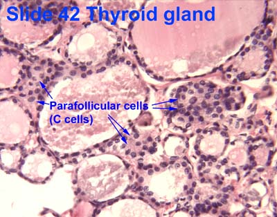
Parafollicular cells, also called C cells, are neuroendocrine cells in the thyroid. The primary function of these cells is to secrete calcitonin. They are located adjacent to the thyroid follicles and reside in the connective tissue. These cells are large and have a pale stain compared with the follicular cells. In teleost and avian species these cells occupy a structure outside the thyroid gland named the ultimopharyngeal body.

Contents

Structure

Parafollicular cells are pale-staining cells found in small number in the thyroid and are typically situated basally in the epithelium, without direct contact with the follicular lumen. They are always situated within the basement membrane, which surrounds the entire follicle.

Development

Parafollicular cells are derived from pharyngeal endoderm. [1] [2] Embryologically, they associate with the ultimopharyngeal body, which is a ventral derivative of the fourth (or fifth) pharyngeal pouch. Parafollicular cells were previously believed to be derived from the neural crest based on a series of experiments in quail-chick chimeras. [3] [4] However, lineage tracing experiments in mice revealed that parafollicular cells are derived from the endoderm origin. [5]

Function

Parafollicular cells secrete calcitonin, a hormone that participates in the regulation of calcium metabolism. Calcitonin lowers blood levels of calcium by inhibiting the resorption of bone by osteoclasts, and its secretion is increased proportionally with the concentration of calcium. [6]

Parafollicular cells are also known to secrete in smaller quantities several neuroendocrine peptides such as serotonin, somatostatin or CGRP. [7] [8] [9] They may also have a role in regulating thyroid hormones production locally, as they express thyrotropin-releasing hormone. [10] [11]

Clinical significance

When parafollicular cells become cancerous, they lead to medullary carcinoma of the thyroid.[ citation needed ]

See also

Related Research Articles

<span class="mw-page-title-main">Endocrine system</span> Hormone-producing glands of a body

The endocrine system is a messenger system in an organism comprising feedback loops of hormones that are released by internal glands directly into the circulatory system and that target and regulate distant organs. In vertebrates, the hypothalamus is the neural control center for all endocrine systems.

<span class="mw-page-title-main">Thyroid</span> Endocrine gland in the neck; secretes hormones that influence metabolism

The thyroid, or thyroid gland, is an endocrine gland in vertebrates. In humans, it is in the neck and consists of two connected lobes. The lower two thirds of the lobes are connected by a thin band of tissue called the isthmus (pl.: isthmi). The thyroid gland is a butterfly-shaped gland located in the neck below the Adam's apple. Microscopically, the functional unit of the thyroid gland is the spherical thyroid follicle, lined with follicular cells (thyrocytes), and occasional parafollicular cells that surround a lumen containing colloid. The thyroid gland secretes three hormones: the two thyroid hormones – triiodothyronine (T3) and thyroxine (T4) – and a peptide hormone, calcitonin. The thyroid hormones influence the metabolic rate and protein synthesis and growth and development in children. Calcitonin plays a role in calcium homeostasis. Secretion of the two thyroid hormones is regulated by thyroid-stimulating hormone (TSH), which is secreted from the anterior pituitary gland. TSH is regulated by thyrotropin-releasing hormone (TRH), which is produced by the hypothalamus.

<span class="mw-page-title-main">Parathyroid gland</span> Endocrine gland

Parathyroid glands are small endocrine glands in the neck of humans and other tetrapods. Humans usually have four parathyroid glands, located on the back of the thyroid gland in variable locations. The parathyroid gland produces and secretes parathyroid hormone in response to a low blood calcium, which plays a key role in regulating the amount of calcium in the blood and within the bones.

Thyroid-stimulating hormone (also known as thyrotropin, thyrotropic hormone, or abbreviated TSH) is a pituitary hormone that stimulates the thyroid gland to produce thyroxine (T4), and then triiodothyronine (T3) which stimulates the metabolism of almost every tissue in the body. It is a glycoprotein hormone produced by thyrotrope cells in the anterior pituitary gland, which regulates the endocrine function of the thyroid.

<span class="mw-page-title-main">Calcitonin</span> Amino acid peptide hormone secreted by the thyroid gland

Calcitonin is a 32 amino acid peptide hormone secreted by parafollicular cells (also known as C cells) of the thyroid (or endostyle) in humans and other chordates in the ultimopharyngeal body. It acts to reduce blood calcium (Ca2+), opposing the effects of parathyroid hormone (PTH).

<span class="mw-page-title-main">Thyroglobulin</span> Protein produced and used by the thyroid

Thyroglobulin (Tg) is a 660 kDa, dimeric glycoprotein produced by the follicular cells of the thyroid and used entirely within the thyroid gland. Tg is secreted and accumulated at hundreds of grams per litre in the extracellular compartment of the thyroid follicles, accounting for approximately half of the protein content of the thyroid gland. Human TG (hTG) is a homodimer of subunits each containing 2768 amino acids as synthesized.

<span class="mw-page-title-main">Thyroid follicular cell</span>

Thyroid follicular cells (also called thyroid epithelial cells or thyrocytes) are the major cell type in the thyroid gland, and are responsible for the production and secretion of the thyroid hormones thyroxine (T4) and triiodothyronine (T3). They form the single layer of cuboidal epithelium that makes up the outer structure of the almost spherical thyroid follicle.

<span class="mw-page-title-main">Pentagastrin</span> Chemical compound

Pentagastrin is a synthetic polypeptide that has effects like gastrin when given parenterally. It stimulates the secretion of gastric acid, pepsin, and intrinsic factor, and has been used as a diagnostic aid as the pentagastrin-stimulated calcitonin test.

<span class="mw-page-title-main">Endocrine gland</span> Glands of the endocrine system that secrete hormones to blood

Endocrine glands are ductless glands of the endocrine system that secrete their products, hormones, directly into the blood. The major glands of the endocrine system include the pineal gland, pituitary gland, pancreas, ovaries, testicles, thyroid gland, parathyroid gland, hypothalamus and adrenal glands. The hypothalamus and pituitary glands are neuroendocrine organs.

Neuroendocrine cells are cells that receive neuronal input and, as a consequence of this input, release messenger molecules (hormones) into the blood. In this way they bring about an integration between the nervous system and the endocrine system, a process known as neuroendocrine integration. An example of a neuroendocrine cell is a cell of the adrenal medulla, which releases adrenaline to the blood. The adrenal medullary cells are controlled by the sympathetic division of the autonomic nervous system. These cells are modified postganglionic neurons. Autonomic nerve fibers lead directly to them from the central nervous system. The adrenal medullary hormones are kept in vesicles much in the same way neurotransmitters are kept in neuronal vesicles. Hormonal effects can last up to ten times longer than those of neurotransmitters. Sympathetic nerve fiber impulses stimulate the release of adrenal medullary hormones. In this way the sympathetic division of the autonomic nervous system and the medullary secretions function together.

The ultimopharyngeal body, or ultimobranchial body or ultimobranchial gland is a small organ found in the neck region of many animals. In humans, it develops from the fourth pharyngeal pouch into the parafollicular cells of the thyroid to produce calcitonin. It may not develop in DiGeorge syndrome.

<span class="mw-page-title-main">Pharyngeal pouch (embryology)</span>

In the embryonic development of vertebrates, pharyngeal pouches form on the endodermal side between the pharyngeal arches. The pharyngeal grooves form the lateral ectodermal surface of the neck region to separate the arches.

<span class="mw-page-title-main">APUD cell</span> Type of endocrine cells

APUD cells (DNES cells) constitute a group of apparently unrelated endocrine cells, which were named by the scientist A.G.E. Pearse, who developed the APUD concept in the 1960s based on calcitonin-secreting parafollicular C cells of dog thyroid. These cells share the common function of secreting a low molecular weight polypeptide hormone. There are several different types which secrete the hormones secretin, cholecystokinin and several others. The name is derived from an acronym, referring to the following:

<span class="mw-page-title-main">HOXA3</span> Protein-coding gene in the species Homo sapiens

Homeobox protein Hox-A3 is a protein that in humans is encoded by the HOXA3 gene.

<span class="mw-page-title-main">Medullary thyroid cancer</span> Malignant thyroid neoplasm originating from C-cells

Medullary thyroid cancer is a form of thyroid carcinoma which originates from the parafollicular cells, which produce the hormone calcitonin. Medullary tumors are the third most common of all thyroid cancers and together make up about 3% of all thyroid cancer cases. MTC was first characterized in 1959.

<span class="mw-page-title-main">Thyroid hormones</span> Hormones produced by the thyroid gland

Thyroid hormones are any hormones produced and released by the thyroid gland, namely triiodothyronine (T3) and thyroxine (T4). They are tyrosine-based hormones that are primarily responsible for regulation of metabolism. T3 and T4 are partially composed of iodine, derived from food. A deficiency of iodine leads to decreased production of T3 and T4, enlarges the thyroid tissue and will cause the disease known as simple goitre.

<span class="mw-page-title-main">Thyroid cancer</span> Medical condition

Thyroid cancer is cancer that develops from the tissues of the thyroid gland. It is a disease in which cells grow abnormally and have the potential to spread to other parts of the body. Symptoms can include swelling or a lump in the neck. Cancer can also occur in the thyroid after spread from other locations, in which case it is not classified as thyroid cancer.

The cranial neural crest is one of the four regions of the neural crest.

The fetal endocrine system is one of the first systems to develop during prenatal development of a human individual. The endocrine system arises from all three embryonic germ layers. The endocrine glands that produce the steroid hormones, such as the gonads and adrenal cortex, arise from the mesoderm. In contrast, endocrine glands that arise from the endoderm and ectoderm produce the amine, peptide, and protein hormones.

<span class="mw-page-title-main">Iain Macintyre</span> British endocrinologist

Iain Macintyre FRS was a British endocrinologist who made important contributions to the understanding of calcium regulation and bone metabolism. Shortly after the hormone calcitonin had been described by Harold Copp, Macintyre's team was the first to isolate and sequence the hormone and to demonstrate its origin in the parafollicular cells of the thyroid gland. He subsequently analysed its physiological actions. Along with H. R. Morris he isolated and sequenced calcitonin gene-related peptide. Later research centred on the role played by nitric oxide on bone metabolism.

References

  1. Nilsson M, Williams D (July 2016). "On the Origin of Cells and Derivation of Thyroid Cancer: C Cell Story Revisited". European Thyroid Journal. 5 (2): 79–93. doi:10.1159/000447333. PMC   4949372 . PMID   27493881.
  2. Johansson, E., Andersson, L., Örnros, J., Carlsson, T., Ingeson-Carlsson, C., Liang, S., … Nilsson, M. (2015). Revising the embryonic origin of thyroid C cells in mice and humans. Development, 142(20), 3519–3528. http://doi.org/10.1242/dev.126581
  3. Le Douarin N, Fontaine J, Le Lièvre C (March 1974). "New studies on the neural crest origin of the avian ultimobranchial glandular cells--interspecific combinations and cytochemical characterization of C cells based on the uptake of biogenic amine precursors". Histochemistry. 38 (4): 297–305. doi:10.1007/bf00496718. PMID   4135055. S2CID   7551942.
  4. Barasch J, Gershon MD, Nunez EA, Tamir H, al-Awqati Q (December 1988). "Thyrotropin induces the acidification of the secretory granules of parafollicular cells by increasing the chloride conductance of the granular membrane". The Journal of Cell Biology. 107 (6 Pt 1): 2137–47. doi:10.1083/jcb.107.6.2137. PMC   2115661 . PMID   2461947.
  5. Johansson E, Andersson L, Örnros J, Carlsson T, Ingeson-Carlsson C, Liang S, Dahlberg J, Jansson S, Parrillo L, Zoppoli P, Barila GO, Altschuler DL, Padula D, Lickert H, Fagman H, Nilsson M (October 2015). "Revising the embryonic origin of thyroid C cells in mice and humans". Development. 142 (20): 3519–28. doi:10.1242/dev.126581. PMC   4631767 . PMID   26395490.
  6. Melmed S, Polonsky KS, Larsen PR, Kronenberg HM (2011). Williams Textbook of Endocrinology (12th ed.). Saunders. pp. 1250–1252. ISBN   978-1437703245.
  7. Zabel M (December 1984). "Ultrastructural localization of calcitonin, somatostatin and serotonin in parafollicular cells of rat thyroid". The Histochemical Journal. 16 (12): 1265–72. doi:10.1007/bf01003725. PMID   6152264. S2CID   7889687.
  8. Barasch JM, Mackey H, Tamir H, Nunez EA, Gershon MD (September 1987). "Induction of a neural phenotype in a serotonergic endocrine cell derived from the neural crest". The Journal of Neuroscience. 7 (9): 2874–83. doi:10.1523/JNEUROSCI.07-09-02874.1987. PMC   6569149 . PMID   3305802.
  9. Bernd P, Gershon MD, Nunez EA, Tamir H (March 1981). "Separation of dissociated thyroid follicular and parafollicular cells: association of serotonin binding protein with parafollicular cells". The Journal of Cell Biology. 88 (3): 499–508. doi:10.1083/jcb.88.3.499. PMC   2112761 . PMID   7217200.
  10. Gkonos PJ, Tavianini MA, Liu CC, Roos BA (December 1989). "Thyrotropin-releasing hormone gene expression in normal thyroid parafollicular cells". Molecular Endocrinology. 3 (12): 2101–9. doi: 10.1210/mend-3-12-2101 . PMID   2516877.
  11. Morillo-Bernal J, Fernández-Santos JM, Utrilla JC, de Miguel M, García-Marín R, Martín-Lacave I (August 2009). "Functional expression of the thyrotropin receptor in C cells: new insights into their involvement in the hypothalamic-pituitary-thyroid axis". Journal of Anatomy. 215 (2): 150–8. doi:10.1111/j.1469-7580.2009.01095.x. PMC   2740962 . PMID   19493188.

Further reading