A basement membrane also surrounds some individual cells, including muscle cells, fat cells, and Schwann cells, separating them from surrounding connective tissue.[1][4] Its composition can vary from tissue to tissue, and even in different regions of the same tissue.[1][4] The other type of ECM is the interstitial matrix.[5] The basement membrane may be described as having two layers or laminae, an external basal lamina, facing the epithelium, and an internal basal lamina that faces the connective tissue.[6] These two laminae are also known as the basal lamina and the reticular lamina.[7]
The basement membrane also acts as a platform for complex cell signaling, for polarization, migration, and differentiation.[2][6] It also regulates the exchange of materials between the epithelium and underlying tissues; binds growth factors from the connective tissue to the epithelium that control the development of epithelium.[3] Epithelial cells are pressed closely together having no blood vessels between them but their basement membrane mostly rests on a bed of loose connective tissue that is rich in blood vessels providing nutrients and removing waste.[3]
Structure
The basement membrane was first described in skeletal muscle tissue in the 1800s. The beginnings of a molecular understanding only came about in the 1970s and 1980s.[2]
Epithelial cells are polarized. The surface of epithelial cells that face the lumen is the apical surface, and the surface facing the basement membrane is the basal surface.[3]
The basement membrane may be described as having two layers or laminae, an external basal lamina, facing the epithelium, and an internal basal lamina that faces the connective tissue.[6] These two laminae are also known as the basal lamina and the reticular lamina.[7] The cells in the internal basal membrane that are closest to the connective tissue show high rates of mitosis, needed to replace skin cell abrasions, and in the GI tract replacement of the cells exposed to digestive enzymes and gastric acid.[3] In the skin the basement membrane is part of a more complex basement membrane zone. In the mucous membrane linings such as the gastric mucosa the basement membrane overlies loose connective tissue known as the lamina propria.
The glomerular basement membrane of the kidney, is an unusually thick basement membrane. It serves as part of a molecular filter that prevents macromolecules from the blood from entering the urine.[1] It is faced by a cell layer on either side, the endothelium, and the podocytes, and has a thicker structure of three laminae. It is thicker by the fusion of the basal lamina from the endothelium of glomerular capillaries and the podocyte basal lamina.[9][10] These layers are known as the central lamina densa, and on each side, a lamina rara – a lamina rara interna facing the endothelium, and a lamina rara externa facing the podocytes.[10]
In the skin the basement membrane that separates, and connects the epidermis and the underlying dermis is part of a complex and specialized structure called the basement membrane zone (BMZ). [12] The BMZ has four distinct layers – the basal cell layer, the lamina lucida, the lamina densa, and the sublaminal densa, and has many functions.[13] Tiny microfilaments called tonofilaments cross the basal cell layer, and extend to the epidermal part of the hemidesmosome. Laminins and other adherence proteins are located in the lamina lucida. The lamina densa is mostly composed of a type IV collagen scaffold. Anchoring fibrils and microfilaments extend and blend with the elastic fibrillary system of the dermis.[12][14]
The components of the BMZ form a complex, functional network that extends from the basal epidermal keratinocytes and their hemidesmosomes, and include anchoring fibrils from the lamina densa, into the extracellular matrix (ECM) of the dermis. In the ECM the anchoring fibrils appear as cross-striated fibrous masses. There are also focal adhesion complexes on the outer cell membrane that bind the cytoskeleton to cell-matrix adhesions.[13]
The basement membrane acts as a mechanical barrier, preventing malignant cells from invading the deeper tissues.[15] Early stages of malignancy that are thus limited to the epithelial layer by the basement membrane are called carcinoma in situ.
The basement membrane is also essential for angiogenesis (development of new blood vessels). Basement membrane proteins have been found to accelerate differentiation of endothelial cells.[16]
Other roles for the basement membrane include blood filtration and muscle homeostasis.[2]Fractones may be a type of basement membrane, serving as a niche for stem cells.[17][18]
Clinical significance
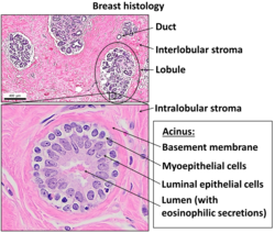
Normal histology of the breast, with basement membrane annotated near center-right.Prostate gland microanatomy, with basement membrane annotated at bottom.
Some diseases result from a poorly functioning basement membrane. The cause can be genetic defects, injuries by the body's own immune system, or other mechanisms.[19] Diseases involving basement membranes at multiple locations include:
Autoimmune diseases targeting basement membranes. Non-collagenous domain basement membrane collagen type IV is autoantigen (target antigen) of autoantibodies in the autoimmune disease Goodpasture's syndrome.[20]
A group of diseases stemming from improper function of the basement membrane zone are united under the name epidermolysis bullosa.[21]
These are only found within diploblastic and homoscleromorphic sponge animals. The homoscleromorph were found to be sister to diploblasts in some studies, making the membrane originate once in the history of life. But more recent studies have disregarded diploblast-homoscleromorph group, so other sponges may have lost it (most probable) or the origin in the two groups may be separate.
References
1234567Alberts, Bruce (2015). Molecular biology of the cell (Sixthed.). New York, NY: Garland Science, Taylor and Francis Group. pp.1068–1071. ISBN9780815344643.
↑Laurila, Pekka; Leivo, Ilmo (1 January 1993). "Basement membrane and interstitial matrix components form separate matrices in heterokaryons of PYS-2 cells and fibroblasts". Journal of Cell Science. 104 (1): 59–68. doi:10.1242/jcs.104.1.59.
↑"Sect. 7, Ch. 4: Basement Membrane". Renal Physiology Glomerular Filtration Rate and Renal Blood Flow. Medical College of Georgia, Robert B. Greenblatt, M.D. Library. 1 April 2008. Archived from the original on 1 April 2008. Retrieved 7 May 2018.{{cite book}}: CS1 maint: bot: original URL status unknown (link)
↑Mercier F, Kitasako JT, Hatton GI (September 2002). "Anatomy of the brain neurogenic zones revisited: fractones and the fibroblast/macrophage network". The Journal of Comparative Neurology. 451 (2): 170–188. doi:10.1002/cne.10342. PMID12209835. S2CID19919800.
↑Bardhan A, Bruckner-Tuderman L, Chapple IL, Fine JD, Harper N, Has C, etal. (September 2020). "Epidermolysis bullosa". Nature Reviews. Disease Primers. 6 (1): 78. doi:10.1038/s41572-020-0210-0. PMID32973163. S2CID221861310.
↑LeBoit PE (October 2000). "A thickened basement membrane is a clue to...lichen sclerosus!". The American Journal of Dermatopathology. 22 (5): 457–458. doi:10.1097/00000372-200010000-00014. PMID11048985.
This page is based on this Wikipedia article Text is available under the CC BY-SA 4.0 license; additional terms may apply. Images, videos and audio are available under their respective licenses.