Epithelial polarity

Last updated

Epithelial polarity is one example of the cell polarity that is a fundamental feature of many types of cells. Epithelial cells feature distinct 'apical', 'lateral' and 'basal' plasma membrane domains. Epithelial cells connect to one another via their lateral membranes to form epithelial sheets that line cavities and surfaces throughout the animal body. Each plasma membrane domain has a distinct protein composition, giving them distinct properties and allowing directional transport of molecules across the epithelial sheet. How epithelial cells generate and maintain polarity remains unclear, but certain molecules have been found to play a key role.

Contents

A variety of molecules are located at the apical membrane, but only a few key molecules act as determinants that are required to maintain the identity of the apical membrane and, thus, epithelial polarity. These molecules are the proteins Cdc42, atypical protein kinase C (aPKC), Par6, Par3/Bazooka/ASIP. [1] Crumbs, "Stardust" and protein at tight junctions (PATJ). These molecules appear to form two distinct complexes: an aPKC-Par3-Par6 "aPKC" (or "Par") complex that also interacts with Cdc42; and a Crumbs-Stardust-PATJ "Crumbs" complex. Of these two complexes, the aPKC complex is the most important for epithelial polarity, being required even when the Crumbs complex is not. Crumbs is the only transmembrane protein in this list and the Crumbs complex serves as an apical cue to keep the aPKC complex apical during complex cellular shape changes.[ citation needed ]

Basolateral membranes

In the context of renal tubule physiology, the term basolateral membrane refers to the cell membrane which is oriented away from the lumen of the tubule, whereas the term apical or luminal membrane refers to the cell membrane which is oriented towards the lumen. The principal function of this basolateral membrane is to take up metabolic waste products into the epithelial cell for disposal into the lumen where it is transported out of the body as urine. A secondary role of the basolateral membrane is to allow the recycling of desirable substrates, such as glucose, that have been rescued from the lumen of the tubule to be secreted into the interstitial fluids. [2]

Basal and lateral membranes share common determinants, the proteins LLGL1, DLG1, and SCRIB. These three proteins all localize to the basolateral domain and are essential for basolateral identity and for epithelial polarity.

Mechanisms of polarity

How epithelial cells polarize is still not fully understood. Some key principles have been proposed to maintain polarity, but the mechanisms behind these principles remain to be discovered.

The first principle is positive feedback. In computer models, a molecule that can be either membrane-associated or cytoplasmic can polarize when its association with the membrane is subject to positive feedback: that membrane localization occurs most strongly where the molecule is already most highly concentrated. In similar models, researchers have shown that epithelial cells can self-assemble into a rich set of robust biological shapes. [3] In the yeast saccharomyces cerevisiae, there is genetic evidence that Cdc42 is subject to positive feedback of this kind and can spontaneously polarize, even in the absence of an external cue. In the fruit fly Drosophila melanogaster, Cdc42 is recruited by the aPKC complex and then promotes the apical localization of the aPKC complex in a probable positive feedback loop. Thus, in the absence of Cdc42 or the aPKC complex, apical determinants cannot be maintained at the apical membrane and consequently, apical identity and polarity is lost.

The second principle is segregation of polarity determinants. The sharp distinction between apical and baso-lateral domains is maintained by an active mechanism that prevents mixing. The nature of this mechanism is not known, but it clearly depends on the polarity determinants. In the absence of the aPKC complex, the baso-lateral determinants spread into the former apical domain. Conversely, in the absence of any of Lgl, Dlg or Scrib, the apical determinants spread into the former baso-lateral domain. Thus, the two determinants behave as if they exert mutual repulsion upon one another.

The third principle is directed exocytosis. Apical membrane proteins are trafficked from the Golgi to the apical, rather than baso-lateral, membrane because apical determinants serve to identify the correct destination for vesicle delivery. A related mechanism is likely to operate for the baso-lateral membranes.

The fourth principle is lipid modification. A component of the lipid bilayer, phosphatidyl inositol phosphate (PIP) can be phosphorylated to form PIP2 and PIP3. In some epithelial cells, PIP2 is apically localised while PIP3 is basolaterally localised. In at least one cultured cell line, the MDCK cell, this system is required for epithelial polarity. The relationship between this system and the polarity determinants in animal tissues remains unclear.

Basal versus lateral

Since basal and lateral membranes share the same determinants, another mechanism must make the difference between the two domains. Cell shape and contacts provide the likely mechanism. Lateral membranes are the site of contact between epithelial cells, whereas basal membranes connect epithelial cells to the basement membrane, an extracellular matrix layer that lies along the basal surface of the epithelium. Certain molecules, such as Integrins, localise specifically to the basal membrane and form connections with the extracellular matrix.

Epithelial cell shape

Epithelial cells come in a variety of shapes that relate to their function in development or physiology. How epithelial cells adopt particular shapes is poorly understood, but it must involve spatial control of the actin cytoskeleton, which is central to cell shape in all plant cells.

Apocrine cells, showing apical snouts towards the lumen. Histology of apocrine cells.png
Apocrine cells, showing apical snouts towards the lumen.

Apical snouts, also called apical blebs, are small protrusions of cytoplasm towards the lumen. They are found normally in apocrine cells, and can also appear in apocrine metaplasia and columnar cell changes in the breast. [4]

Epithelial cadherin

All epithelial cells express the transmembrane adhesion molecule E-cadherin, a cadherin which localises most prominently to the junction between the apical and lateral membranes. The extra-cellular domains of E-cadherin molecules from neighbouring cells bind to one another via a homotypic interaction. The intra-cellular domains of E-cadherin molecules bind to the actin cytoskeleton via the adaptor proteins alpha-catenin and beta-catenin. [5] Thus, E-cadherin forms adherens junctions that connect the actin cytoskeletons of neighbouring cells. Adherens junctions are the primary force-bearing junctions between epithelial cells and are fundamentally important for maintaining epithelial cell shape and for dynamic changes in shape during tissue development. How E-cadherin localizes to the boundary between apical and lateral membranes is not known, but polarized membranes are essential for maintaining E-cadherin at adherens junctions.

See also

Related Research Articles

<span class="mw-page-title-main">Cell adhesion</span> Process of cell attachment

Cell adhesion is the process by which cells interact and attach to neighbouring cells through specialised molecules of the cell surface. This process can occur either through direct contact between cell surfaces such as cell junctions or indirect interaction, where cells attach to surrounding extracellular matrix, a gel-like structure containing molecules released by cells into spaces between them. Cells adhesion occurs from the interactions between cell-adhesion molecules (CAMs), transmembrane proteins located on the cell surface. Cell adhesion links cells in different ways and can be involved in signal transduction for cells to detect and respond to changes in the surroundings. Other cellular processes regulated by cell adhesion include cell migration and tissue development in multicellular organisms. Alterations in cell adhesion can disrupt important cellular processes and lead to a variety of diseases, including cancer and arthritis. Cell adhesion is also essential for infectious organisms, such as bacteria or viruses, to cause diseases.

<span class="mw-page-title-main">Cadherin</span>

Cadherins (named for "calcium-dependent adhesion") are cell adhesion molecules important in forming adherens junctions that let cells adhere to each other. Cadherins are a class of type-1 transmembrane proteins, and they depend on calcium (Ca2+) ions to function, hence their name. Cell-cell adhesion is mediated by extracellular cadherin domains, whereas the intracellular cytoplasmic tail associates with numerous adaptors and signaling proteins, collectively referred to as the cadherin adhesome.

<span class="mw-page-title-main">Cell junction</span> Multiprotein complex that forms a point of contact or adhesion in animal cells

Cell junctions or junctional complexes, are a class of cellular structures consisting of multiprotein complexes that provide contact or adhesion between neighboring cells or between a cell and the extracellular matrix in animals. They also maintain the paracellular barrier of epithelia and control paracellular transport. Cell junctions are especially abundant in epithelial tissues. Combined with cell adhesion molecules and extracellular matrix, cell junctions help hold animal cells together.

<span class="mw-page-title-main">Tight junction</span> Structure preventing inter-cell leakage

Tight junctions, also known as occluding junctions or zonulae occludentes, are multiprotein junctional complexes whose canonical function is to prevent leakage of solutes and water and seals between the epithelial cells. They also play a critical role maintaining the structure and permeability of endothelial cells. Tight junctions may also serve as leaky pathways by forming selective channels for small cations, anions, or water. The corresponding junctions that occur in invertebrates are septate junctions.

<span class="mw-page-title-main">Catenin</span>

Catenins are a family of proteins found in complexes with cadherin cell adhesion molecules of animal cells. The first two catenins that were identified became known as α-catenin and β-catenin. α-Catenin can bind to β-catenin and can also bind filamentous actin (F-actin). β-Catenin binds directly to the cytoplasmic tail of classical cadherins. Additional catenins such as γ-catenin and δ-catenin have been identified. The name "catenin" was originally selected because it was suspected that catenins might link cadherins to the cytoskeleton.

<span class="mw-page-title-main">Adherens junction</span>

Adherens junctions are protein complexes that occur at cell–cell junctions and cell–matrix junctions in epithelial and endothelial tissues, usually more basal than tight junctions. An adherens junction is defined as a cell junction whose cytoplasmic face is linked to the actin cytoskeleton. They can appear as bands encircling the cell or as spots of attachment to the extracellular matrix.

<span class="mw-page-title-main">Catenin beta-1</span> Mammalian protein found in Homo sapiens

Catenin beta-1, also known as β-catenin (beta-catenin), is a protein that in humans is encoded by the CTNNB1 gene.

α-Catenin Primary protein link between cadherins and the actin cytoskeleton

α-Catenin (alpha-catenin) functions as the primary protein link between cadherins and the actin cytoskeleton. It has been reported that the actin binding proteins vinculin and α-actinin can bind to alpha-catenin. It has been suggested that alpha-catenin does not bind with high affinity to both actin filaments and the E-cadherin-beta-catenin complex at the same time. It has been observed that when α-catenin is not in a molecular complex with β-catenin, it dimerizes and functions to regulate actin filament assembly, possibly by competing with Arp2/3 protein. α-Catenin exhibits significant protein dynamics. However, a protein complex including a cadherin, actin, β-catenin and α-catenin has not been isolated.

<span class="mw-page-title-main">SCRIB</span> Protein-coding gene in the species Homo sapiens

SCRIB, also known as Scribble, SCRIBL, or Scribbled homolog (Drosophila), is a scaffold protein which in humans is encoded by the SCRIB gene. It was originally isolated in Drosophila melanogaster in a pathway (also known as the Scribble complex) with DLGAP5 (Discs large) and LLGL1 (Lethal giant larvae) as a tumor suppressor. In humans, SCRIB is found as a membrane protein and is involved in cell migration, cell polarity, and cell proliferation in epithelial cells. There is also strong evidence that SCRIB may play a role in cancer progression because of its strong homology to the Drosophila protein.

<span class="mw-page-title-main">PARD6A</span> Protein-coding gene in the species Homo sapiens

Partitioning defective 6 homolog alpha is a protein that in humans is encoded by the PARD6A gene.

<span class="mw-page-title-main">Stress fiber</span> Contractile actin bundles found in non-muscle cells

Stress fibers are contractile actin bundles found in non-muscle cells. They are composed of actin (microfilaments) and non-muscle myosin II (NMMII), and also contain various crosslinking proteins, such as α-actinin, to form a highly regulated actomyosin structure within non-muscle cells. Stress fibers have been shown to play an important role in cellular contractility, providing force for a number of functions such as cell adhesion, migration and morphogenesis.

<span class="mw-page-title-main">Cadherin-1</span> Human protein-coding gene

Cadherin-1 or Epithelial cadherin(E-cadherin), is a protein that in humans is encoded by the CDH1 gene. Mutations are correlated with gastric, breast, colorectal, thyroid, and ovarian cancers. CDH1 has also been designated as CD324. It is a tumor suppressor gene.

<span class="mw-page-title-main">Intestinal epithelium</span> Single-cell layer lining the intestines

The intestinal epithelium is the single cell layer that form the luminal surface (lining) of both the small and large intestine (colon) of the gastrointestinal tract. Composed of simple columnar epithelial cells, it serves two main functions: absorbing useful substances into the body and restricting the entry of harmful substances. As part of its protective role, the intestinal epithelium forms an important component of the intestinal mucosal barrier. Certain diseases and conditions are caused by functional defects in the intestinal epithelium. On the other hand, various diseases and conditions can lead to its dysfunction which, in turn, can lead to further complications.

<span class="mw-page-title-main">Catenin alpha-1</span> Protein-coding gene in the species Homo sapiens

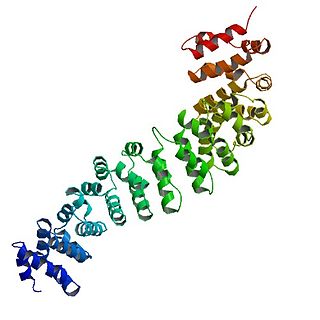
αE-catenin, also known as Catenin alpha-1 is a protein that in humans is encoded by the CTNNA1 gene. αE-catenin is highly expressed in cardiac muscle and localizes to adherens junctions at intercalated disc structures where it functions to mediate the anchorage of actin filaments to the sarcolemma. αE-catenin also plays a role in tumor metastasis and skin cell function.

<span class="mw-page-title-main">VEZT</span> Protein-coding gene in the species Homo sapiens

VEZT is a gene located on chromosome 12 and encodes for the protein vezatin. Vezatin is a major component of the cadherin-catenin complex that is critical to the formation and maintenance of adherens junctions. The protein is expressed in most epithelial cells and is crucial to the formation of cell-cell contact junctions. Mutations of the gene can lead to upregulation or downregulation of the protein which can have detrimental effects on physiological systems, particularly those involved in development.

<span class="mw-page-title-main">Cell polarity</span> Polar morphology of a cell, a specific orientation of the cell structure

Cell polarity refers to spatial differences in shape, structure, and function within a cell. Almost all cell types exhibit some form of polarity, which enables them to carry out specialized functions. Classical examples of polarized cells are described below, including epithelial cells with apical-basal polarity, neurons in which signals propagate in one direction from dendrites to axons, and migrating cells. Furthermore, cell polarity is important during many types of asymmetric cell division to set up functional asymmetries between daughter cells.

Cell–cell interaction refers to the direct interactions between cell surfaces that play a crucial role in the development and function of multicellular organisms. These interactions allow cells to communicate with each other in response to changes in their microenvironment. This ability to send and receive signals is essential for the survival of the cell. Interactions between cells can be stable such as those made through cell junctions. These junctions are involved in the communication and organization of cells within a particular tissue. Others are transient or temporary such as those between cells of the immune system or the interactions involved in tissue inflammation. These types of intercellular interactions are distinguished from other types such as those between cells and the extracellular matrix. The loss of communication between cells can result in uncontrollable cell growth and cancer.

<span class="mw-page-title-main">PLEKHA7</span> Protein-coding gene in the species Homo sapiens

PLEKHA7 is an adherens junction (AJ) protein, involved in the junction's integrity and stability.

The internal surface of the uterus is lined by uterine epithelial cells which undergo dramatic changes during pregnancy. The role of the uterine epithelial cells is to selectively allow the blastocyst to implant at a specific time. All other times of the cycle, these uterine epithelial cells are refractory to blastocyst implantation. Uterine epithelial cells have a similar structure in most species and the changes which occur in the uterine epithelial cells at the time of blastocyst implantation are also conserved among most species.

Barry James Thompson is an Australian and British developmental biologist and cancer biologist. Thompson is known for identifying genes, proteins and mechanisms involved in epithelial polarity, morphogenesis and cell signaling via the Wnt and Hippo signaling pathways, which have key roles in human cancer.

References

Bruce Alberts; Alexander Johnson; Julian Lewis; Martin Raff; Keith Roberts; Peter Walter, eds. (2002). Molecular Biology of the Cell (4th ed.). Garland Science. ISBN   978-0-8153-3218-3.

  1. Izumi Y, Hirose T, Tamai Y, Hirai S, Nagashima Y, Fujimoto T, Tabuse Y, Kemphues KJ, Ohno S (1998). "An Atypical PKC Directly Associates and Colocalizes at the Epithelial Tight Junction with ASIP, a Mammalian Homologue of Caenorhabditis elegans Polarity Protein PAR-3". J Cell Biol. 143 (1): 95–106. doi:10.1083/jcb.143.1.95. PMC   2132825 . PMID   9763423.
  2. Sekine T, Miyazaki H, Endou H (February 2006). "Molecular physiology of renal organic anion transporters". Am. J. Physiol. Renal Physiol. 290 (2): F251–61. doi:10.1152/ajprenal.00439.2004. PMID   16403838.
  3. Nissen, Silas Boye; Rønhild, Steven; Trusina, Ala; Sneppen, Kim (November 27, 2018). "Theoretical tool bridging cell polarities with development of robust morphologies". eLife. 7: e38407. doi: 10.7554/eLife.38407 . PMC   6286147 . PMID   30477635.
  4. Logullo, A.F., Nimir, C. (2019). "Columnar cell lesions of the breast: a practical review for the pathologist". Surg Exp Pathol. 2 (2). doi: 10.1186/s42047-018-0027-2 .{{cite journal}}: CS1 maint: multiple names: authors list (link)
  5. Knudsen KA, Soler AP, Johnson KR, Wheelock MJ (July 1995). "Interaction of alpha-actinin with the cadherin/catenin cell-cell adhesion complex via alpha-catenin". J. Cell Biol. 130 (1): 67–77. doi:10.1083/jcb.130.1.67. PMC   2120515 . PMID   7790378.