Mucous membrane

Last updated
Mucous membrane
Normal gastric mucosa intermed mag.jpg
Histological section taken from the gastric antrum, showing the mucosa of the stomach
Details
Identifiers
Latin tunica mucosa
MeSH D009092
TA98 A05.4.01.015
A05.3.01.029
A05.5.01.029
A05.6.01.009
A05.6.01.010
A05.7.01.006
A05.7.01.007
A05.8.02.009
A06.1.02.017
A06.2.09.019
A06.3.01.010
A06.4.02.029
A08.1.05.011
A08.2.01.007
A08.3.01.023
A09.1.02.013
A09.1.04.011
A09.2.03.012
A09.3.05.010
A09.3.06.004
A09.4.02.015
A09.4.02.020
A09.4.02.029
A15.3.02.083
Anatomical terms of microanatomy

A mucous membrane or mucosa is a membrane that lines various cavities in the body of an organism and covers the surface of internal organs. It consists of one or more layers of epithelial cells overlying a layer of loose connective tissue. It is mostly of endodermal origin and is continuous with the skin at body openings such as the eyes, eyelids, ears, inside the nose, inside the mouth, lips, the genital areas, the urethral opening and the anus. Some mucous membranes secrete mucus, a thick protective fluid. The function of the membrane is to stop pathogens and dirt from entering the body and to prevent bodily tissues from becoming dehydrated.

Contents

Structure

The mucosa is composed of one or more layers of epithelial cells that secrete mucus, and an underlying lamina propria of loose connective tissue. [1] The type of cells and type of mucus secreted vary from organ to organ and each can differ along a given tract. [2] [3]

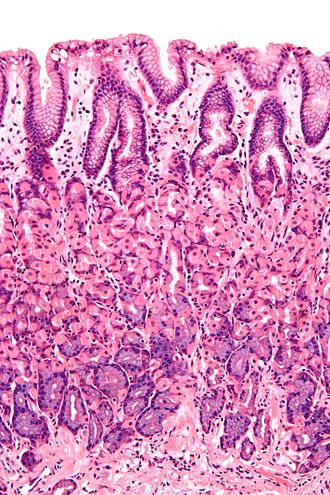
Mucous membranes line the digestive, respiratory and reproductive tracts and are the primary barrier between the external world and the interior of the body; in an adult human the total surface area of the mucosa is about 400 square meters while the surface area of the skin is about 2 square meters. [4] :1 Along with providing a physical barrier, they also contain key parts of the immune system and serve as the interface between the body proper and the microbiome. [2] :437

Examples

Some examples include:

Development

Developmentally, the majority of mucous membranes are of endodermal origin. [5] Exceptions include the palate, cheeks, floor of the mouth, gums, lips and the portion of the anal canal below the pectinate line, which are all ectodermal in origin. [6] [7]

Function

One of its functions is to keep the tissue moist (for example in the respiratory tract, including the mouth and nose). [2] :480 It also plays a role in absorbing and transforming nutrients. [2] :5,813 Mucous membranes also protect the body from itself. For instance, mucosa in the stomach protects it from stomach acid, [2] :384,797 and mucosa lining the bladder protects the underlying tissue from urine. [8] In the uterus, the mucous membrane is called the endometrium, and it swells each month and is then eliminated during menstruation. [2] :1019

Nutrition

Niacin [2] :876 and vitamin A are essential nutrients that help maintain mucous membranes. [9]

See also

Related Research Articles

<span class="mw-page-title-main">Salivary gland</span> Exocrine glands that produce saliva through a system of ducts

The salivary glands in many vertebrates including mammals are exocrine glands that produce saliva through a system of ducts. Humans have three paired major salivary glands, as well as hundreds of minor salivary glands. Salivary glands can be classified as serous, mucous, or seromucous (mixed).

<span class="mw-page-title-main">Lamina propria</span> Thin connective layer forming part of the mucous membranes

The lamina propria is a thin layer of connective tissue that forms part of the moist linings known as mucous membranes or mucosae, which line various tubes in the body, such as the respiratory tract, the gastrointestinal tract, and the urogenital tract.

<span class="mw-page-title-main">Immunoglobulin A</span> Antibody that plays a crucial role in the immune function of mucous membranes

Immunoglobulin A is an antibody that plays a role in the immune function of mucous membranes. The amount of IgA produced in association with mucosal membranes is greater than all other types of antibody combined. In absolute terms, between three and five grams are secreted into the intestinal lumen each day. This represents up to 15% of total immunoglobulins produced throughout the body.

<span class="mw-page-title-main">Mucus</span> Secretion produced by mucous membranes

Mucus is a slippery aqueous secretion produced by, and covering, mucous membranes. It is typically produced from cells found in mucous glands, although it may also originate from mixed glands, which contain both serous and mucous cells. It is a viscous colloid containing inorganic salts, antimicrobial enzymes, immunoglobulins, and glycoproteins such as lactoferrin and mucins, which are produced by goblet cells in the mucous membranes and submucosal glands. Mucus serves to protect epithelial cells in the linings of the respiratory, digestive, and urogenital systems, and structures in the visual and auditory systems from pathogenic fungi, bacteria and viruses. Most of the mucus in the body is produced in the gastrointestinal tract.

<span class="mw-page-title-main">Leukoplakia</span> Medical condition

Oral leukoplakia is a potentially malignant disorder affecting the oral mucosa. It is defined as "essentially an oral mucosal white lesion that cannot be considered as any other definable lesion." Oral leukoplakia is a white patch or plaque that develops in the oral cavity and is strongly associated with smoking. Leukoplakia is a firmly attached white patch on a mucous membrane which is associated with increased risk of cancer. The edges of the lesion are typically abrupt and the lesion changes with time. Advanced forms may develop red patches. There are generally no other symptoms. It usually occurs within the mouth, although sometimes mucosa in other parts of the gastrointestinal tract, urinary tract, or genitals may be affected.

<span class="mw-page-title-main">Mucin</span> Glycoprotein

Mucins are a family of high molecular weight, heavily glycosylated proteins (glycoconjugates) produced by epithelial tissues in most animals. Mucins' key characteristic is their ability to form gels; therefore they are a key component in most gel-like secretions, serving functions from lubrication to cell signalling to forming chemical barriers. They often take an inhibitory role. Some mucins are associated with controlling mineralization, including nacre formation in mollusks, calcification in echinoderms and bone formation in vertebrates. They bind to pathogens as part of the immune system. Overexpression of the mucin proteins, especially MUC1, is associated with many types of cancer.

<span class="mw-page-title-main">Goblet cell</span> Epithelial cells that secrete mucins

Goblet cells are simple columnar epithelial cells that secrete gel-forming mucins, like mucin 5AC. The goblet cells mainly use the merocrine method of secretion, secreting vesicles into a duct, but may use apocrine methods, budding off their secretions, when under stress. The term goblet refers to the cell's goblet-like shape. The apical portion is shaped like a cup, as it is distended by abundant mucus laden granules; its basal portion lacks these granules and is shaped like a stem.

The oral mucosa is the mucous membrane lining the inside of the mouth. It comprises stratified squamous epithelium, termed "oral epithelium", and an underlying connective tissue termed lamina propria. The oral cavity has sometimes been described as a mirror that reflects the health of the individual. Changes indicative of disease are seen as alterations in the oral mucosa lining the mouth, which can reveal systemic conditions, such as diabetes or vitamin deficiency, or the local effects of chronic tobacco or alcohol use. The oral mucosa tends to heal faster and with less scar formation compared to the skin. The underlying mechanism remains unknown, but research suggests that extracellular vesicles might be involved.

<span class="mw-page-title-main">Simple columnar epithelium</span> Tissue type

Simple columnar epithelium is a single layer of columnar epithelial cells which are tall and slender with oval-shaped nuclei located in the basal region, attached to the basement membrane. In humans, simple columnar epithelium lines most organs of the digestive tract including the stomach, and intestines. Simple columnar epithelium also lines the uterus.

The mucosa-associated lymphoid tissue (MALT), also called mucosa-associated lymphatic tissue, is a diffuse system of small concentrations of lymphoid tissue found in various submucosal membrane sites of the body, such as the gastrointestinal tract, nasopharynx, thyroid, breast, lung, salivary glands, eye, and skin. MALT is populated by lymphocytes such as T cells and B cells, as well as plasma cells, dendritic cells and macrophages, each of which is well situated to encounter antigens passing through the mucosal epithelium. In the case of intestinal MALT, M cells are also present, which sample antigen from the lumen and deliver it to the lymphoid tissue. MALT constitute about 50% of the lymphoid tissue in human body. Immune responses that occur at mucous membranes are studied by mucosal immunology.

<span class="mw-page-title-main">Gastric glands</span> Glands in lining of the human stomach

The gastric glands are glands in the lining of the stomach that play an essential role in the process of digestion. All of the glands have mucus-secreting foveolar cells. Mucus lines the entire stomach, and protects the stomach lining from the effects of hydrochloric acid released from other cells in the glands.

<span class="mw-page-title-main">Respiratory epithelium</span> Mucosa that serves to moisten and protect the airways

Respiratory epithelium, or airway epithelium, is a type of ciliated columnar epithelium found lining most of the respiratory tract as respiratory mucosa, where it serves to moisten and protect the airways. It is not present in the vocal cords of the larynx, or the oropharynx and laryngopharynx, where instead the epithelium is stratified squamous. It also functions as a barrier to potential pathogens and foreign particles, preventing infection and tissue injury by the secretion of mucus and the action of mucociliary clearance.

<span class="mw-page-title-main">Submucosa</span> Thin layer of tissue in various organs

The submucosa is a thin layer of tissue in various organs of the gastrointestinal, respiratory, and genitourinary tracts. It is the layer of dense irregular connective tissue that supports the mucosa and joins it to the muscular layer, the bulk of overlying smooth muscle.

<span class="mw-page-title-main">Intestinal epithelium</span> Single-cell layer lining the intestines

The intestinal epithelium is the single cell layer that form the luminal surface (lining) of both the small and large intestine (colon) of the gastrointestinal tract. Composed of simple columnar epithelial cells, it serves two main functions: absorbing useful substances into the body and restricting the entry of harmful substances. As part of its protective role, the intestinal epithelium forms an important component of the intestinal mucosal barrier. Certain diseases and conditions are caused by functional defects in the intestinal epithelium. On the other hand, various diseases and conditions can lead to its dysfunction which, in turn, can lead to further complications.

<span class="mw-page-title-main">Mucosal immunology</span> Field of study

Mucosal immunology is the study of immune system responses that occur at mucosal membranes of the intestines, the urogenital tract, and the respiratory system. The mucous membranes are in constant contact with microorganisms, food, and inhaled antigens. In healthy states, the mucosal immune system protects the organism against infectious pathogens and maintains a tolerance towards non-harmful commensal microbes and benign environmental substances. Disruption of this balance between tolerance and deprivation of pathogens can lead to pathological conditions such as food allergies, irritable bowel syndrome, susceptibility to infections, and more.

<span class="mw-page-title-main">Nasal mucosa</span> Part of the mucus membrane lining the nasal cavity

The nasal mucosa lines the nasal cavity. It is part of the respiratory mucosa, the mucous membrane lining the respiratory tract. The nasal mucosa is intimately adherent to the periosteum or perichondrium of the nasal conchae. It is continuous with the skin through the nostrils, and with the mucous membrane of the nasal part of the pharynx through the choanae. From the nasal cavity its continuity with the conjunctiva may be traced, through the nasolacrimal and lacrimal ducts; and with the frontal, ethmoidal, sphenoidal, and maxillary sinuses, through the several openings in the nasal meatuses. The mucous membrane is thickest, and most vascular, over the nasal conchae. It is also thick over the nasal septum where increased numbers of goblet cells produce a greater amount of nasal mucus. It is very thin in the meatuses on the floor of the nasal cavities, and in the various sinuses. It is one of the most commonly infected tissues in adults and children. Inflammation of this tissue may cause significant impairment of daily activities, with symptoms such as stuffy nose, headache, mouth breathing, etc.

<span class="mw-page-title-main">Human digestive system</span> Digestive system in humans

The human digestive system consists of the gastrointestinal tract plus the accessory organs of digestion. Digestion involves the breakdown of food into smaller and smaller components, until they can be absorbed and assimilated into the body. The process of digestion has three stages: the cephalic phase, the gastric phase, and the intestinal phase.

<span class="mw-page-title-main">Intestinal mucosal barrier</span>

The intestinal mucosal barrier, also referred to as intestinal barrier, refers to the property of the intestinal mucosa that ensures adequate containment of undesirable luminal contents within the intestine while preserving the ability to absorb nutrients. The separation it provides between the body and the gut prevents the uncontrolled translocation of luminal contents into the body proper. Its role in protecting the mucosal tissues and circulatory system from exposure to pro-inflammatory molecules, such as microorganisms, toxins, and antigens is vital for the maintenance of health and well-being. Intestinal mucosal barrier dysfunction has been implicated in numerous health conditions such as: food allergies, microbial infections, irritable bowel syndrome, inflammatory bowel disease, celiac disease, metabolic syndrome, non-alcoholic fatty liver disease, diabetes, and septic shock.

<span class="mw-page-title-main">Anatomical terms of microanatomy</span> Anatomical terminology is used to describe microanatomical (or histological) structures

Anatomical terminology is used to describe microanatomical structures. This helps describe precisely the structure, layout and position of an object, and minimises ambiguity. An internationally accepted lexicon is Terminologia Histologica.

A tissue membrane is a thin layer or sheet of cells that covers the outside of the body, the organs, internal passageways that lead to the exterior of the body, and the lining of the moveable joint cavities. There are two basic types of tissue membranes: connective tissue and epithelial membranes.

References

  1. "Mucous membrane". Encyclopædia Britannica. Retrieved 1 August 2015.
  2. 1 2 3 4 5 6 7 Guyton, Arthur C.; Hall, John E. (2006). Textbook of Medical Physiology, 11th Edition. Philadelphia: Elsevier Saunders. ISBN   9780721602400. OCLC   56661571.
  3. Stuart-Low, William (1905). Mucous Membranes Normal & Abnormal Including Mucin & Malignancy. Bailliére, Tindall & Cox. OCLC   643969757.
  4. Sompayrac, Lauren (30 January 2012). How the Immune System Works, 4th Edition. Chichester, England: Wiley Publishing. ISBN   9780470657294. OCLC   1036250938.
  5. "Chapter 25. Germ Layers and Their Derivatives - Review of Medical Embryology Book - LifeMap Discovery". discovery.lifemapsc.com. Archived from the original on 2017-01-09. Retrieved 2017-02-18.
  6. Squier, Christopher; Brogden, Kim (2010-12-29). "Chapter 7, Development and aging of the oral mucosa". Human Oral Mucosa: Development, Structure and Function. John Wiley & Sons. p. 81. ISBN   9780470959732.
  7. Schoenwolf, Gary C.; Bleyl, Steven B.; Brauer, Philip R.; Francis-West, Philippa H. (2014-12-01). Larsen's Human Embryology. Elsevier Health Sciences. p. 372. ISBN   9781455727919.
  8. Fry, CH; Vahabi, B (October 2016). "The Role of the Mucosa in Normal and Abnormal Bladder Function". Basic & Clinical Pharmacology & Toxicology. 119 Suppl 3 (Suppl 3): 57–62. doi:10.1111/bcpt.12626. PMC   5555362 . PMID   27228303.
  9. "Vitamin A". MedlinePlus Medical Encyclopedia. February 2, 2015. Retrieved 16 February 2017.