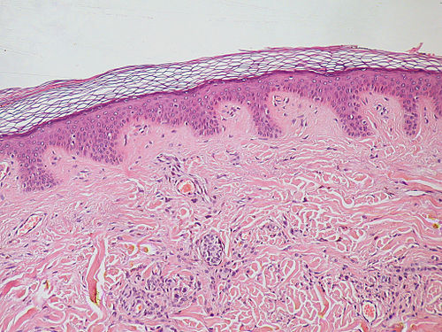
Rete pegs

Last updated
Skin epithelium (purple) with lamina propria (underlying connective tissue) (pink) -- the epithelium exhibits rete pegs. Rete pegs protect the tissue from shearing. Normal Epidermis and Dermis with Intradermal Nevus 10x.JPG
Skin epithelium (purple) with lamina propria (underlying connective tissue) (pink) -- the epithelium exhibits rete pegs. Rete pegs protect the tissue from shearing.

Rete pegs (or rete processes, or rete ridges) are the epithelial extensions that project into the underlying connective tissue in both skin and mucous membranes.

Mucous membrane the protective layer, which lines the interior of hollow organs

A mucous membrane or mucosa is a membrane that lines various cavities in the body and covers the surface of internal organs. It consists of one or more layers of epithelial cells overlying a layer of loose connective tissue. It is mostly of endodermal origin and is continuous with the skin at various body openings such as the eyes, ears, inside the nose, inside the mouth, lip, vagina, the urethral opening and the anus. Some mucous membranes secrete mucus, a thick protective fluid. The function of the membrane is to stop pathogens and dirt from entering the body and to prevent bodily tissues from becoming dehydrated.

In the epithelium of the mouth, the attached gingiva exhibit rete pegs, while the sulcular [2] and junctional epithelia do not. [3] Scar tissue lacks rete pegs and scars tend to shear off more easily than normal tissue as a result. [1]

Sulcular epithelium

The sulcular epithelium is that epithelium which lines the gingival sulcus. It is apically bounded by the junctional epithelium and meets the epithelium of the oral cavity at the height of the free gingival margin. The sulcular epithelium is nonkeratinized.

The junctional epithelium (JE) is that epithelium which lies at, and in health also defines, the base of the gingival sulcus. The probing depth of the gingival sulcus is measured by a calibrated periodontal probe. In a healthy-case scenario, the probe is gently inserted, slides by the sulcular epithelium (SE), and is stopped by the epithelial attachment (EA). However, the probing depth of the gingival sulcus may be considerably different from the true histological gingival sulcus depth.

Scar area of fibrous tissue that replaces normal skin after an injury

A scar is an area of fibrous tissue that replaces normal skin after an injury. Scars result from the biological process of wound repair in the skin, as well as in other organs and tissues of the body. Thus, scarring is a natural part of the healing process. With the exception of very minor lesions, every wound results in some degree of scarring. An exception to this are animals with complete regeneration, which regrow tissue without scar formation.

Also known as papillae, they are downward thickenings of the epidermis between the dermal papillae.

Epidermis outermost layers of the skin

The epidermis is the outermost of the three layers that make up the skin, the inner layers being the dermis and hypodermis. The epidermis layer provides a barrier to infection from environmental pathogens and regulates the amount of water released from the body into the atmosphere through transepidermal water loss. The epidermis is composed of multiple layers of flattened cells that overlie a base layer composed of columnar cells arranged perpendicularly.

Related Research Articles

Dermis layer of skin between the epidermis (with which it makes up the cutis) and subcutaneous tissues, that primarily consists of dense irregular connective tissue and cushions the body from stress and strain

The dermis or corium is a layer of skin between the epidermis and subcutaneous tissues, that primarily consists of dense irregular connective tissue and cushions the body from stress and strain. It is divided into two layers, the superficial area adjacent to the epidermis called the papillary region and a deep thicker area known as the reticular dermis. The dermis is tightly connected to the epidermis through a basement membrane. Structural components of the dermis are collagen, elastic fibers, and extrafibrillar matrix. It also contains mechanoreceptors that provide the sense of touch and thermoreceptors that provide the sense of heat. In addition, hair follicles, sweat glands, sebaceous glands, apocrine glands, lymphatic vessels, nerves and blood vessels are present in the dermis. Those blood vessels provide nourishment and waste removal for both dermal and epidermal cells.

Gums mucosal tissue that lies over the mandible and maxilla inside the mouth

The gums or gingiva, consist of the mucosal tissue that lies over the mandible and maxilla inside the mouth. Gum health and disease can have an effect on general health.

Papilla generally meaning a nipple-like structure, may refer to:

Cementoenamel junction

The cementoenamel junction, frequently abbreviated as the CEJ, is a slightly visible anatomical border identified on a tooth. It is the location where the enamel, which covers the anatomical crown of a tooth, and the cementum, which covers the anatomical root of a tooth, meet. Informally it is known as the neck of the tooth. The border created by these two dental tissues has much significance as it is usually the location where the gingiva attaches to a healthy tooth by fibers called the gingival fibers.

The oral mucosa is the mucous membrane lining the inside of the mouth and consists of stratified squamous epithelium termed oral epithelium and an underlying connective tissue termed lamina propria. The oral cavity has sometimes been described as a mirror that reflects the health of the individual. Changes indicative of disease are seen as alterations in the oral mucosa lining the mouth, which can reveal systemic conditions, such as diabetes or vitamin deficiency, or the local effects of chronic tobacco or alcohol use. The oral mucosa tends to heal faster and with less scar formation compared to the skin. The underlying mechanism remains unknown but research suggest that extracellular vesicles might be involved.

Desquamative gingivitis is an erythematous (red), desquamatous (shedding) and ulcerated appearance of the gums. It is a descriptive term and can be caused by several different disorders.

Crown lengthening

Crown lengthening is a surgical procedure performed by a dentist, or more frequently a specialist periodontist. There are a number of reasons for considering crown lengthening in a treatment plan. Commonly, the procedure is used to expose a greater amount of tooth structure for the purpose of subsequently restoring the tooth prosthetically. However, other indications include accessing subgingival caries, accessing perforations and to treat aesthetic disproportions such as a gummy smile. There are a number of procedures used to achieve an increase in crown length.

Gingival and periodontal pocket

Gingival and periodontal pockets are dental terms indicating the presence of an abnormal depth of the gingival sulcus near the point at which the gingival tissue contacts the tooth.

Gingival enlargement Human disease

Gingival enlargement is an increase in the size of the gingiva (gums). It is a common feature of gingival disease. Gingival enlargement can be caused by a number of factors, including inflammatory conditions and the side effects of certain medications. The treatment is based on the cause. A closely related term is epulis, denoting a localized tumor on the gingiva.

A mucogingival junction is an anatomical feature found on the intraoral mucosa. The mucosa of the cheeks and floor of the mouth are freely moveable and fragile, whereas the mucosa around the teeth and on the palate are firm and keratinized. Where the two tissue types meet is known as a mucogingival junction.

Stippling (dentistry)

The gingiva often possess a textured surface that is referred to as being stippled. Stippling only presents on the attached gingiva bound to underlying alveolar bone, not the freely moveable alveolar mucosa. Stippling used to be thought to indicate health, but it has since been shown that smooth gingiva is not an indication of disease, unless it is smooth due to a loss of previously existing stippling.

The gingival fibers are the connective tissue fibers that inhabit the gingival tissue adjacent to teeth and help hold the tissue firmly against the teeth. They are primarily composed of type I collagen, although type III fibers are also involved.

Gingivectomy is a dental procedure in which a dentist or oral surgeon cuts away part of the gums in the mouth.

The interdental papilla, also known as the interdental gingiva, is the part of the gums (gingiva) that exists coronal to the free gingival margin on the buccal and lingual surfaces of the teeth. The interdental papillae fill in the area between the teeth apical to their contact areas to prevent food impaction; they assume a conical shape for the anterior teeth and a blunted shape buccolingually for the posterior teeth.

Epulis is any tumor like enlargement situated on the gingival or alveolar mucosa. The word literally means "(growth) on the gingiva", and describes only the location of the mass and has no further implications on the nature of the lesion. There are three types: fibromatous, ossifying and acanthomatous. The related term parulis refers to a mass of inflamed granulation tissue at the opening of a draining sinus on the alveolus over the root of an infected tooth. Another closely related term is gingival enlargement, which tends to be used where the enlargement is more generalized over the whole gingiva rather than a localized mass.

Debridement (dental) removal of plaque and calculus

In dentistry, debridement refers to the removal by dental cleaning of accumulations of plaque and calculus (tartar) in order to maintain dental health.

Periodontal abscess

A periodontal abscess, is a localized collection of pus within the tissues of the periodontium. It is a type of dental abscess. A periodontal abscess occurs alongside a tooth, and is different from the more common periapical abscess, which represents the spread of infection from a dead tooth. To reflect this, sometimes the term "lateral (periodontal) abscess" is used. In contrast to a periapical abscess, periodontal abscesses are usually associated with a vital (living) tooth. Abscesses of the periodontium are acute bacterial infections classified primarily by location.

Necrotizing periodontal diseases are a type of inflammatory periodontal (gum) disease caused by bacteria. The diseases appear to represent different severities or stages of the same disease process, although this is not completely certain. These diseases usually have a sudden onset, and so the term acute is often added to the diagnosis. The mildest on the spectrum is necrotizing ulcerative gingivitis (NUG), followed by the successively more severe conditions necrotizing ulcerative periodontitis (NUP), necrotizing stomatitis and finally cancrum oris (noma), which is frequently fatal.

Clinical attachment loss (CAL) is the predominant clinical manifestation and determinant of periodontal disease.

References

  1. 1 2 Ira D. Papel (2011). Facial Plastic and Reconstructive Surgery (Third ed.). USA: Thieme Medical Publishers. p. 7. ISBN   9781588905154.
  2. Itoiz, ME; Carranza, FA: The Gingiva. In Newman, MG; Takei, HH; Carranza, FA; editors: Carranza’s Clinical Periodontology, 9th Edition. Philadelphia: W.B. Saunders Company, 2002. pages 23.
  3. Page, RC; Schroeder, HE. "Pathogenesis of Inflammatory Periodontal Disease: A Summary of Current Work." Lab Invest 1976;34(3):235-249