Renal fascia

Last updated
Renal fascia
Anterior and posterior renal fascia.jpg
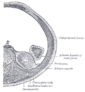
Transverse plane through the kidneys, showing anterior and posterior renal fascia
Details
System Urinary system
Identifiers
Latin fascia renalis
TA98 A08.1.01.010
TA2 3819
FMA 18104
Anatomical terminology

The renal fascia is a dense, elastic connective tissue envelope enclosing the kidney and adrenal gland, together with the layer of perirenal fat surrounding these two. [1]

Contents

The renal fascia separates the adipose capsule of kidney from the overlying pararenal fat. The deeper layers deep to the renal fascia are, in order, the adipose capsule (or perirenal fat), the renal capsule and finally the parenchyma of the renal cortex. [2] At the renal hilum, the renal capsule extends into the renal sinus. [1]

The renal fascia was originally described as consisting of two distinct structures: the anterior renal fascia (Gerota's fascia), and posterior renal fascia (Zuckerkandl's fascia); these two fasciae were said to fuse laterally to form the lateroconal fascia. Understanding of the structure of the renal fascia has subsequently evolved. [1]

Additional images

Related Research Articles

<span class="mw-page-title-main">Inguinal canal</span> Human abdominal anatomy

The inguinal canals are the two passages in the anterior abdominal wall of humans and animals which in males convey the spermatic cords and in females the round ligament of the uterus. The inguinal canals are larger and more prominent in males. There is one inguinal canal on each side of the midline.

<span class="mw-page-title-main">Retroperitoneal space</span> Anatomical space in the abdominal cavity behind the peritoneum

The retroperitoneal space (retroperitoneum) is the anatomical space behind (retro) the peritoneum. It has no specific delineating anatomical structures. Organs are retroperitoneal if they have peritoneum on their anterior side only. Structures that are not suspended by mesentery in the abdominal cavity and that lie between the parietal peritoneum and abdominal wall are classified as retroperitoneal.

<span class="mw-page-title-main">Renal calyx</span> Anatomical structure in the kidneys

The renal calyces are chambers of the kidney through which urine passes. The minor calyces surround the apex of the renal pyramids. Urine formed in the kidney passes through a renal papilla at the apex into the minor calyx; two or three minor calyces converge to form a major calyx, through which urine passes before continuing through the renal pelvis into the ureter.

<span class="mw-page-title-main">Renal capsule</span>

The renal capsule is a tough fibrous layer surrounding the kidney and covered in a layer of perirenal fat known as the adipose capsule of kidney. The adipose capsule is sometimes included in the structure of the renal capsule. It provides some protection from trauma and damage. The renal capsule is surrounded by the renal fascia. Overlying the renal fascia and between this and the transverse fascia is a region of pararenal fat.

<span class="mw-page-title-main">Renal vein</span> Short thick veins which return blood from the kidneys to the vena cava

The renal veins in the renal circulation, are large-calibre veins that drain blood filtered by the kidneys into the inferior vena cava. There is one renal vein draining each kidney.

<span class="mw-page-title-main">Superior mesenteric artery</span> Artery which supplies blood to the intestines and pancreas

In human anatomy, the superior mesenteric artery (SMA) is an artery which arises from the anterior surface of the abdominal aorta, just inferior to the origin of the celiac trunk, and supplies blood to the intestine from the lower part of the duodenum through two-thirds of the transverse colon, as well as the pancreas.

<span class="mw-page-title-main">Transverse abdominal muscle</span> Muscle of the abdominal area

The transverse abdominal muscle (TVA), also known as the transverse abdominis, transversalis muscle and transversus abdominis muscle, is a muscle layer of the anterior and lateral abdominal wall which is deep to the internal oblique muscle. It is thought by most fitness instructors to be a significant component of the core.

<span class="mw-page-title-main">Inferior mesenteric artery</span>

In human anatomy, the inferior mesenteric artery, often abbreviated as IMA, is the third main branch of the abdominal aorta and arises at the level of L3, supplying the large intestine from the distal transverse colon to the upper part of the anal canal. The regions supplied by the IMA are the descending colon, the sigmoid colon, and part of the rectum.

<span class="mw-page-title-main">External iliac artery</span> Arteries of the pelvis

The external iliac arteries are two major arteries which bifurcate off the common iliac arteries anterior to the sacroiliac joint of the pelvis.

<span class="mw-page-title-main">Lesser omentum</span>

The lesser omentum is the double layer of peritoneum that extends from the liver to the lesser curvature of the stomach, and to the first part of the duodenum. The lesser omentum is usually divided into these two connecting parts: the hepatogastric ligament, and the hepatoduodenal ligament.

<span class="mw-page-title-main">Inferior epigastric artery</span> Blood vessel

In human anatomy, the inferior epigastric artery is an artery that arises from the external iliac artery. It is accompanied by the inferior epigastric vein; inferiorly, these two inferior epigastric vessels together travel within the lateral umbilical fold The inferior epigastric artery then traverses the arcuate line of rectus sheath to enter the rectus sheath, then anastomoses with the superior epigastric artery within the rectus sheath.

<span class="mw-page-title-main">Erector spinae muscles</span> Human muscle group

The erector spinae or spinal erectors is a set of muscles that straighten and rotate the back. The spinal erectors work together with the glutes to maintain stable posture standing or sitting.

<span class="mw-page-title-main">Transversalis fascia</span> Aponeurosis between the transverse abdominal muscle and the extraperitoneal fat

The transversalis fascia is a thin aponeurotic membrane of the abdomen. It lies between the inner surface of the transverse abdominal muscle and the parietal peritoneum.

<span class="mw-page-title-main">Greater omentum</span> Fat sheath under abdominal wall

The greater omentum is a large apron-like fold of visceral peritoneum that hangs down from the stomach. It extends from the greater curvature of the stomach, passing in front of the small intestines and doubles back to ascend to the transverse colon before reaching to the posterior abdominal wall. The greater omentum is larger than the lesser omentum, which hangs down from the liver to the lesser curvature. The common anatomical term "epiploic" derives from "epiploon", from the Greek epipleein, meaning to float or sail on, since the greater omentum appears to float on the surface of the intestines. It is the first structure observed when the abdominal cavity is opened anteriorly.

<span class="mw-page-title-main">Fascia of Colles</span>

The membranous layer of the superficial fascia of the perineum is the deeper layer of the superficial perineal fascia. It is thin, aponeurotic in structure, and of considerable strength, serving to bind down the muscles of the root of the penis. Colles' fascia emerges from the perineal membrane, which divides the base of the penis from the prostate. Colles' fascia emerges from the inferior side of the perineal membrane and continues along the ventral (inferior) penis without covering the scrotum. It separates the skin and subcutaneous fat from the superficial perineal pouch.

<span class="mw-page-title-main">Fascia of Camper</span>

The fascia of Camper is a thick superficial layer of the anterior abdominal wall.

<span class="mw-page-title-main">Inferior suprarenal artery</span> Artery that supplies the adrenal gland

The inferior suprarenal artery is a paired artery that supplies the adrenal gland. It usually originates at the trunk of the renal artery before its terminal division, but with many common variations. It supplies the adrenal gland parenchyma, the ureter, and the surrounding cellular tissue and muscles.

<span class="mw-page-title-main">Medial umbilical ligament</span>

The medial umbilical ligament is a paired structure found in human anatomy. It is on the deep surface of the anterior abdominal wall, and is covered by the medial umbilical folds. It is different from the median umbilical ligament, a structure that represents the remnant of the embryonic urachus.

<span class="mw-page-title-main">Superficial perineal pouch</span>

The superficial perineal pouch is a compartment of the perineum.

<span class="mw-page-title-main">Rectus sheath</span> Laminas around abdominal muscles

The rectus sheath, also called the rectus fascia, is formed by the aponeuroses of the transverse abdominal and the internal and external oblique muscles. It contains the rectus abdominis and pyramidalis muscles.

References

PD-icon.svgThis article incorporates text in the public domain from page 1220 of the 20th edition of Gray's Anatomy (1918)

  1. 1 2 3 Standring, Susan (2020). Gray's Anatomy: The Anatomical Basis of Clinical Practice (42nd ed.). New York. pp. 1260–1261. ISBN   978-0-7020-7707-4. OCLC   1201341621.
  2. Netter FH (2014). Atlas of Human Anatomy Including Student Consult Interactive Ancillaries and Guides (6th ed.). Philadelphia, Penn.: W B Saunders Co. p. 315. ISBN   978-1-4557-0418-7.