The rib cage or thoracic cage is an endoskeletal enclosure in the thorax of most vertebrates that comprises the ribs, vertebral column and sternum, which protect the vital organs of the thoracic cavity, such as the heart, lungs and great vessels and support the shoulder girdle to form the core part of the axial skeleton.
Articles related to anatomy include:

The lumbar vertebrae are located between the thoracic vertebrae and pelvis. They form the lower part of the human back in humans, and the tail end of the back in quadrupeds. In humans, there are five lumbar vertebrae. The term is used to describe the anatomy of humans and quadrupeds, such as horses, pigs, or cattle. These bones are found in particular cuts of meat, including tenderloin or sirloin steak.

In human anatomy, the abdominal aorta is the largest artery in the abdominal cavity. As part of the aorta, it is a direct continuation of the descending aorta.

In tetrapods, cervical vertebrae are the vertebrae of the neck, immediately below the skull. Truncal vertebrae lie caudal of cervical vertebrae. In sauropsid species, the cervical vertebrae bear cervical ribs. In lizards and saurischian dinosaurs, the cervical ribs are large; in birds, they are small and completely fused to the vertebrae. The vertebral transverse processes of mammals are homologous to the cervical ribs of other amniotes. Most mammals have seven cervical vertebrae, with the only three known exceptions being the manatee with six, the two-toed sloth with five or six, and the three-toed sloth with nine.

In vertebrates, thoracic vertebrae compose the middle segment of the vertebral column, between the cervical vertebrae and the lumbar vertebrae. In humans, there are twelve thoracic vertebrae of intermediate size between the cervical and lumbar vertebrae; they increase in size going towards the lumbar vertebrae. They are distinguished by the presence of facets on the sides of the bodies for articulation with the heads of the ribs, as well as facets on the transverse processes of all, except the eleventh and twelfth, for articulation with the tubercles of the ribs. By convention, the human thoracic vertebrae are numbered T1–T12, with the first one (T1) located closest to the skull and the others going down the spine toward the lumbar region.

The mediastinum is the central compartment of the thoracic cavity. Surrounded by loose connective tissue, it is a region that contains vital organs and structures within the thorax, namely the heart and its vessels, the esophagus, the trachea, the vagus, phrenic and cardiac nerves, the thoracic duct, the thymus and the lymph nodes of the central chest.

The sagittal plane is an anatomical plane that divides the body into right and left sections. It is perpendicular to the transverse and coronal planes. The plane may be in the center of the body and divide it into two equal parts (mid-sagittal), or away from the midline and divide it into unequal parts (para-sagittal).

In anatomy, the left and right common carotid arteries (carotids) are arteries that supply the head and neck with oxygenated blood; they divide in the neck to form the external and internal carotid arteries.

The abdomen is the front part of the torso between the thorax (chest) and pelvis in humans and in other vertebrates. The area occupied by the abdomen is called the abdominal cavity. In arthropods, it is the posterior tagma of the body; it follows the thorax or cephalothorax.

A lower transverse plane midway between the upper transverse and the upper border of the pubic symphysis; this is termed the intertubercular plane, since it practically corresponds to that passing through the iliac tubercles; behind, its plane cuts the body of the fifth lumbar vertebra.
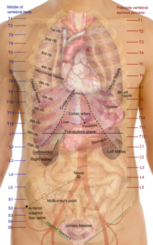
The transpyloric plane, also known as Addison's plane, is an imaginary horizontal plane, located halfway between the suprasternal notch of the manubrium and the upper border of the symphysis pubis at the level of the first lumbar vertebrae, L1. It lies roughly a hand's breadth beneath the xiphisternum or midway between the xiphisternum and the umbilicus. The plane in most cases cuts through the pylorus of the stomach, the tips of the ninth costal cartilages and the lower border of the first lumbar vertebra.

The iliolumbar ligament is a strong ligament which attaches medially to the transverse process of the 5th lumbar vertebra, and laterally to back of the inner lip of the iliac crest.
In anatomy, arterial tree is used to refer to all arteries and/or the branching pattern of the arteries. This article regards the human arterial tree. Starting from the aorta:

The following outline is provided as an overview of and topical guide to human anatomy:
Supracristal plane is an anatomical transverse plane lying at the upper most part of the pelvis, the iliac crest. This is usually at the level of the L4 vertebrae. It passes through the umbilical region and the left and right lumbar regions.
An anatomical plane is a hypothetical plane used to transect the body, in order to describe the location of structures or the direction of movements. In human and non-human anatomy, three principal planes are used:

This article describes anatomical terminology that is used to describe the central and peripheral nervous systems - including the brain, brainstem, spinal cord, and nerves.