Transpyloric plane

Last updated
Transpyloric plane
Gray1220.png
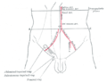
Surface lines of the front of the thorax and abdomen. (Transpyloric is top horizontal line.)
Gray1225.png
Front of abdomen, showing surface markings for duodenum, pancreas, and kidneys.
Details
Identifiers
Latin planum transpyloricum
TA98 A01.2.00.007
TA2 53
FMA 14608
Anatomical terminology

The transpyloric plane, also known as Addison's plane, is an imaginary horizontal plane, located halfway between the suprasternal notch of the manubrium and the upper border of the symphysis pubis at the level of the first lumbar vertebrae, L1. It lies roughly a hand's breadth beneath the xiphisternum [1] or midway between the xiphisternum and the umbilicus. [2] The plane in most cases cuts through the pylorus of the stomach, the tips of the ninth costal cartilages and the lower border of the first lumbar vertebra. [2]

Contents

Structures crossed

The transpyloric plane is clinically notable because it passes through several important abdominal structures. It also divides the supracolic and infracolic compartments, with the liver, spleen and gastric fundus above it and the small intestine and colon below it. [2]

Lumbar vertebra and spinal cord

The lower border of first lumbar vertebra lies at the level of the transpyloric plane. [3] Despite the conus medullaris, the end of the spinal cord, being understood to typically terminate at the level of the transpyloric plane, there is significant variability. Up to 40% of people have spinal cords ending below the transpyloric plane. [2] [4]

Stomach

The transpyloric plane passes through the pylorus of the stomach, despite it being suspended by the lesser and greater omentum and being relatively mobile. [2] [5]

Duodenum

The horizontal part of the duodenum slopes upwards to the left of the vertical midline, following which the vertical ascending part of the duodenum reaches the transpyloric plane. [6] It ends in the duodenojejunal junction, which lies approximately 2.5 cm to the left of the midline and just below the transpyloric plane. [1]

Pancreas

The neck of pancreas lies on the transpyloric plane, whilst the body and tail are to the left and above it. [3]

Gallbladder

The fundus of the gallbladder projects from the liver's inferior border at the intersection of the transpyloric plane and the right lateral midline. [7]

Kidneys

Despite the right kidney lying 1 cm lower than the left (right just below and the left just above the plane), [2] to be practical, the surface markings are taken the same way. The hilum of the kidney on the left and right is taken as 5 cm from the vertical midline and is on the transpyloric plane. [6]

Vasculature

The superior mesenteric artery arises from the aorta at the level of the transpyloric plane and emerges between the head and neck of the pancreas. [8]

The superior mesenteric vein is joined by the splenic vein to form the portal vein at the level of the transpyloric plane. [2] [8]

Spleen

The lower border of the spleen lies near the transpyloric plane. [8]

Other structures

History

The transpyloric plane relates to the three-dimensional mapping of the abdomen founded on more than 10,000 measurements completed on 40 bodies, that surgeon Viscount Addison took at the turn of the 20th century. [9]

Addison reported his findings in a paper titled, "On the anatomical topography of the abdominal viscera in man, especially the gastro-intestinal canal" in which he established a baseline for the anatomy of the abdomen based on the arrangement of the map of the Earth. Using the suprasternal notch as the North Pole of the trunk, and the upper border of the pubic symphysis as the South Pole, he drew a vertical line joining these two points as his meridian. At the meridian's midpoint, he then drew a perpendicular line corresponding to the Equator. As this transverse plane crossed the pylorus, he called it the transpyloric plane. [10]

Images

See also

Related Research Articles

<span class="mw-page-title-main">Duodenum</span> First section of the small intestine

The duodenum is the first section of the small intestine in most higher vertebrates, including mammals, reptiles, and birds. In mammals it may be the principal site for iron absorption. The duodenum precedes the jejunum and ileum and is the shortest part of the small intestine.

<span class="mw-page-title-main">Small intestine</span> Organ in the gastrointestinal tract

The small intestine or small bowel is an organ in the gastrointestinal tract where most of the absorption of nutrients from food takes place. It lies between the stomach and large intestine, and receives bile and pancreatic juice through the pancreatic duct to aid in digestion. The small intestine is about 5.5 metres long and folds many times to fit in the abdomen. Although it is longer than the large intestine, it is called the small intestine because it is narrower in diameter.

<span class="mw-page-title-main">Inferior vena cava</span> One of two veinous trunks bringing deoxygenated blood back to the heart

The inferior vena cava is a large vein that carries the deoxygenated blood from the lower and middle body into the right atrium of the heart. It is formed by the joining of the right and the left common iliac veins, usually at the level of the fifth lumbar vertebra.

<span class="mw-page-title-main">Mesentery</span> Contiguous fold of tissues that supports the intestines

The mesentery is an organ that attaches the intestines to the posterior abdominal wall and is formed by the double fold of peritoneum. It helps in storing fat and allowing blood vessels, lymphatics, and nerves to supply the intestines, among other functions.

<span class="mw-page-title-main">Abdominal aorta</span> Largest artery in the abdomen

In human anatomy, the abdominal aorta is the largest artery in the abdominal cavity. As part of the aorta, it is a direct continuation of the descending aorta.

<span class="mw-page-title-main">Epigastrium</span> Upper central region of the abdomen

In anatomy, the epigastrium is the upper central region of the abdomen. It is located between the costal margins and the subcostal plane. Pain may be referred to the epigastrium from damage to structures derived from the foregut.

<span class="mw-page-title-main">Celiac artery</span> First major branch of the abdominal aorta

The celiacartery, also known as the celiac trunk or truncus coeliacus, is the first major branch of the abdominal aorta. It is about 1.25 cm in length. Branching from the aorta at thoracic vertebra 12 (T12) in humans, it is one of three anterior/ midline branches of the abdominal aorta.

<span class="mw-page-title-main">Superior mesenteric artery</span> Artery which supplies blood to the intestines and pancreas

In human anatomy, the superior mesenteric artery (SMA) is an artery which arises from the anterior surface of the abdominal aorta, just inferior to the origin of the celiac trunk, and supplies blood to the intestine from the lower part of the duodenum through two-thirds of the transverse colon, as well as the pancreas.

<span class="mw-page-title-main">Inferior mesenteric artery</span>

In human anatomy, the inferior mesenteric artery (IMA) is the third main branch of the abdominal aorta and arises at the level of L3, supplying the large intestine from the distal transverse colon to the upper part of the anal canal. The regions supplied by the IMA are the descending colon, the sigmoid colon, and part of the rectum.

<span class="mw-page-title-main">Gastroduodenal artery</span>

In anatomy, the gastroduodenal artery is a small blood vessel in the abdomen. It supplies blood directly to the pylorus and proximal part of the duodenum. It also indirectly supplies the pancreatic head.

<span class="mw-page-title-main">Ascending colon</span> Section of the large intestine

In the anatomy of humans and homologous primates, the ascending colon is the part of the colon located between the cecum and the transverse colon.

<span class="mw-page-title-main">Abdomen</span> Part of the body between the chest and pelvis

The abdomen is the part of the body between the thorax (chest) and pelvis, in humans and in other vertebrates. The abdomen is the front part of the abdominal segment of the torso. The area occupied by the abdomen is called the abdominal cavity. In arthropods, it is the posterior tagma of the body; it follows the thorax or cephalothorax.

<span class="mw-page-title-main">Greater omentum</span> Fat sheath under abdominal wall

The greater omentum is a large apron-like fold of visceral peritoneum that hangs down from the stomach. It extends from the greater curvature of the stomach, passing in front of the small intestines and doubles back to ascend to the transverse colon before reaching to the posterior abdominal wall. The greater omentum is larger than the lesser omentum, which hangs down from the liver to the lesser curvature. The common anatomical term "epiploic" derives from "epiploon", from the Greek epipleein, meaning to float or sail on, since the greater omentum appears to float on the surface of the intestines. It is the first structure observed when the abdominal cavity is opened anteriorly.

<span class="mw-page-title-main">Duodenojejunal flexure</span> Border between the duodenum and the jejunum

The duodenojejunal flexure or duodenojejunal junction, also known as the angle of Treitz, is the border between the duodenum and the jejunum.

<span class="mw-page-title-main">Intertubercular plane</span>

A lower transverse plane midway between the upper transverse and the upper border of the pubic symphysis; this is termed the intertubercular plane, since it practically corresponds to that passing through the iliac tubercles; behind, its plane cuts the body of the fifth lumbar vertebra.

<span class="mw-page-title-main">Hepatic plexus</span>

The hepatic plexus is a sympathetic and parasympathetic nerve plexus that provides innervation to the parenchyma of the liver as well as contributing innervation to some other abdominal structures.

<span class="mw-page-title-main">Umbilical region</span>

The umbilical region is one of the nine regions of the abdomen. It is the region that surrounds the area around the umbilicus and is placed approximately half way between the xiphoid process and the pubic symphysis. This region of the abdomen contains part of the stomach, the head of the pancreas, the duodenum, a section of the transverse colon and the lower aspects of the left and right kidney. The upper three regions, from left to right, are the left hypochondriac, epigastric, and right hypochondriac regions. The middle three regions, from left to right, are the left lumbar, umbilical, and right lumbar regions. The bottom three regions, from left to right, are the left inguinal, hypogastric, and right inguinal regions.

<span class="mw-page-title-main">Omental foramen</span> Part of the human abdomen

In human anatomy, the omental foramen is the passage of communication, or foramen, between the greater sac, and the lesser sac.

<span class="mw-page-title-main">Anatomical terminology</span> Scientific terminology used by anatomists, zoologists, and health professionals

Anatomical terminology is a form of scientific terminology used by anatomists, zoologists, and health professionals such as doctors, physicians, and pharmacists.

<span class="mw-page-title-main">Quadrants and regions of abdomen</span> Anatomical subdivision scheme

The human abdomen is divided into quadrants and regions by anatomists and physicians for the purposes of study, diagnosis, and treatment. The division into four quadrants allows the localisation of pain and tenderness, scars, lumps, and other items of interest, narrowing in on which organs and tissues may be involved. The quadrants are referred to as the left lower quadrant, left upper quadrant, right upper quadrant and right lower quadrant. These terms are not used in comparative anatomy, since most other animals do not stand erect.

References

  1. 1 2 Singh, Vishram. (2014). Textbook of Anatomy Abdomen and Lower Limb Volume 2 (2nd ed.). Elsevier. p. 26. ISBN   978-81-312-3626-0.
  2. 1 2 3 4 5 6 7 8 9 Sinnatamby, Chummy S. (2011). "5". Last's Anatomy e-Book: Regional and Applied (12th ed.). Churchill Livingstone Elsevier. ISBN   978-0-7020-4839-5 . Retrieved 16 March 2018.
  3. 1 2 Snell, Richard S. (2012). Clinical Anatomy by Regions (9th ed.). Lippincott Williams & Wilkins, Wolters Kluwer. ISBN   978-1-60913-446-4.
  4. Elliott L. Mancall; David G. Brock (2011). Gray's Clinical Neuroanatomy E-Book. Elsevier Health Sciences. p. 111. ISBN   978-1-4377-3580-2.
  5. Drake, Richard L.; Wayne, Vogl A.; Mitchell, Adam W.M. (2015). Gray's Anatomy for Students (3rd ed.) Philadelphia: Churchill Livingstone Elsevier. p. 311.
  6. 1 2 Gray, Henry; Lewis, Warren Harmon (1918). Anatomy of the Human Body (20th ed.). Philadelphia: Lea & Febiger. pp. 1320–1321.
  7. Constantine C. Karaliotas; Christoph E. Broelsch; Nagy A. Habib (Eds.) (2008). Liver and Biliary Tract Surgery: Embryological Anatomy to 3D-Imaging and Transplant Innovations. Springer Wien New York. pp. 14–15. ISBN   978-3-211-49277-2.
  8. 1 2 3 4 Herbert H, Srebnik (2002). Concepts in Anatomy. USA: Kluwer Academic Publishers. pp. 174–175. ISBN   978-0-7923-7539-5 . Retrieved 16 March 2018.
  9. Addison, Sir Christopher, Viscount Addison of Stallingborough (1869 - 1951). Royal College of Surgeons: Plarr's Lives of the Fellows Online. Retrieved 16 March 2018.
  10. Keith, Arthur (1 March 1952). "Lord Addison as Anatomist". British Journal of Surgery . 39 (157): 385–387. doi:10.1002/bjs.18003915702. ISSN   1365-2168. PMID   14925320.(subscription required)

PD-icon.svgThis article incorporates text in the public domain from page 1315 of the 20th edition of Gray's Anatomy (1918)