External links
| | This anatomy article is a stub. You can help Wikipedia by expanding it. |
| Anterior median line | |
|---|---|
| Details | |
| Identifiers | |
| Latin | linea mediana anterior |
| TA98 | A01.2.00.012 |
| TA2 | 60 |
| FMA | 20310 |
| Anatomical terminology | |
The anterior median line is a sagittal line on the anterior of the head and torso running at midline.
| | This anatomy article is a stub. You can help Wikipedia by expanding it. |

In anatomy, the atlas (C1) is the most superior (first) cervical vertebra of the spine and is located in the neck. It is named for Atlas of Greek mythology because, just as Atlas supported the globe, it supports the entire head.

The neck is the part of the body on many vertebrates that connects the head with the torso and provides the mobility and movements of the head. The structures of the human neck are anatomically grouped into four compartments; vertebral, visceral and two vascular compartments. Within these compartments, the neck houses the cervical vertebrae and cervical part of the spinal cord, upper parts of the respiratory and digestive tracts, endocrine glands, nerves, arteries and veins. Muscles of the neck are described separately from the compartments. They bound the neck triangles.

The humerus is a long bone in the arm that runs from the shoulder to the elbow. It connects the scapula and the two bones of the lower arm, the radius and ulna, and consists of three sections. The humeral upper extremity consists of a rounded head, a narrow neck, and two short processes. The body is cylindrical in its upper portion, and more prismatic below. The lower extremity consists of 2 epicondyles, 2 processes, and 3 fossae. As well as its true anatomical neck, the constriction below the greater and lesser tubercles of the humerus is referred to as its surgical neck due to its tendency to fracture, thus often becoming the focus of surgeons.

The tibia, also known as the shinbone or shankbone, is the larger, stronger, and anterior (frontal) of the two bones in the leg below the knee in vertebrates, and it connects the knee with the ankle bones. The tibia is found on the medial side of the leg next to the fibula and closer to the median plane or centre-line. The tibia is connected to the fibula by the interosseous membrane of the leg, forming a type of fibrous joint called a syndesmosis with very little movement. The tibia is named for the flute tibia. It is the second largest bone in the human body next to the femur. The leg bones are the strongest long bones as they support the rest of the body.

The thorax or chest is a part of the anatomy of humans, mammals, other tetrapod animals located between the neck and the abdomen. In insects, crustaceans, and the extinct trilobites, the thorax is one of the three main divisions of the creature's body, each of which is in turn composed of multiple segments.
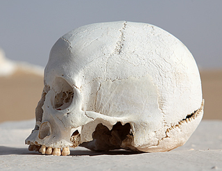
The occipital bone is a cranial dermal bone and the main bone of the occiput. It is trapezoidal in shape and curved on itself like a shallow dish. The occipital bone overlies the occipital lobes of the cerebrum. At the base of skull in the occipital bone, there is a large oval opening called the foramen magnum, which allows the passage of the spinal cord.

The gluteus medius, one of the three gluteal muscles, is a broad, thick, radiating muscle. It is situated on the outer surface of the pelvis.

The digastric muscle is a small muscle located under the jaw. The term "digastric muscle" refers to this specific muscle. However, other muscles that have two separate muscle bellies include the suspensory muscle of duodenum, omohyoid, occipitofrontalis.

The pectineus muscle is a flat, quadrangular muscle, situated at the anterior (front) part of the upper and medial (inner) aspect of the thigh. The pectineus muscle is the most anterior adductor of the hip. The muscle does adduct and internally rotate the thigh but its primary function is hip flexion.

In vertebrate anatomy, hip refers to either an anatomical region or a joint.

The abdominal external oblique muscle is the largest and outermost of the three flat abdominal muscles of the lateral anterior abdomen.

The anterior chamber (AC) is the aqueous humor-filled space inside the eye between the iris and the cornea's innermost surface, the endothelium. Hyphema, anterior uveitis and glaucoma are three main pathologies in this area. In hyphema, blood fills the anterior chamber as a result of a hemorrhage, most commonly after a blunt eye injury. Anterior uveitis is an inflammatory process affecting the iris and ciliary body, with resulting inflammatory signs in the anterior chamber. In glaucoma, blockage of the trabecular meshwork prevents the normal outflow of aqueous humour, resulting in increased intraocular pressure, progressive damage to the optic nerve head, and eventually blindness.

The squamous part of temporal bone, or temporal squama, forms the front and upper part of the temporal bone, and is scale-like, thin, and translucent.

The anterior cranial fossa is a depression in the floor of the cranial base which houses the projecting frontal lobes of the brain. It is formed by the orbital plates of the frontal, the cribriform plate of the ethmoid, and the small wings and front part of the body of the sphenoid; it is limited behind by the posterior borders of the small wings of the sphenoid and by the anterior margin of the chiasmatic groove. The lesser wings of the sphenoid separate the anterior and middle fossae.

The wing of ilium is the large expanded portion which bounds the greater pelvis laterally. It presents for examination two surfaces—an external and an internal—a crest, and two borders—an anterior and a posterior.

The arcuate line of rectus sheath, the linea semicircularis, the arcuate line, or the semicircular line of Douglas, is a horizontal line that demarcates the lower limit of the posterior layer of the rectus sheath. It is commonly known simply as the arcuate line. It is also where the inferior epigastric vessels perforate the rectus abdominis.

The axillary lines are the anterior axillary line, midaxillary line and the posterior axillary line.
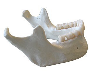
In anatomy, the mandible, lower jaw or jawbone is the largest, strongest and lowest bone in the human facial skeleton. It forms the lower jaw and holds the lower teeth in place. The mandible sits beneath the maxilla. It is the only movable bone of the skull. It is connected to the temporal bone by the temporomandibular joint.

The pelvis is either the lower part of the trunk of the human body between the abdomen and the thighs or the skeleton embedded in it.
The gluteal lines are three curved lines outlined from three bony ridges on the exterior surface of the ilium in the gluteal region. They are the anterior gluteal line; the inferior gluteal line, and the posterior gluteal line.