Intertubercular plane

Last updated
Intertubercular plane
Gray1220.png
Surface lines of the front of the thorax and abdomen. (Transtubercular is bottom horizontal line.)
Gray1225.png
Front of abdomen, showing surface markings for duodenum, pancreas, and kidneys.
Details
Identifiers
Latin planum intertuberculare
TA98 A01.2.00.010
TA2 57
FMA 74566
Anatomical terminology

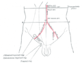
A lower transverse plane midway between the upper transverse and the upper border of the pubic symphysis; this is termed the intertubercular plane (or transtubercular), since it practically corresponds to that passing through the iliac tubercles; behind, its plane cuts the body of the fifth lumbar vertebra.

Contents

Additional images

See also

Related Research Articles

<span class="mw-page-title-main">Rib cage</span> Bone structure of the thorax

The rib cage or thoracic cage is an endoskeletal enclosure in the thorax of most vertebrates that comprises the ribs, vertebral column and sternum, which protect the vital organs of the thoracic cavity, such as the heart, lungs and great vessels and support the shoulder girdle to form the core part of the axial skeleton.

<span class="mw-page-title-main">Clavicle</span> Long bone that serves as a strut between the scapula and the sternum

The clavicle, collarbone, or keybone is a slender, S-shaped long bone approximately 6 inches (15 cm) long that serves as a strut between the shoulder blade and the sternum (breastbone). There are two clavicles, one on each side of the body. The clavicle is the only long bone in the body that lies horizontally. Together with the shoulder blade, it makes up the shoulder girdle. It is a palpable bone and, in people who have less fat in this region, the location of the bone is clearly visible. It receives its name from Latin clavicula 'little key' because the bone rotates along its axis like a key when the shoulder is abducted. The clavicle is the most commonly fractured bone. It can easily be fractured by impacts to the shoulder from the force of falling on outstretched arms or by a direct hit.

<span class="mw-page-title-main">Sacrum</span> Bone of the spine

The sacrum, in human anatomy, is a large, triangular bone at the base of the spine that forms by the fusing of the sacral vertebrae (S1–S5) between ages 18 and 30.

<span class="mw-page-title-main">Tibia</span> Leg bone in vertebrates

The tibia, also known as the shinbone or shankbone, is the larger, stronger, and anterior (frontal) of the two bones in the leg below the knee in vertebrates ; it connects the knee with the ankle. The tibia is found on the medial side of the leg next to the fibula and closer to the median plane. The tibia is connected to the fibula by the interosseous membrane of leg, forming a type of fibrous joint called a syndesmosis with very little movement. The tibia is named for the flute tibia. It is the second largest bone in the human body, after the femur. The leg bones are the strongest long bones as they support the rest of the body.

<span class="mw-page-title-main">Lumbar vertebrae</span> Five bones of the spine in the lower back

The lumbar vertebrae are located between the thoracic vertebrae and pelvis. They form the lower part of the back in humans, and the tail end of the back in quadrupeds. In humans, there are five lumbar vertebrae. The term is used to describe the anatomy of humans and quadrupeds, such as horses, pigs, or cattle. These bones are found in particular cuts of meat, including tenderloin or sirloin steak.

<span class="mw-page-title-main">Occipital bone</span> Bone of the neurocranium

The occipital bone is a cranial dermal bone and the main bone of the occiput. It is trapezoidal in shape and curved on itself like a shallow dish. The occipital bone overlies the occipital lobes of the cerebrum. At the base of the skull in the occipital bone, there is a large oval opening called the foramen magnum, which allows the passage of the spinal cord.

<span class="mw-page-title-main">Thoracic vertebrae</span> Vertebrae between the cervical vertebrae and the lumbar vertebrae

In vertebrates, thoracic vertebrae compose the middle segment of the vertebral column, between the cervical vertebrae and the lumbar vertebrae. In humans, there are twelve thoracic vertebrae of intermediate size between the cervical and lumbar vertebrae; they increase in size going towards the lumbar vertebrae. They are distinguished by the presence of facets on the sides of the bodies for articulation with the heads of the ribs, as well as facets on the transverse processes of all, except the eleventh and twelfth, for articulation with the tubercles of the ribs. By convention, the human thoracic vertebrae are numbered T1–T12, with the first one (T1) located closest to the skull and the others going down the spine toward the lumbar region.

<span class="mw-page-title-main">Mediastinum</span> Central part of the thoracic cavity

The mediastinum is the central compartment of the thoracic cavity. Surrounded by loose connective tissue, it is a region that contains vital organs and structures within the thorax, namely the heart and its vessels, the esophagus, the trachea, the vagus, phrenic and cardiac nerves, the thoracic duct, the thymus and the lymph nodes of the central chest.

<span class="mw-page-title-main">Superior mesenteric artery</span> Artery which supplies blood to the intestines and pancreas

In human anatomy, the superior mesenteric artery (SMA) is an artery which arises from the anterior surface of the abdominal aorta, just inferior to the origin of the celiac trunk, and supplies blood to the intestine from the lower part of the duodenum through two-thirds of the transverse colon, as well as the pancreas.

<span class="mw-page-title-main">Sagittal plane</span> Anatomical plane dividing the body into left and right

The sagittal plane is an anatomical plane that divides the body into right and left sections. It is perpendicular to the transverse and coronal planes. The plane may be in the center of the body and divide it into two equal parts (mid-sagittal), or away from the midline and divide it into unequal parts (para-sagittal).

<span class="mw-page-title-main">Abdomen</span> Part of the body between the chest and pelvis

The abdomen is the front part of the torso between the thorax (chest) and pelvis in humans and in other vertebrates. The area occupied by the abdomen is called the abdominal cavity. In arthropods, it is the posterior tagma of the body; it follows the thorax or cephalothorax.

<span class="mw-page-title-main">Hypochondrium</span> Upper left and right sections of the abdomen

In anatomy, the division of the abdomen into regions can employ a nine-region scheme. The hypochondrium refers to the two hypochondriac regions in the upper third of the abdomen; the left hypochondrium and right hypochondrium. They are located on the lateral sides of the abdominal wall respectively, inferior to (below) the thoracic cage, being separated by the epigastrium.

<span class="mw-page-title-main">Duodenojejunal flexure</span> Border between the duodenum and the jejunum

The duodenojejunal flexure or duodenojejunal junction, also known as the angle of Treitz, is the border between the duodenum and the jejunum.

<span class="mw-page-title-main">Transpyloric plane</span> Anatomical plane

The transpyloric plane, also known as Addison's plane, is an imaginary horizontal plane, located halfway between the suprasternal notch of the manubrium and the upper border of the symphysis pubis at the level of the first lumbar vertebrae, L1. It lies roughly a hand's breadth beneath the xiphisternum or midway between the xiphisternum and the umbilicus. The plane in most cases cuts through the pylorus of the stomach, the tips of the ninth costal cartilages and the lower border of the first lumbar vertebra.

The terminal sulcus is a groove on the outer surface of the right atrium of the heart marking the transition between the sinus venarum cavarum and the rest of the right atrium. The terminal sulcus corresponds to the position of the terminal crest on the inner surface of the right atium. The terminal sulcus indicate the position of the sinoatrial node.

The antebrachial fascia continuous above with the brachial fascia, is a dense, membranous investment, which forms a general sheath for the muscles in this region; it is attached, behind, to the olecranon and dorsal border of the ulna, and gives off from its deep surface numerous intermuscular septa, which enclose each muscle separately.

<span class="mw-page-title-main">Transverse perineal muscles</span> Muscles in the perineum

The transverse perineal muscles are the superficial and the deep transverse perineal muscles.

<span class="mw-page-title-main">Anatomical plane</span> Anatomy method to describe locations

An anatomical plane is a hypothetical plane used to transect the body, in order to describe the location of structures or the direction of movements. In human and non-human anatomy, three principal planes are used:

<span class="mw-page-title-main">Spinal column</span> Bony structure found in vertebrates

The spinal column, also known as the vertebral column, spine or backbone, is the core part of the axial skeleton in vertebrates. The vertebral column is the defining and eponymous characteristic of the vertebrate. The spinal column is a segmented column of vertebrae that surrounds and protects the spinal cord. The vertebrae are separated by intervertebral discs in a series of cartilaginous joints. The dorsal portion of the spinal column houses the spinal canal, an elongated cavity formed by the alignment of the vertebral neural arches that encloses and protects the spinal cord, with spinal nerves exiting via the intervertebral foramina to innervate each body segment.

<span class="mw-page-title-main">Anatomical terminology</span> Scientific terminology used by anatomists, zoologists, and health professionals

Anatomical terminology is a specialized system of terms used by anatomists, zoologists, and health professionals, such as doctors, surgeons, and pharmacists, to describe the structures and functions of the body.

References

    PD-icon.svgThis article incorporates text in the public domain from page 1315 of the 20th edition of Gray's Anatomy (1918)