The labia majora are two prominent longitudinal cutaneous folds that extend downward and backward from the mons pubis to the perineum. Together with the labia minora they form the labia of the vulva.

Standard anatomical terms of location are used to unambiguously describe anatomical locations on or within limbs, organs, and other bodily structures. The terms, typically derived from Latin or Greek roots, refer to a body part in its standard anatomical position. Since the meanings of terms can change depending on whether a body is bipedal or quadrupedal, international organisations have formulated standard vocabularies for different disciplines, such as the Terminologia Anatomica for physicians and the Nomina Anatomica Veterinaria for veterinarians.

The femur, or thigh bone, is the proximal bone of the hindlimb in tetrapod vertebrates. The head of the femur articulates with the acetabulum in the pelvic bone forming the hip joint, while the distal part of the femur articulates with the tibia (shinbone) and patella (kneecap), forming the knee joint. By most measures the two femurs are the strongest bones of the body, and in humans, the largest and thickest.

The humerus is a long bone in the arm that runs from the shoulder to the elbow. It connects the scapula and the two bones of the lower arm, the radius and ulna, and consists of three sections. The humeral upper extremity consists of a rounded head, a narrow neck, and two short processes. The body is cylindrical in its upper portion, and more prismatic below. The lower extremity consists of 2 epicondyles, 2 processes, and 3 fossae. As well as its true anatomical neck, the constriction below the greater and lesser tubercles of the humerus is referred to as its surgical neck due to its tendency to fracture, thus often becoming the focus of surgeons.

The sacrum, in human anatomy, is a large, triangular bone at the base of the spine that forms by the fusing of sacral vertebrae S1–S5 between 18 and 30 years of age.

The tibia, also known as the shinbone or shankbone, is the larger, stronger, and anterior (frontal) of the two bones in the leg below the knee in vertebrates, and it connects the knee with the ankle bones. The tibia is found on the medial side of the leg next to the fibula and closer to the median plane or centre-line. The tibia is connected to the fibula by the interosseous membrane of the leg, forming a type of fibrous joint called a syndesmosis with very little movement. The tibia is named for the flute tibia. It is the second largest bone in the human body next to the femur. The leg bones are the strongest long bones as they support the rest of the body.

The occipital bone is a cranial dermal bone and the main bone of the occiput. It is trapezoidal in shape and curved on itself like a shallow dish. The occipital bone overlies the occipital lobes of the cerebrum. At the base of skull in the occipital bone, there is a large oval opening called the foramen magnum, which allows the passage of the spinal cord.

The digastric muscle is a small muscle located under the jaw. The term "digastric muscle" refers to this specific muscle. However, other muscles that have two separate muscle bellies include the suspensory muscle of duodenum, omohyoid, occipitofrontalis.

In anatomy, the sagittal plane, or longitudinal plane, is an anatomical plane which divides the body into right and left parts. The plane may be in the center of the body and split it into two halves (mid-sagittal) or away from the midline and split it into unequal parts (para-sagittal).The anatomical term sagittal was coined by Gerard of Cremona.
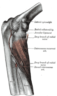
In human anatomy, the supinator is a broad muscle in the posterior compartment of the forearm, curved around the upper third of the radius. Its function is to supinate the forearm.

The squamous part of occipital bone, is situated above and behind the foramen magnum, and is curved from above downward and from side to side.

This glossary of entomology describes terms used in the formal study of insect species by entomologists.
Fish measurement is the measuring of the length of individual fish and of various parts of their anatomy. These data are used in many areas of ichthyology, including taxonomy and fisheries biology.
This is a list of definitions of commonly used terms of location and direction in dentistry. This set of terms provides orientation within the oral cavity, much as anatomical terms of location provide orientation throughout the body.
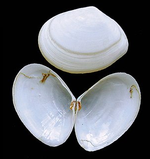
A bivalve shell is part of the body, the exoskeleton or shell, of a bivalve mollusk. In life, the shell of this class of mollusks is composed of two hinged parts or valves. Bivalves are very common in essentially all aquatic locales, including saltwater, brackish water, and freshwater. The shells of bivalves commonly wash up on beaches and along the edges of lakes, rivers, and streams. Bivalves by definition possess two shells or valves, a "right valve" and a "left valve", that are joined by a ligament. The two valves usually articulate with one another using structures known as "teeth" which are situated along the hinge line. In many bivalve shells, the two valves are symmetrical along the hinge line—when truly symmetrical, such an animal is said to be equivalved; if the valves vary from each other in size or shape, inequivalved. If symmetrical front-to-back, the valves are said to be equilateral, and are otherwise considered inequilateral.

A cephalic presentation or head presentation or head-first presentation is a situation at childbirth where the fetus is in a longitudinal lie and the head enters the pelvis first; the most common form of cephalic presentation is the vertex presentation, where the occiput is the leading part. All other presentations are abnormal (malpresentations) and are either more difficult to deliver or not deliverable by natural means.
In dentistry, overeruption is the physiological movement of a tooth lacking an opposing partner in the dental occlusion. Because of the lack of opposing force and the natural eruptive potential of the tooth there is a tendency for the tooth to erupt out of the line of the occlusion.
An anatomical plane is a hypothetical plane used to transect the body, in order to describe the location of structures or the direction of movements. In human and animal anatomy, three principal planes are used:

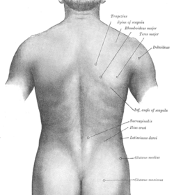
The vertebral column, also known as the backbone or spine, is part of the axial skeleton. The vertebral column is the defining characteristic of a vertebrate in which the notochord found in all chordates has been replaced by a segmented series of bone: vertebrae separated by intervertebral discs. The vertebral column houses the spinal canal, a cavity that encloses and protects the spinal cord.

The human abdomen is divided into quadrants and regions by anatomists and physicians for the purposes of study, diagnosis, and treatment. The division into four quadrants allows the localisation of pain and tenderness, scars, lumps, and other items of interest, narrowing in on which organs and tissues may be involved. The quadrants are referred to as the left lower quadrant, left upper quadrant, right upper quadrant and right lower quadrant. These terms are not used in comparative anatomy, since most other animals do not stand erect.